
Abstract
Chickens were infected under experimental conditions with Mycoplasma gallisepticum and low pathogenic avian influenza (LPAI) strain A/mallard/Hungary/19616/07 (H3N8). Two groups of chickens were aerosol challenged with M. gallisepticum strain 1226. Seven days later, one of these groups and one mycoplasma-free group was challenged with LPAI H3N8 virus; one group without challenge remained as negative control. Eight days later, the birds were euthanized and examined for gross pathologic and histologic lesions. The body weight was measured, and the presence of antimycoplasma and antiviral antibodies was tested before the mycoplasma challenge, before the virus challenge, and at the end of the study to confirm both infections. Chickens in the mycoplasma-infected group developed antibodies against M. gallisepticum but not against the influenza virus. Chickens of the group infected with the influenza virus became serologically positive only against the virus, while the birds in the coinfected group developed antibodies against both agents. The LPAI H3N8 virus strain did not cause decrease in body weight and clinical signs, and macroscopic pathological lesions were not present in the chickens. The M. gallisepticum infection caused respiratory signs, airsacculitis, and peritonitis characteristic of mycoplasma infection. However, the clinical signs and pathologic lesions and the reduction in weight gain were much more significant in the group challenged with both M. gallisepticum and LPAI H3N8 virus than in the group challenged with M. gallisepticum alone.
Considerable information on the epidemiology of low pathogenicity avian influenza (LPAI) and high pathogenicity avian influenza (HPAI) viruses in poultry and wild birds has been published in recent years as a result of influenza surveillance schemes throughout the world. 5 HPAI viruses have been rarely isolated from wild bird populations, while LPAI viruses have been frequently isolated. Only viruses of the H5 and H7 subtypes have been shown to cause HPAI in susceptible species, but not all subtype H5 and H7 avian influenza viruses are virulent.5,42,51 Numerous influenza viral subtypes have been identified as circulating among migrating birds, particularly in water fowl, and can thereby act as a significant wildlife reservoir for the disease.18,31,32,53,55,65
Typically, LPAI virus causes an asymptomatic infection in wild birds. However, several recent publications have shown that LPAI strains can have a measurable effect on bird physiology in certain species. Examples of this include reports of the LPAI virus multiplying in the respiratory tract of the developing mallard embryo, 44 while infection of mallards has a negative effect on total body mass, predominantly in younger birds. There was also a negative correlation on staging periods (stopover duration) of birds during autumn migration in association with LPAI. 31 Furthermore, other publications describe LPAI as potentially causing a delayed spring migration, 67 combined with a slight and transient increase of body temperature 19 and a decrease of egg production. 2
LPAI subtypes H13N2, H9N8, H6N2, and H9N2 have been reported to cause clinical signs such as moderate respiratory distress, depression, mucopurulent tracheitis, and airsacculitis in immunocompetent chickens,17,26,33 while in immunocompromised chickens, H9N2 has been shown to result in clinical disease with a decrease of body weights and a pathogenicity index value of 1.22, including severe atrophy of follicles with fibrosis in the bursa fabricius, diffuse atrophy of the cortex in the thymus, severe tubular necrosis in the kidneys, and even 25% mortality on day 7 postinfection. 25 Since conditions in which the immune system is negatively affected may occur at any time in commercial poultry flocks due to preexisting infections such as Mycoplasma gallisepticum, we decided to study the effect of a LPAI virus on chickens previously challenged with M. gallisepticum. For the purpose of this study, we selected the LPAI H3N8 virus, which has been isolated from mallards in Hungary. 66
Materials and Methods
Experimental Design
Forty 1-day-old chicks of Ross 308 breed, originating from Mycoplasma synoviae– and M. gallisepticum–free parent stock, were maintained in the animal facility of the Central Veterinary Institute and kept in isolation. The animal study protocol was approved by the Animal Research and Care Committee of the Central Veterinary Institute, and animal care and experimentation were carried out in accordance with institutional and national guidelines.
The birds were divided into 4 equal groups designated as K, V, M, and M+V. At 15 days of age, the K and V groups were treated with an aerosol spray of sterile broth (10 ml). M and M+V groups were challenged with M. gallisepticum consisting of a challenge aerosol containing 10 ml of broth culture of the strain 1226 10 (titer: 2.1 × 10⁷ colony-forming units per ml). Subsequent to manipulation, each group was placed in an isolation unit. The feeding and cleaning of the units were performed by different caretakers to avoid cross contamination; however, the management procedures for all groups were the same. The birds were fed with a standard feed containing an anticoccidial agent but no antibacterial agents.
Challenge With Influenza Virus
At 22 days of age, groups V and M+V were challenged with influenza virus strain A/mallard/Hungary/19616/07 (H3N8). 66 Ten milliliters of virus suspension, propagated in the allantois of chicken embryos having a titer of 1 × 10⁵ median chicken embryo infectious dose (EID50), were sprayed in the challenge chamber for 20 minutes. The groups were then placed in their isolation units and observed for clinical signs of disease for an additional 8 days.
Before challenge and at the end of the study, sera of chickens were tested for presence of specific antibodies to M. gallisepticum, 8 using the MyGa blocking ELISA test kit (Diagnosztikum Rt, Budapest, Hungary), as well as for influenza virus, using hemagglutination inhibition employing 4 hemagglutination units. At 8 days postviral challenge, all birds were euthanized and examined for gross pathologic and histologic lesions as well as presence of M. gallisepticum in various organs (trachea, lung, air sacs, heart, kidney, spleen, and liver). Mycoplasma culture was confirmed by species-specific polymerase chain reaction (PCR) 3 as well as for presence of antibodies against M. gallisepticum in sera. Lung lavage and lung samples were also collected and tested for presence of LPAI H3N8 virus by isolation and PCR.
Pathologic Examination
At the end of the study, all birds were euthanized and examined for presence of pathologic lesions. The lesions of trachea were scored according to the following: 0, no redness of mucosal membrane and no mucus; 1, reddish mucosal membrane, congestion, and small amount of mucus; 2, intensive redness, congestion of mucosal membrane, and significant amount of mucus. The sum of the scores of the trachea’s lesions of each bird in one group was used for statistical comparison of the severity of tracheal lesions between the groups.
Lesions of air sacs were scored grossly for severity as described by several authors.12,18,23,34,36,69 For example, the right anterior and posterior thoracic air sacs were scored together; the left anterior and thoracic air sacs were scored together; and the abdominal air sacs on both sides were scored together, all on a scale of 0 to 3: 0, no lesions; l, cloudy air sac walls; 2, thickened air sac walls and small amounts of serofibrinous exudates in the air sacs; and 3, thickened air sac walls and meaty in consistency, with large accumulation of cheesy (fibrinous) exudates. These lesions are characteristic of M. gallisepticum infection. The obtained scores were summed to give an overall lesion score for each bird. The maximum score for a bird was 9, if all 3 groups of air sacs were given a score of 3. The sum of scores in one group was used for statistical comparison of the severity of the lesions between the groups.
For histologic examination of trachea and lung, a piece of the trachea (1 cm) was taken 5 cm away from the syrinx, and about half the right lung was taken from each chicken. These were placed in a specimen container with 10% buffered formalin solution. Sections of organs were stained using hematoxylin and eosin. Histologic lesions were evaluated and scored.
In trachea and primary bronchial samples, thickness of the mucosal membrane was measured in micrometers, with consideration of size of objective and scale in the ocular of the microscope.18,47,48 Histologic lesions of the respiratory epithelium in trachea and primary bronchi were evaluated and scored for the presence or absence of ciliated columnar cells, goblet cells, mucus glands, edema, hyperplasia or metaplasia of epithelial cells, and lymphocytes infiltration, based on the following scoring system:9,45,47 0, presence of ciliated columnar epithelium, normal goblet cells, many mucus glands filled with mucus, and no lymphocyte infiltration in respiratory epithelium; 1, presence of focal lesions, some degenerative or necrotic epithelial cells, small focal areas lacking cilia, many mucus glands, no lymphocyte infiltration in respiratory epithelium: 2, multifocal areas lacking cilia accompanied by edema, degenerative and hyperplastic changes in epithelial cells, locally disrupted epithelial layer, reduced number of mucus glands, infiltration of lymphocytes; 3, diffuse or severe lesions showing replacement of normal ciliated columnar epithelial lining by the squamous to cuboidal epithelium without cilia, disrupted epithelial layer in many places, depletion of goblet cells and mucus glands, and infiltration of lymphocytes into respiratory epithelium. The sums of scores of lesions in trachea or primary bronchi for each bird in one group were summed, and this figure was used for statistical comparison of the severity lesions between the groups.
Histologic lesions of pneumonia were scored based on the presence of lesions of epithelial cells and their distribution in the secondary bronchi and parabronchi, the presence of lymphoid nodules consisting of 5 to 7 lymphocyte-like cells (germinal centers) within areas of secondary bronchi and blood vessels, the intensity of dispersed lymphocyte infiltration in the interatrial septum and interparabronchial septum and around the secondary bronchi as well as around the blood vessels,11,59 either with the accumulation of serous exudates and neutrophils in the parabronchi, the atrium, and the infundibulum (catarrhal pneumonia) or without (interstitial pneumonia): 0, normal nonciliated epithelial flat cells in the secondary bronchi and parabronchi, no lymphocyte accumulation in interatrial septum and interparabronchial septum, and very few germinal centers around secondary bronchi and blood vessels; 1, normal nonciliated epithelial cells in the secondary bronchi and parabronchi and in some areas of the interatrial septum and the interparabronchial septum, accumulation of a few dispersed lymphocytes without the increase of the number of germinal centers; 2, scattered hyperplasia of nonciliated epithelial cells in secondary bronchi and parabronchi, moderate accumulation of lymphocytes in interatrial septum and interparabronchial septum, increased size of germinal centers (group of 10–13 cells) around blood vessels; 3, thickened interatrial septum in a large area of the lung, interparabronchial septum infiltrated with histiocytes and lymphocytes, and increased number of germinal centers. The sum of scores of the birds in one group was used for statistical comparison of the severity of lung lesions between the groups.
The challenge study was performed twice in parallel.
Statistical Analysis
Scores of gross pathologic lesions of the trachea, the left and right thoracic air sacs, and abdominal air sacs, as well as histopathologic scoring of lesions within the trachea, primary bronchi, and other pulmonary tissues of the chickens in the different groups, were compared by the χ 2 test. Thickness of the mucosal membranes of trachea and primary bronchi was compared by the t test.
Results
Before challenge, the sera from chickens did not contain antibodies against M. gallisepticum and LPAI H3N8 virus. At the end of the study, chickens in groups K and V remained consistently negative for M. gallisepticum infections, while birds in groups M and M+V were positive for M. gallisepticum infection by serology and culture, with confirmation by PCR. Birds in the K and M groups remained negative for the presence of antibodies against LPAI H3N8 virus. Chickens in the V and M+V groups became serologically positive for LPAI H3N8 virus, and virus was detected in pulmonary tissue and the lung lavage by isolation and PCR. These results confirm effective challenge and no cross infection between groups.
No respiratory signs were observed in the control group, and the LPAI H3N8 strains challenge alone did not cause any clinical signs. Respiratory rales and difficult breathing were noticed in 5 of 10 birds in group M, while in the M+V group signs of respiratory disease such coughing, breathing through a partly open beak, mild nasal exudates, and conjunctivitis were more pronounced and seen more frequently (in 9 of 10 chicken) than in case of monoinfection with M. gallisepticum.
Gross Pathologic Lesions
Animals within K (aerosol control) and V (LPAI control) groups were euthanized, and necropsy did not reveal any macroscopic lesions in any of the examined organs. In group M, the macroscopic pathologic lesions were characterized as catarrhal exudates of various intensity in nasal passages, trachea, and bronchi. The tracheal mucosal membrane was generally erythematous and covered by a small quantity of mucus. The walls of thoracic air sacs were noted to be cloudy and thickened. Accumulation of serofibrinous exudates of various quantities was noticed in thoracic air sacs. In group M+V, the gross pathologic lesions consisted of large quantities of catarrhal exudates within the trachea. The tracheal mucosal membrane of these chickens was erythomatous and thickened, overlain with a thick layer by mucus. The thoracic air sacs on both sides, as well as the abdominal air sacs, were frequently filled with serofibrinous exudates (Fig. 1).
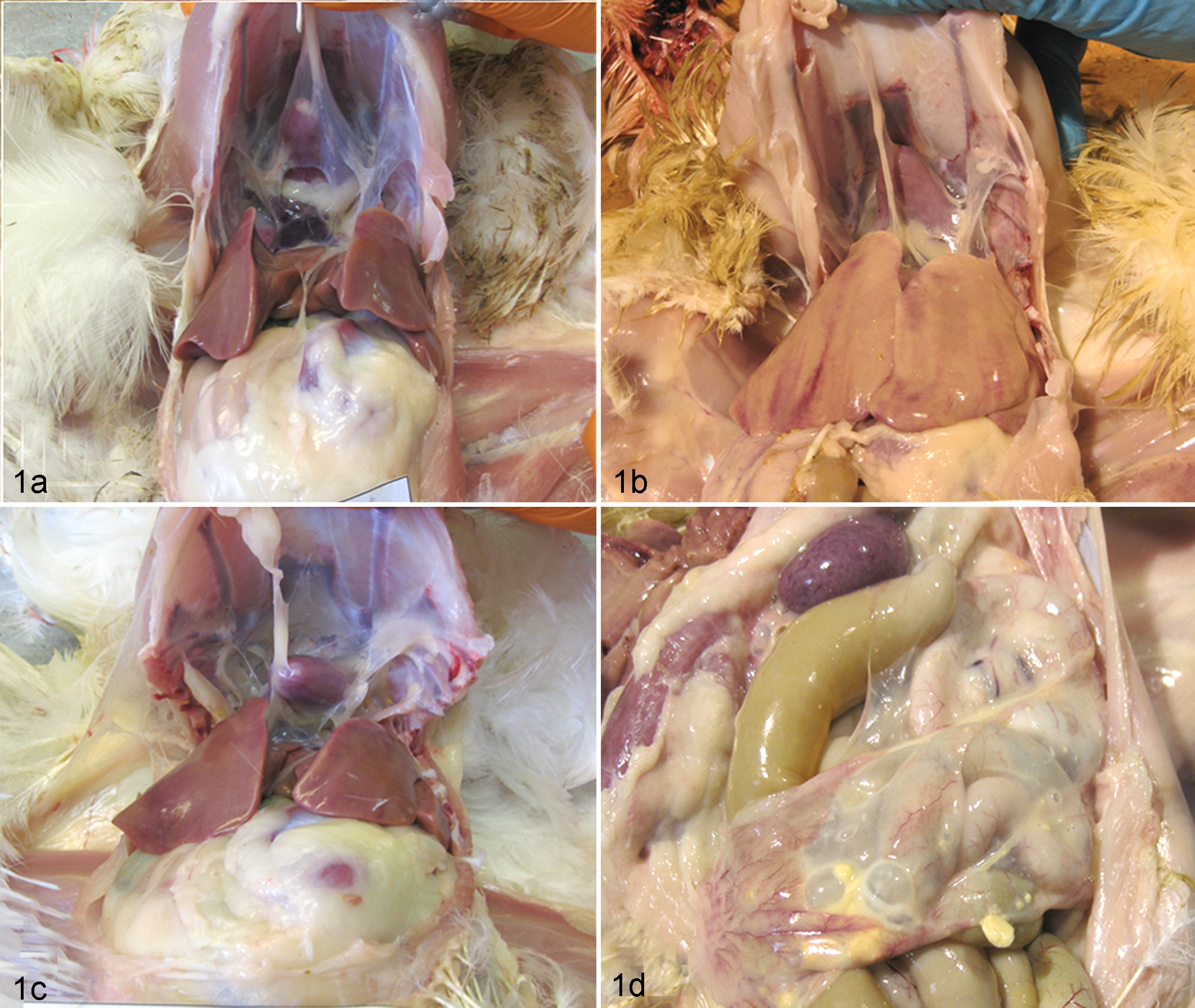
Gross pathologic lesions of experimentally infected chickens. a, Control chicken, not challenged with Mycoplasma gallisepticum or low pathogenicity avian influenza (LPAI) H3N8 virus. Walls of the thoracic air sacs are clear, and there is no accumulation of exudates in air sacs. b, Chicken challenged with LPAI H3N8. As with the control, walls of the thoracic air sacs are clear, and there is no accumulation of exudates. c, Chicken challenged with M. gallisepticum, showing airsacculitis. The walls of the thoracic air sacs are thickened and covered by fibrin, and there is accumulation of small quantities of serofibrinous exudates. d, Chicken challenged with M. gallisepticum and, 7 days later, challenged with LPAI H3N8 virus. Cloudy and thickened walls of thoracic air sacs are apparent, especially in the posterior thoracic air sacs, along with accumulation of fibrinous exudates in the cavity.
Comparison of pathologic lesion scoring of tracheitis, airsacculitis (inflammation of thoracic air sacs), and peritonitis (inflammation of abdominal air sacs) found in these chickens is presented in Figure 2. Scoring of lesions in group V did not differ significantly from those of nonchallenged control birds, except for a mild erythema of the tracheal mucosa. The pathologic scores of airsacculitis as well as total lesion scores of chickens in group M were statistically significantly higher (P < .001) than those of the control birds. The pathologic lesion scores of airsacculitis and peritonitis in chickens of group M+V had statistically significant increased values compared to group M (P < .001).
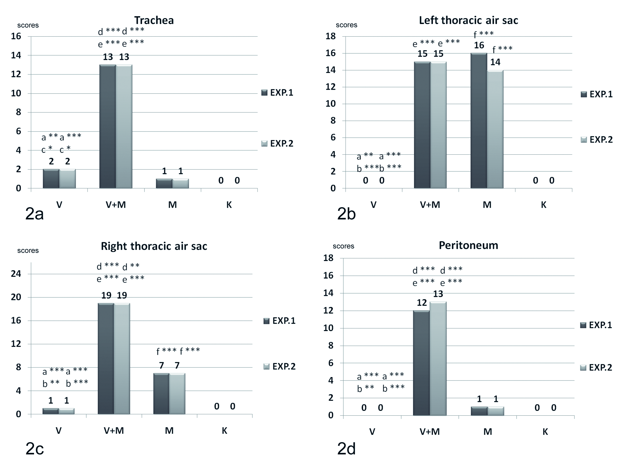
Scores of macroscopic lesions in trachea, left and right thoracic air sacs, and peritoneum of chickens challenged with Mycoplasma gallisepticum and low pathogenicity avian influenza (LPAI) H3N8 virus. V, group challenged with LPAI H3N8 virus; M, group challenged with M. gallisepticum; V+M, group challenged with M. gallisepticum and LPAI H3N8 virus; K, group nonchallenged. a, V – V+M; b, V – M; c, V – K; d, V+M – M; e, V+M – K; f, M – K.* P < .05. ** P < .01. *** P < .001.
Histologic Lesions
The thickness of respiratory epithelium of tracheal and primary bronchi in the control group was an average of 50 μm. In group V, the thickness of the respiratory epithelium of trachea and primary bronchi did not differ statistically from the control chickens (Fig. 3). However, the thicknesses of respiratory epithelium of trachea and primary bronchi in group M were generally statistically significantly larger (between 85 and 99 μm, P < .01–.05) than in groups K and V. Furthermore, the respiratory epithelium of the trachea and primary bronchi of the chickens in group M+V was significantly thicker than those of birds in groups K (P < .001), V (P < .001–.05), and M (P < .001–.05), reaching sizes of 170 to 175 μm (Fig. 4).
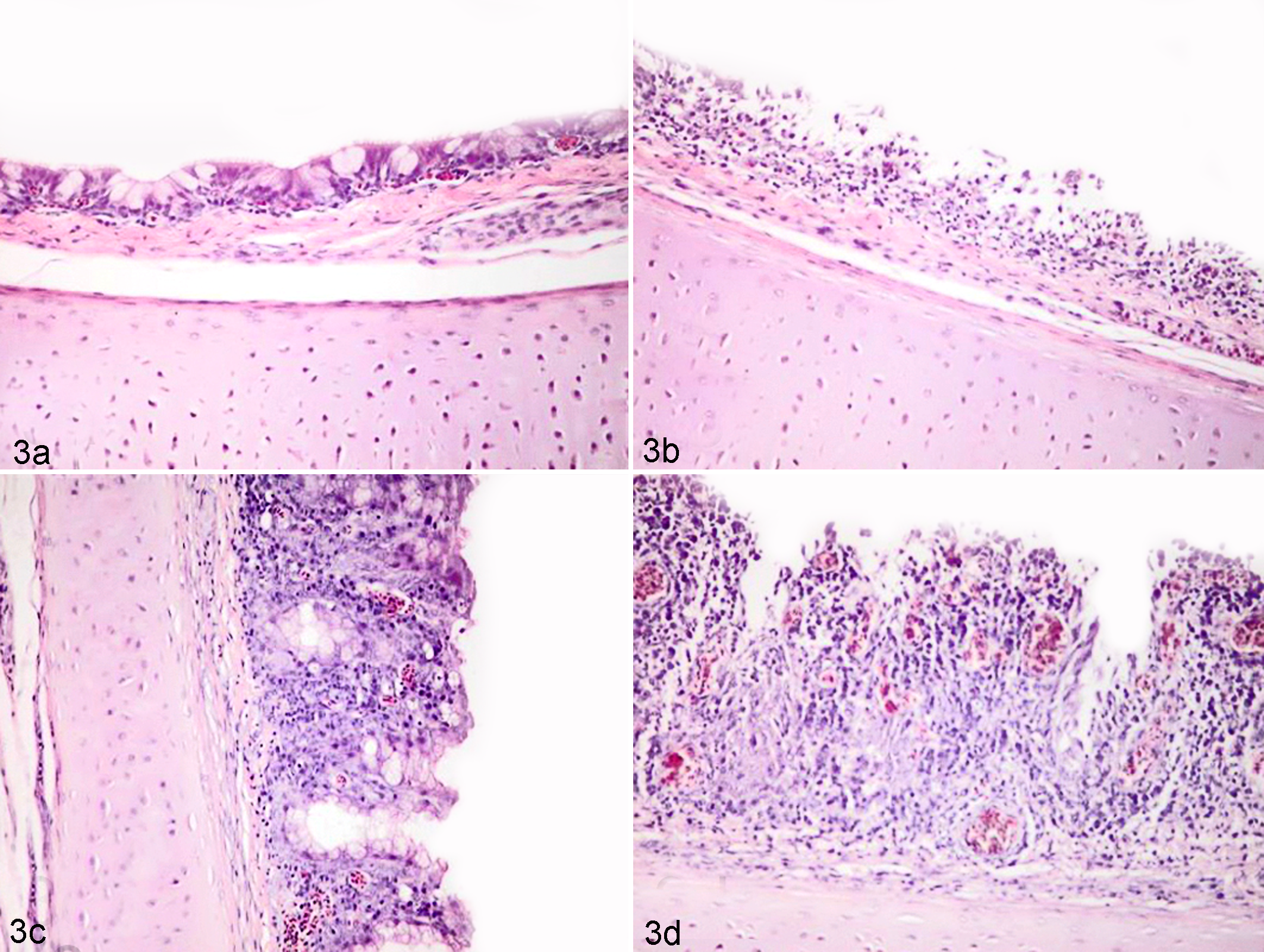
Histologic section of the trachea of the experimentally infected chickens. a, Nonchallenged chicken. The mucosa is covered by a monolayer of epithelial cells. b, Chicken challenged with low pathogenicity avian influenza (LPAI) H3N8 virus. In some areas, the mucosa is thickened and infiltrated by lymphocytes and histiocytes, with degeneration and desquamation of epithelial cells. c, Chicken challenged with Mycoplasma gallisepticum. The mucosa is thickened, and there is diffuse infiltration with lymphocytes and histiocytes, loss of cilia, and degeneration and metaplasia of epithelial cells. d, Chicken challenged with M. gallisepticum and LPAI H3N8 virus. Thickening of the mucosa is evident, including diffuse infiltration of the mucosa with lymphocytes and histiocytes, along with degeneration and metaplasia of epithelial cells. HE.
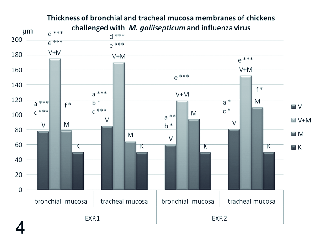
Thickness of tracheal and bronchial mucosal membranes of chickens challenged with Mycoplasma gallisepticum and low pathogenicity avian influenza (LPAI) H3N8 virus. V, group challenged with LPAI H3N8 virus; M, group challenged with M. gallisepticum; V+M, group challenged with M. gallisepticum and LPAI H3N8 virus; K, group nonchallenged. a, V – V+M; b, V – M; c, V – K; d, V+M – M; e, V+M – K; f, M – K.
The respiratory epithelium of trachea and primary bronchi in chickens from the control group were covered by ciliated columnar epithelial cells, and many goblet cells filled with mucus; no histiocyte and lymphocyte infiltration was noticed. In the birds of group V, the respirator epithelium of the trachea and primary bronchi was disrupted on some places; the epithelial cells on these parts lost cilia; and desquamation of epithelial cells was observed (Fig. 3). The histologic score was significantly higher than that of the control group (P < .001–.05). In chickens challenged with M. gallisepticum, the epithelial cells in many parts of the trachea and primary bronchi lost their cilia; they were desquamated and degenerated; and the number of the mucosal glands was decreased. The mucosal glands were scattered irregularly in the edematous respiratory epithelium, which was slightly infiltrated by histiocytes and lymphocytes (Fig. 3). The histologic scores were higher (P < .001) in comparison with those observed in group V. The respiratory epithelium of the trachea and primary bronchi of chickens infected with both agents showed degeneration of epithelial cells. The majority of these cells showed the loss of their cilia, which were thrown off on several places. The epithelium showed reduction of mucosal glands and intensive and diffuse infiltration with lymphocytes and histiocytes. These lesions were present along the entire respiratory epithelium (Fig. 3). Scores of lesions in this group were significantly higher than in groups V (P < .001–.05) and M (P < .001) (see below).
Histologic examination of the lung in group K chickens showed normal, nonciliated epithelial flat cells in the secondary bronchi and parabronchi. No lymphocytes accumulation was seen in the interatrial septum and interparabronchial septum as well as around the blood of vessels. Few small-size germinal centers were present. The lung of the birds in the group V was similar to the birds in the group K (Fig. 5). In the lung of group M birds, scattered degeneration or hyperplasia of nonciliated epithelial cell layer was observed in the secondary bronchi and parabronchi, along with the accumulation of significant numbers of lymphocytes in the interartrial septum and interparabronchial septum. The size of the germinal centers was increased around secondary bronchi and blood vessels. In chickens challenged with both agents, a thickened interatrial septum and interparabronchial septum were present in a large area of lung section. The accumulation of dispersed lymphocytes around the secondary bronchi and blood vessels is mixed with a significant number of germinal centers (Fig. 5). These lesions are characteristic of interstitial pneumonia. The accumulation of serous exudates, cellular debris, and neutrophils in the atrium, infundibulum, and parabronchi and secondary bronchi (catarrhal pneumonia) was observed relatively rarely in groups M and V+M.
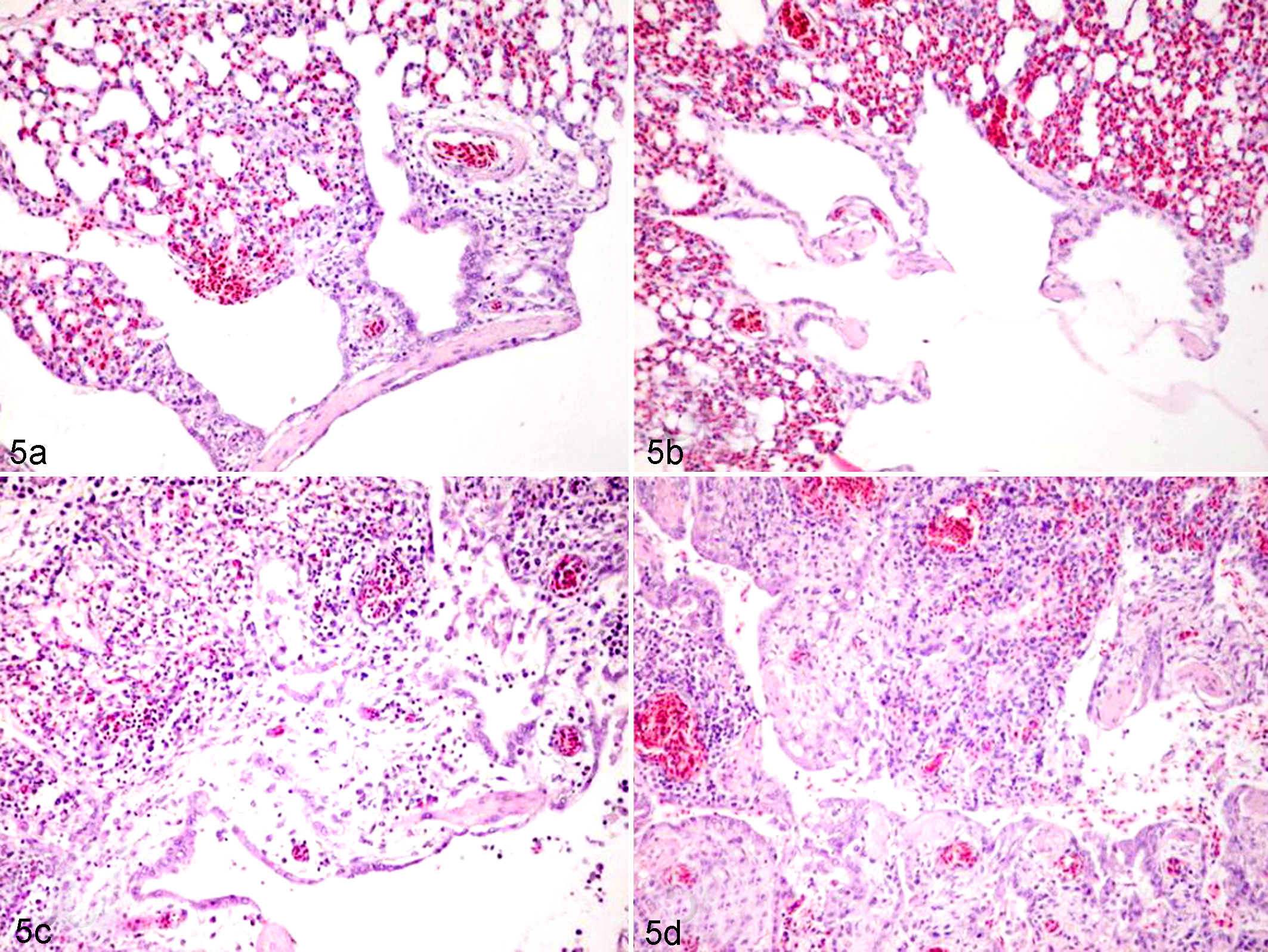
Histologic sections of lungs of experimentally infected chickens. a, Nonchallenged chicken. The histologic structure of parabronchi and air capillaries is normal. b, Chicken challenged with low pathogenicity avian influenza (LPAI) H3N8 virus. The walls of parabronchi and air capillaries are slightly infiltrated with lymphocytes and histiocytes. c, Chicken challenged with Mycoplasma gallisepticum. The walls of parabronchi and air capillaries are strongly infiltrated with lymphocytes and histiocytes. d, Chicken challenged with M. gallisepticum and LPAI H3N8 virus. There is severe interstitial and catarrhal pneumonia. HE.
The scores of interstitial pneumoniae in group V were significantly higher (P < .05) than in group K. Scores of these lesions were higher in group M than in group K. However, scores of interstitial pneumonia were significantly higher in group M+V, compared with the group challenged with M. gallisepticum alone (P < .001) (Fig. 6). Scores of catarrhal pneumonia were higher in group M or V+M than in group V or K.
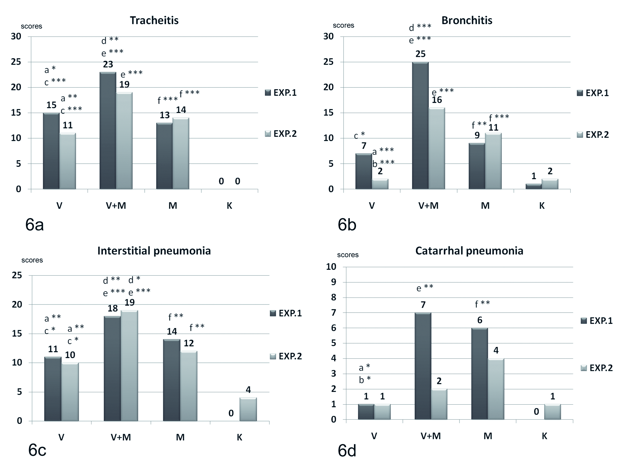
Severity scores of histologic lesions of trachea, bronchi, interstitial pneumonia, and catarrhal pneumonia in chickens challenged with Mycoplasma gallisepticum and low pathogenicity avian influenza (LPAI) H3N8 virus. V, group challenged with LPAI H3N8 virus; M, group challenged with M. gallisepticum; V+M, group challenged with M. gallisepticum and LPAI H3N8 virus; K, group nonchallenged. a, V – V+M; b, V – M; c, V – K; d, V+M – M; e, V+M – K; f, M – K.
Discussion
The coinfection of avian mycoplasmas and influenza viruses in turkeys and ducks56,60 has been observed in field conditions. LPAI virus has been reported to cause only minimal lymphocytic infiltration in the trachea, bronchi, and lungs, as recognized in cases of LPAI viral infection with serotypes H7N2 and H6N2. 27 Challenging broiler and specific-pathogen-free (SPF) chickens with LPAI H9N2 resulted in mild tracheal rales, reduced body weight, and severe congestions and lymphoplasmacytic inflammation of trachea, air sacs, and lung, more significantly in broilers compared with the SPF chickens. 15 This was probably due to coinfection of broiler chickens with other pathogens. 1 LPAI H9N2 can produce severe disease dependant on the type of secondary pathogen present in birds under field conditions. Mixed infections of influenza virus (LPAI H9N2) and other respiratory pathogens, such as infectious bronchitis virus and M. gallisepticum, caused mortality between 20% and 60% on affected farms.45,46,62 However, this statement was not confirmed by challenged studies. Other authors have performed intravenous or intravenous and oculonasal challenge of M. gallisepticum–infected chickens with LPAI H9N2 and could demonstrate mild respiratory signs and severe congestion of mucosa in the trachea and lung and formation of exudative (fibrinous) cast in the tracheal bifurcation in some birds. 54 However, the respiratory challenge model with M. gallisepticum and LPAI is more relevant to field conditions. Coinfection of infectious bronchitis live vaccine and H9N2 avian influenza virus has also been shown to lead to increasing severity of clinical signs and mortality rates in chickens. 20 Additionally, it has been reported that the presence of LPAI H9N2 virus predisposes chickens to secondary Escherichia coli infection, causing slight clinical signs in broiler chickens but not in SPF chickens. 2 Furthermore, studies have shown that LPAI H9N2 can cause immune system modulation in chickens. 71 Therefore, it is important to understand the pathomechanism of coinfections with LPAI viruses.
Mycoplasma infection is widespread in poultry flocks.21,22,24,35,49,63 Prevalence of M. gallisepticum and M. synoviae infections in many countries can be as high as 50% to 70%.2,49,50,63
It is well known that M. gallisepticum, through its adhesins, attaches to the surface of the epithelial cells.* It can also fuse with host cells. 61 Mycoplasmas cause depletion of amino acids, fatty acids, and nucleic acid precursors from the host cells, and they are responsible for the induction of various substances, such as hydrogen peroxide and nitric oxide, which result in damage to host cells. M. gallisepticum can inhibit phagocytosis and affect the functions of B and T lymphocytes. 6,64 It induces expression of various enzymes and cytokines (lymphoactin, CXCL13, CXCL14, RANTES, MIP-1β, IL-1β, and IFN-γ) that contribute to development of local tissue lesions.28–30,41 Mycoplasma-variable lipoproteins interact with the host’s immune system, 7 and M. gallisepticum has been reported to penetrate into nonphagocytic cells, such as the erythrocytes of chickens.43,68,70
Chickens infected with mycoplasmas are sensitive to secondary bacterial infections. M. gallisepticum predisposes chickens to other infections. It has been demonstrated that E. coli can spread faster and can cause bacteremia, severe airsacculitis, and peritonitis in conjunction with mycoplasma infection,39,40 and M gallisepticum is reported to suppress the immune response of chickens to Haemophilus gallinarum. 38
It has also been demonstrated that in immunocompromised chickens, in which T-cell-mediated immunity is decreased, there is increased sensitivity to infection with LPAI virus. This was observed with H9N2 25 when cyclosporine A treatment of chickens led to suppression of CD8 T-cell activity and reduced expression of IFN-γ production.
In our study, we have demonstrated that coinfection of chickens with M. gallisepticum and LPAI H3N8 virus resulted in severe tracheitis, bronchitis, airsacculitis, and pneumonia, which in commercial production can lead to significant economic losses due to poor weight gain, decreased egg production, and mortality. With consideration of the significant spread of mycoplasma infections as well as LPAI virus in poultry, a surveillance of LPAI infection and regular diagnosis of mycoplasma infection and antimycoplasmal treatment of flocks may help to prevent development of severe clinical disease and economic losses due to such coinfections.
Footnotes
Acknowledgement
This study was supported by the Hungarian National Scientific Research Fund grant (OTKA K68156).
The authors declared no potential conflicts of interest with respect to the research, authorship, and/or publication of this article.
The authors received no financial support for the research, authorship, and/or publication of this article.