
Abstract
Mammary Paget disease, characterized by diffuse infiltration of the nipple and areolar epidermis by carcinoma cells, develops in 1% to 3% of human mammary carcinomas. The purpose of this article is to present 2 cases of intraepidermal adenocarcinoma that resembled human mammary Paget disease, histologically and immunohistochemically, in dogs with underlying mammary carcinoma.
Mammary Paget disease (MPD) of the human nipple is an uncommon neoplastic condition characterized by intraepithelial (usually intraepidermal) infiltration by neoplastic cells with glandular differentiation. 3,5–7,10 It develops in 1% to 3% of human breast cancer cases, 3,6,7 beginning in the nipple–areola complex from where it may spread to surrounding skin. Most, if not all, cases of MPD are thought to originate by the migration of neoplastic cells of invasive carcinoma or ductal carcinoma in situ from mammary tissue into the overlying epidermis via lactiferous ducts. 5–7
To our knowledge, mammary Paget-like disease has not been reported in the dog, but a cutaneous lesion resembling human extramammary Paget disease was reported in 2 cats. 5 Extramammary Paget disease resembles MPD in that intraepidermal neoplastic cells are derived from an underlying in situ or invasive cutaneous carcinoma, usually apocrine gland in origin, or from a noncutaneous carcinoma (eg, anal, rectal, urothelial, ovarian, endometrial). 6 We document intraepidermal adenocarcinoma in a male and a female dog that histologically and immunohistochemically resembled human MPD.
Case 1
A 9-year-old intact male Rottweiler dog had a 2-month history of erythema, thickening, and drainage from the left inguinal nipple skin. A firm, painless nodule was palpable in the underlying tissue. Both testes were palpably normal. The mammary nodule was resected with the nipple and overlying skin. Macroscopically, a well-demarcated, bilobulated solid mass (3.5 cm in diameter) was in the subcutis. Histologically, this mammary mass was composed mainly of papillary projections covered by neoplastic epithelial cells with focal squamous differentiation (Figs. 1, 2). Neoplastic cells were pleomorphic with clear cytoplasm, a large round-to-oval nucleus, prominent nucleolus, and ≥ 3 mitotic figures per 400× field. Similar neoplastic cells were in the epithelium of terminal ductulolobular units and lactiferous ducts. The histologic diagnosis was invasive papillary carcinoma.
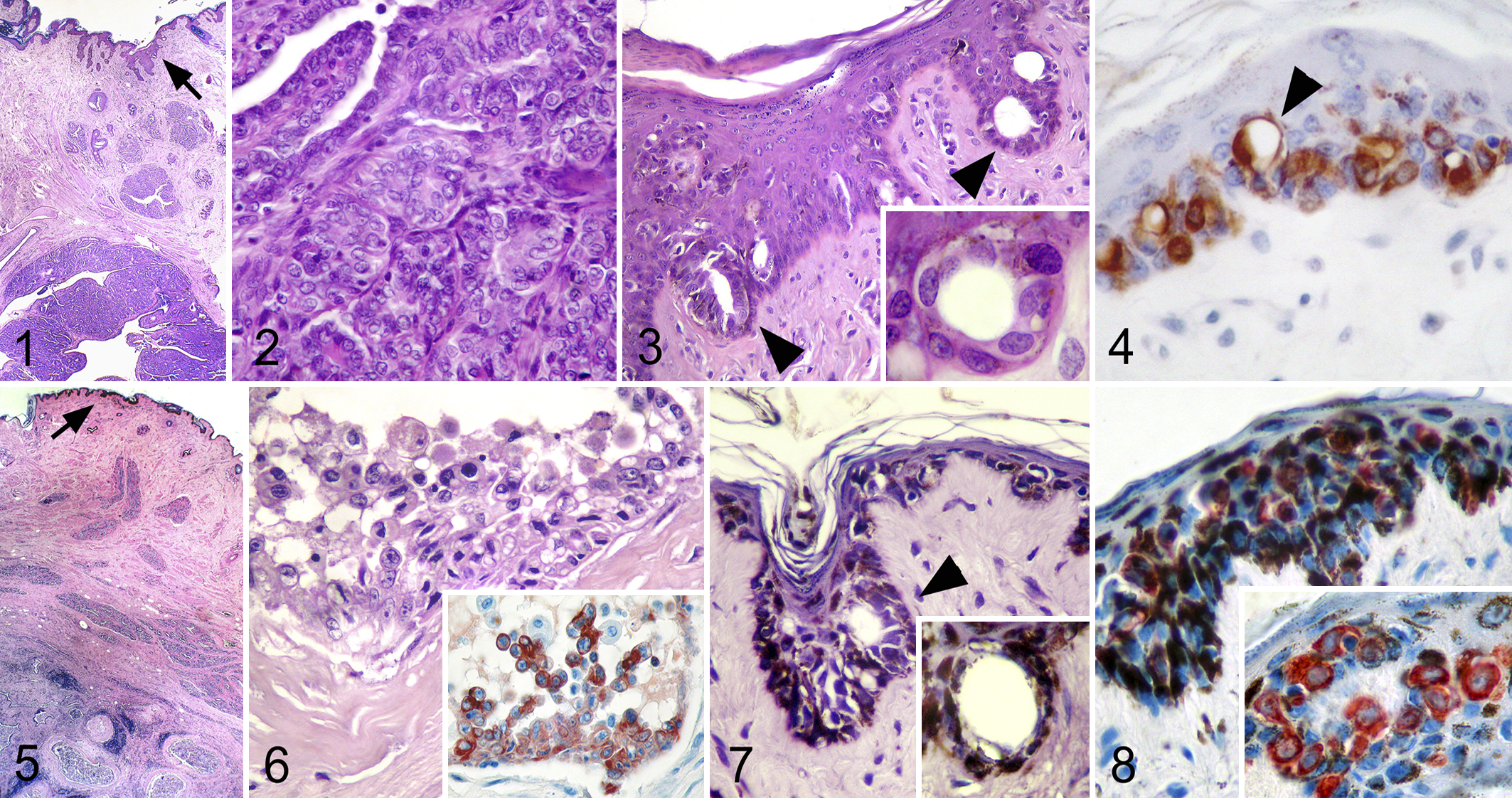
The acanthotic epidermis of the nipple was infiltrated by cells that were twice as large as adjacent keratinocytes and dispersed singly or in clusters, acinar structures, or solid nests (Fig. 3). The cells had abundant pale eosinophilic or amphophilic cytoplasm and a large round-to-oval central nucleus with finely granular chromatin (Fig. 3 inset); some cells had a prominent nucleolus. Nuclear atypia was moderate. Signet ring cells were present in small numbers. The differential diagnosis included intraepidermal carcinoma of squamous, basal, or glandular cell origin; epitheliotropic lymphoma; and melanoma. Formalin-fixed paraffin-embedded samples were serially sectioned and reacted immunohistochemically for cytokeratins (CKs) AE1/AE3 (Dako, Denmark), 5/8 (RCK102 antibody, Eurodiagnostica, Netherlands), 7 (Dako), 8/18 (NCL5D3 antibody, Eurodiagnostica), 20 (Dako), neuron-specific enolase (Dako), and S100 protein (Dako). The intraepidermal neoplastic cells (Fig. 4), the apocrine sweat gland epithelium, and the normal and neoplastic mammary epithelium were strongly positive for CK7 and CK8/18. Keratinocytes were negative for these markers. CK20 was restricted to a few normal and neoplastic mammary cells. Neoplastic cells were negative for neuron-specific enolase and S100 protein.
Case 2
A 5-year-old spayed female crossbred dog was presented for an ill-defined mass in the right inguinal mammary gland. The nipple skin had heterogeneous dark pigmentation extending to its base. The mastectomy specimen contained an irregularly shaped, firm mass (2.2 cm in diameter). Histologically, the mammary tumor was multinodular with cystic cavities filled with necrotic debris, cholesterol clefts, and desquamated neoplastic epithelial cells. The ductal epithelium had chaotically arranged polyhedral or anaplastic cells with enlarged and hyperchromatic nuclei; mitotic figures were rare. Micropapillae (without fibrovascular cores) of atypical cells projected into some ductal lumina (Figs. 5, 6). Squamous differentiation was common. Periductal fibroplasia was pronounced and combined with moderate-to-intense lymphoid cell infiltration with fewer melanophages. Histologic diagnosis was multifocal intraductal micropapillary carcinoma.
Histologically, the nipple skin had numerous intraepidermal atypical cells with enlarged, pleomorphic, and hyperchromatic eccentric nuclei and abundant pale or vacuolated cytoplasm, giving a signet ring appearance to several cells. Some cells contained cytoplasmic melanin granules. The cells were distributed individually and as small aggregates with acinar formation through and above the stratum basale (Fig. 7). Cells were also scattered individually through the infundibular epithelium of a hair follicle. Results of immunohistochemistry on formalin-fixed paraffin-embedded sections were similar to those in dog No. 1, indicating a simple (glandular) epithelial origin for the atypical intraepidermal cells (Fig. 8).
The diagnosis of human MPD is based on clinical appearance and distribution, histopathologic features, and immunohistochemistry results, as well as a thorough examination for underlying neoplasia. 3,6 About 98% of human Paget disease of the breast nipple–areola complex are associated with underlying mammary carcinomas, approximately 42% of which are ductal carcinoma in situ and 58% are invasive carcinomas. 6,7 In dogs with palpable mammary tumors, ductal carcinoma in situ was the most common mammary intraepithelial lesion. 1 In our series, case No. 2 had an intraductal micropapillary carcinoma with squamous metaplasia, periductal fibroplasia, and inflammation—histologic features that are more prevalent in ductal carcinomas than in other mammary carcinomas. 2,3 Mammary tumors in male dogs have been described as rare, usually benign neoplasms. 11 However, dog No. 1 had an infiltrating papillary carcinoma with atypical ductal hyperplasia in lactiferous ducts. Atypical ductal hyperplasia is usually associated with malignant mammary tumors. 1,3
In situ or invasive tumors that arise from mammary ducts have been well documented in the dog 1,2 and share many features with human mammary ductal carcinomas 1 —in particular, a higher fatality rate than that of other mammary carcinomas. 2 It is not surprising that MPD would develop in the dog; however, it may go undiagnosed, if skin overlying a mammary tumor is not evaluated. A small proportion of human cases of MPD are clinically occult and detected only histologically. 6,7 The lesions in the dogs of this report were strictly limited to the skin of the nipple, and serial sections were necessary to detect them. The typical clinical presentation of human MPD includes thickening, itching, eczema, excoriation, erythema, ulceration, and nipple discharge or bleeding. 3,6,10 Some of these signs were observed in dog No. 1. In contrast, dog No. 2 had only an increase in nipple pigmentation. Hyperpigmented MPD is a rare human condition in which hyperpigmented patches or plaques involving the areola and nipple can be the only clinical feature. 7,10 However, hyperpigmentation alone is not a distinctive or reliable sign of MPD, because lentigo (melanocytic hyperplasia) may be restricted to the nipple skin in the dog and would have the same clinical characteristics. 4
The differential diagnosis for Paget disease includes squamous cell carcinoma in situ or Bowen disease, epitheliotropic cutaneous lymphoma, and malignant melanoma (MM). Also, Merkel cells are normal constituents of the epidermis that resemble Paget cells histologically. 3,9 Signet ring cells and acinar structures, which are features of MPD and were found in the dogs of this report, are absent in squamous cell carcinoma or MM. 6,7,10 In cases without morphologic features of glandular differentiation, immunohistochemistry is required. 6,10 As in our cases, Paget cells do not express high molecular weight cytokeratins. Furthermore, the strong CK7 and CK8/18 labeling of the intraepidermal neoplastic cells (and negative reaction in epidermal basal cells and keratinocytes) was consistent with simple (glandular) epithelial origin. Individual intraepidermal neoplastic lymphocytes in epitheliotropic cutaneous lymphoma often have a clear halo and may resemble Paget cells; intraepidermal aggregates of neoplastic lymphocytes (Pautrier microabscesses) could be confused with glandular clusters. 4,6 However, epitheliotropic neoplastic lymphocytes characteristically have convoluted nuclei, which are not seen in Paget cells. Furthermore, neoplastic lymphocytes should not express CKs. 4,6 MPD and MM cells can both contain melanin granules; 7,10 however, the neoplastic cells in superficial MM are nested along the dermoepidermal junction with extension into the superficial dermis, whereas those in MPD are usually distributed more diffusely in the epidermis. 4,7,10 Melanocytes are immunohistochemically positive for S100 protein and neuron-specific enolase in most cases but do not react with most of the antibodies used to identify Paget cells. 4,6,7,10
Merkel cells are a clear-cell population ubiquitously dispersed in the epidermis and in follicular and mucosal epithelium of humans and dogs. 8,9 They could be confused with Paget cells if hyperplastic and forming aggregates in the superficial layers of the epithelium. 8 CK20 is considered Merkel cell specific in humans 8 and dogs. 9 The intraepidermal neoplastic cells in our cases did not express CK20, effectively excluding Merkel cell origin. A few authors have described occasional CK20 labeling in cells of MPD. 5,6 Possibly, these cells were Merkel cells in the nipple epidermis and not Paget cells. CK7 has been described as a sensitive marker for MPD cells; 6 however, CK7 is also present in human and canine Merkel cells. 8,9 Therefore, CK20 is recommended as a marker to differentiate Merkel cells from Paget cells.
Reportedly, human patients, male and female, with MPD and underlying invasive carcinoma have a much greater incidence of multifocal lesions, lymph node metastasis, and shorter survival than do patients in whom the tumor does not spread to epidermis. 3,6 Canine mammary carcinomas of ductal origin metastasize more frequently than do adenocarcinomas. 2 Therefore, diagnosis of Paget-like disease in dogs, even if subclinical, may have negative prognostic implications. Unfortunately, outcome was unknown in our cases.
The authors declared no potential conflicts of interest with respect to the research, authorship, and/or publication of this article.
The authors received no financial support for the research, authorship, and/or publication of this article.