
Abstract
In horses, African horsesickness virus (AHSV) exhibits marked tropism for certain microvascular endothelia and components of the mononuclear phagocyte system. In this study, the tropism of a field isolate of AHSV serotype 5 was studied in 24 chicken embryos. Histopathology on embryonic tissues harvested with 12 hour intervals revealed progressive changes associated with endothelial damage. Immunolabeling demonstrated viral antigens in the microvascular endothelium of the spleen, lungs, and the mesenchymal connective tissue at the base of the neck, from 24 hours post inoculation. Subsequently, specific immunolabeling increased steadily in endothelia of these and other tissues such as skeletal and cardiac muscle, gastrointestinal smooth muscle, mesonephric glomeruli, liver, subcutis and feathers. Positive immunolabeling was also occasionally observed in circulating mononuclear cells and in Kupffer cells in the liver. It was concluded, that this isolate of AHSV displayed similar tissue tropism in the chicken embryo as in the horse.
Keywords
African horsesickness (AHS) is a viral disease of horses transmitted by biting midges of the genus Culicoides. The causative agent, African horsesickness virus (AHSV), is a nonenveloped, double-stranded RNA virus classified in the genus Orbivirus, family Reoviridae. In horses, AHSV infection may result in disease characterized by 4 somewhat overlapping clinical syndromes: a peracute form characterized by fulminant pulmonary edema; a subacute form characterized by severe edema of the head and neck; an acute form typified by characteristics of both the peracute form and of the subacute form; and a mild form characterized by subtle clinical signs of which the transient increase in rectal temperature is the most readily detected. 3
Factors determining the outcome of infection and the development of the different clinical syndromes in horses are not fully understood. Host factors, such as the level of immunity against AHSV, definitely play a role. 3 However, the tissue tropism of viral quasispecies within the virus population may also influence the outcome of infection. The effect of tissue tropism of viral quasispecies was eloquently demonstrated by serial passage, in susceptible horses, of AHSV obtained from the spleen and from the lung of a horse that suffered from the mixed form of AHS. Passaging of “splenic virus” resulted in progressively longer incubation periods and milder disease on each successive passage. With the third serial passage the disease was no longer fatal, although the animal developed a protracted viremia. In contrast, every horse that received “lung virus” succumbed to peracute AHS with marked pulmonary involvement. 5
Tissue tropism of AHSV in the horse is well-documented. Virus is present in the lungs, lymphoid tissues (spleen, lymph nodes, cecum, and pharynx), and choroid plexus as early as 2 days following experimental infection, before the onset of the secondary viremia. Virus has also been detected during the early stages of the disease in the pancreas, adrenal glands, intestines, and salivary glands. 5 Histological changes within these tissues are nonspecific but compatible with endothelial damage. 3 Depending on the clinical syndrome, severe pulmonary edema and/or prominent edema of the intermuscular and subcutaneous connective tissues of the head, neck and shoulders may be present. Perivascular accumulation of lymphocytes and plasma cells, fewer macrophages, and occasional neutrophils in the tunica adventitia of pulmonary blood vessels may be detected. Separation of type I pneumocytes and myocardial degeneration and necrosis can occasionally be observed. 3 Varying degrees of lymphoid depletion and necrosis (or follicular hyperplasia) may occur in the spleen and other lymphoid tissues.3,19 Immunohistochemical, in situ hybridization and ultrastructural studies have confirmed the endotheliotropism of AHSV, particularly for endothelia of pulmonary, cardiac, and splenic microvasculature.18,17,19 African horsesickness viral RNA was also demonstrated in various other microvascular endothelia, including endothelia from the neck muscles and the supraorbital fat. 19 However, AHSV is not limited to endothelial cells in affected horses. Immunohistochemical techniques have also revealed antigen in mononuclear cells such as circulating monocytes, macrophages associated with lymphoid tissue, splenic reticular cells, and follicular dendritic cells.1,19
Little has been published on the tissue tropism of Orbiviruses in laboratory hosts. Strong circumstantial evidence exists that intracerebral cultivation of AHSV in adult mice may drive the selection of AHSV quasispecies toward a strong neurotropism.15,16,18 Some of the old vaccine strains that were attenuated by extensive serial passage in adult mouse brain are purported to cause severe viral encephalitis in guinea pigs, dogs, horses, and primates (including humans).3,4,15,16,18 In this study, we investigated the tissue tropism and target cell predilection of AHSV in the chicken embryo.
Methods
Infection of Chicken Embryos
Thirty embryonated chicken eggs (incubated at 37°C) were obtained from the specific pathogen–free (SPF) White Leghorn flock managed by Avifarms (Pty) Ltd (Lyttelton, South Africa). Twenty-four embryonated chicken eggs (embryosAHS) were infected with a low-passage (2 generations) field strain of virulent AHSV serotype 5 (AHSV5). This virus was isolated in embryonated chicken eggs (intravascular route) during April 2005 from heparinized horse blood obtained from a clinical case of AHS (acute form) in the Kwazulu-Natal province of South Africa. Before inoculation, the AHSV-rich homogenized embryo supernatant was diluted 1:1000 in sterile phosphate-buffered saline (PBS) (pH = 7.2) to obtain a working dilution of 1 × 102.36 ELD50 (50% egg lethal dose) as calculated with the Reed-Muench method. 13 Six eggs (embryosPBS) were inoculated with sterile PBS to serve as negative controls. The eggs were candled twice a day at 12-hour intervals, starting 12 hours post inoculation (PI). At each candling session, 3 embryosAHS and 1 embryoPBS were sacrificed and harvested. The last embryos were harvested 60 hours PI when all the embryosAHS were dead. After fixation, 3 sets of tissue sections were prepared from the harvested embryos. One set of sections was stained routinely with hematoxylin and eosin (HE) and evaluated for histological changes. In the second set, the distribution of labeled AHSV antigens was studied immunohistochemically. The third set was labeled with an irrelevant antibody to function as a negative reagent control. Relevant tissues of embryosAHS, harvested at different stages of viral replication, were compared to identify predilection sites.
Immunoglobulins Used for Immunodetection
A group-specific, polyclonal, rabbit anti-AHSV serum routinely used for diagnostic purposes by the Section of Pathology in the Department of Paraclinical Sciences, Faculty of Veterinary Science, University of Pretoria, Onderstepoort (referred to as Section Veterinary Pathology, UP), functioned as the primary antibody for immunoperoxidase labeling. This antibody (designated anti-AHSVPB) was supplied to the University by Hamblin (Pirbright Laboratories, UK), which raised it in rabbits using sucrose gradient-purified AHSV9 as antigen. A commercially available biotinylated, goat, anti-rabbit serum (DAKO Norden A/S Produktionsvej 42 DK-2600, Glostrup, Denmark) was used as secondary antibody. For negative reagent control purposes, the primary antibody was replaced by an irrelevant, polyclonal antibody raised in rabbits against purified rabies ribonucleoprotein. This antibody (originating from the Health of Animals Laboratory, Guelph, Ontario, Canada) was also supplied by the Section of Veterinary Pathology, UP.
Virus Cultivation
The embryonated chicken eggs were inoculated intravascularly using an established method.6,7 Briefly, using a hobby drill, a small window was created in the egg shell overlying a suitably sized chorio-allantoic blood vessel. Liquid paraffin was painted onto the exposed shell membrane to increase visibility of the underlying blood vessels. An inoculum of 0.05 ml was injected by means of an insulin syringe (Terumo Medicinal Products, Somerset, NJ, USA) with a fixed 27-gauge needle. The windows were sealed with a drop of Ponal wood and paper glue (Henkel South Africa, Alrode, Alberton, RSA) and incubated at 33°C in a Labcon (Laboratory Marketing Services Roodepoort, RSA) FSIE Incubator. Embryos that died within 12 hours of intravascular inoculation were regarded as nonspecific deaths and were discarded.
Harvesting and Fixation
The live embryos sacrificed at each candling session were killed by hypothermia (4°C in a refrigerator for 12 hours) before removal from the egg. The embryos were separated from allantoic, amniotic, and yolk sacs by tearing off the umbilical cord. Before fixation in 10% neutral buffered formalin, openings were created in the cranial and abdominal cavities with a scalpel blade to facilitate penetration of the fixative. Fixation time was restricted to 24 hours at room temperature.9,12
Sampling and Tissue Processing
To support and protect the soft, friable embryonal tissues during cutting, the embryos were first embedded in 1.5% bacteriological agar (Merck 111925, South Africa, Modderfontein, RSA). Two-millimeter slices were cut from the agar blocks with new 130-mm trimming blades (Tissue Tek Accu Edge 4785 Sakura, Torrance, CA 90501, USA), placed in cassettes, and processed using standard operating procedures for formalin-fixed paraffin-embedded tissue. To optimize the likelihood of finding representative tissue of all the major organs (including small organs such as the spleen) in a set of sections from the 3 embryosAHS harvested at each candling session, the head and body of 2 embryosAHS were sliced sagittally, whereas the remaining embryoAHS was cut transversely at 3 different levels through the peritoneal cavity. The sagittal slices were taken slightly to the left of the midline, attempting to include organs positioned laterally to the central axis of the embryo. Three sets of 4-μm-thick sections were mounted on glass slides. The first set, intended for routine HE staining, was mounted on normal Menzel-Glaser (Thermo-Fischer, Kempton Park, RSA) slides, whereas the remaining 2 sets, intended for immunohistochemistry, were mounted on Superfrost Plus Thermo-Fischer (Kempton Park, RSA) slides.
Immunohistochemistry
The avidin–biotin immunoperoxidase complex (ABC) method described by Wohlsein et al 19 was adapted and standardized for routine use on formalin-fixed paraffin-embedded equine tissues in the Veterinary Pathology section, UP. 2 A similar method was followed for immunolabeling of AHSV antigens in the embryonic avian tissues. Briefly, this method entailed inactivation of endogenous peroxidase activity using a 3.0% (wt/vol) H2O2 solution (Associated Chemical Enterprises, H1142CC, ACE, Johannesburg, RSA) in methanol (Labchem, LC16800 Labchem, Edenvale, RSA) for 5 minutes at room temperature, followed by enzymatic epitope retrieval with 0.05% (wt/vol) protease XIV in PBS containing 0.1% bovine serum albumin (Sigma-Aldrich A7030, Sigma-Aldrich South Africa, Kempton Park, RSA) at 37°C for 30 minutes. Nonspecific immunoglobulin binding was blocked with normal goat serum diluted 1:10 in PBS for 20 minutes at room temperature. The sections were incubated overnight at room temperature with the respective primary antibodies at a dilution of 1:2000 in PBS. The biotinylated secondary antibody (at a dilution of 1:500) and the avidin–biotin-peroxidase complex (Vectastain Elite ABC Kit, Catalogue nr PK-6100, Vector Laboratories) were both applied for 30 minutes at room temperature. The sections were exposed to freshly prepared chromogen (Vector NovaRed Substrate Kit for Peroxidase, catalogue no. SK-4800, Vector Laboratories, Southern Cross Biotechnology, Claremont, Cape Town, RSA) until the desired level of labeling was reached (usually between 2 and 15 minutes) and were counterstained with Mayer’s hematoxylin.
Immunohistochemistry Interpretation
Positive immunolabeling of viral antigens were observed under 400× to 1000× magnification in the cytoplasm of intact cells (as opposed to necrotic cells). Positive immunolabeling was recognized as discreet, granular to bead-like deposits with a brick-red or burnt-sienna color (Figs. 7, 8 ) in the tissues of embryosAHS labeled with the anti-AHSV primary antiserum. Such deposits were absent from both sets of control sections (the set prepared from the embryosPBS as well as the negative reagent control set).
Immunohistochemistry Scoring System
Positive immunolabeling was scored subjectively according to the following scheme: – No positive immunolabeling was detected. (+) On average, less than 1 well-defined focus of positive immunolabeling per 5 high-power (400×) fields occurred in the target cells within the tissue of the relevant organ. 1+ At least 2 of 5 high-power (400×) fields contained 1 or more well-defined foci of positive immunolabeling. 2+ Five of 5 high-power fields contained 1 or more well-defined foci of positive immunolabeling. 3+ Five of 5 high-power fields contained numerous foci of positive immunolabeling, which in some areas coalesced to such an extent that it became difficult to count the individual foci. 4+ Five of 5 high-power fields contained numerous extensively coalescing foci too numerous to count.
Results
EmbryosAHS Harvested 12 Hours PI
No macroscopic changes, histopathological changes, or positive immunolabeling were detected.
EmbryosAHS Harvested 24 Hours PI
No macroscopic or histopathological changes were observed. A few scattered foci of positive immunolabeling occurred in the cytoplasm of endothelial cells in the microvascular network surrounding the parabronchi in lung tissue, in the parenchyma of the spleen, and in the small blood vessels of the mesenchymal connective tissue supporting the trachea, esophagus, and jugular veins in the caudal cervical region.
EmbryosAHS Harvested 36 Hours PI
No macroscopic changes were observed. Histologically, multifocal areas of necrosis, with an abundance of macrophages and granulocytic leukocytes, were observed in the spleen. The amount of positively labeled antigens in the spleen and lungs increased noticeably (reaching a subjective score of 2+). Small amounts (up to 1+) of antigen were also detectable in a variety of other organs (see Table 1). In the lungs, like most tissues, positively labeled antigen was clearly limited to the endothelial lining of the microvasculature, but in the spleen the exact location of the antigen was obscured by the densely packed nature of the cellular elements.
Distribution of AHSV-Specific Staining in the Tissues Collected From EmbryosAHS Harvested at 12-Hour Intervals

MCT, mesenchymal connective tissue at the base of the neck; NA, tissue not available for evaluation.
EmbryosAHS Harvested 48 Hours PI
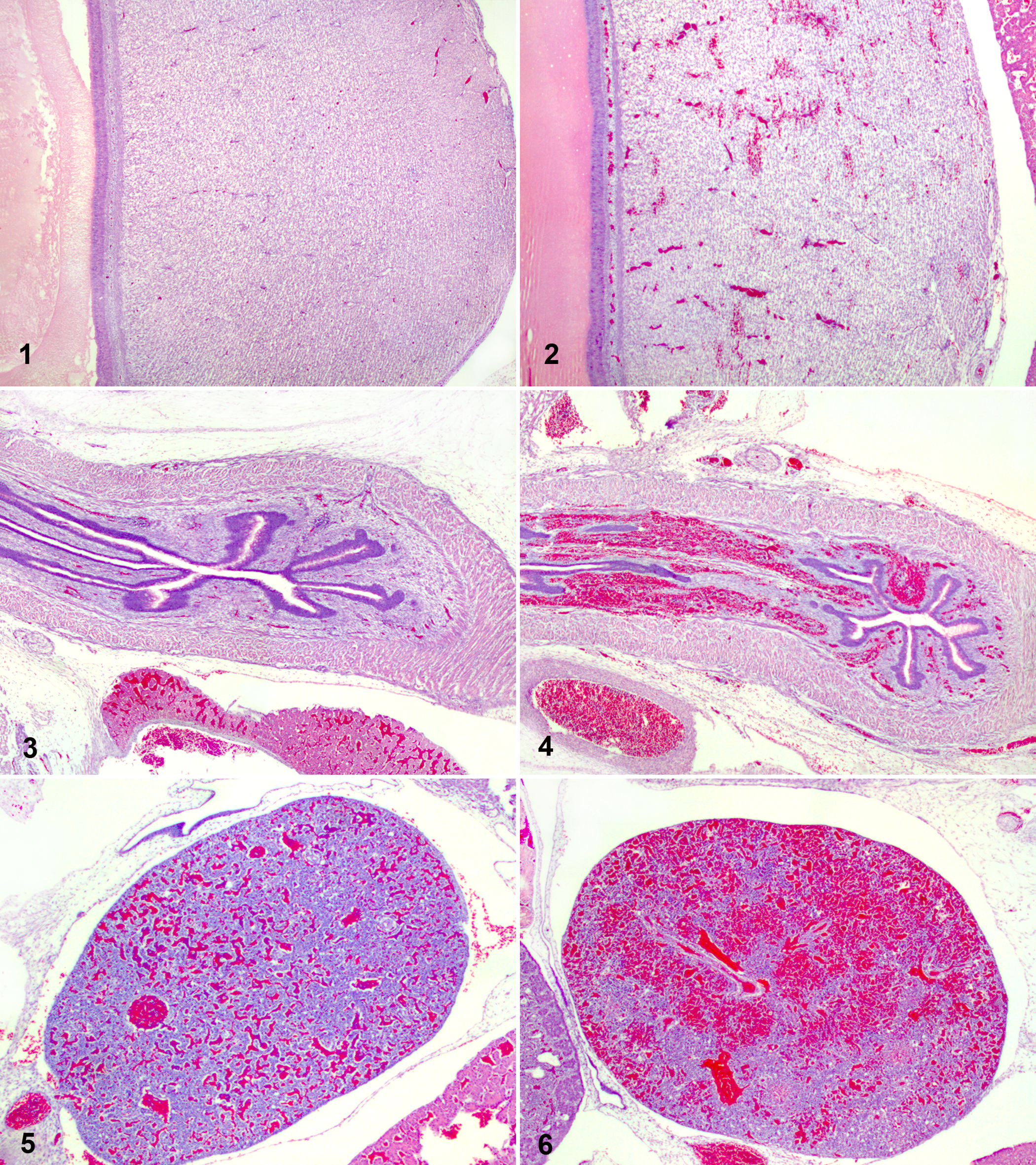
Histologically, most tissues were moderately to severely congested and contained numerous petechial and ecchymotic hemorrhages. The hemorrhage and congestion were particularly prominent in skeletal muscle and to a lesser extent in the thick layers of smooth muscle in the tunica muscularis of the ventriculus (Figs. 1, 2 ). Large sugillations obliterated portions of the mesenchymal connective tissue in the esophageal mucosa (Figs. 3, 4 ). The well-vascularized adipose tissue situated lateral to the cranial thoracic wall was also prominently affected. Parenchymatous organs of the peritoneal cavity were only mildly congested and contained few inconspicuous petechiae, except for the spleen, which showed striking congestion and hemorrhage (Figs. 5, 6 ). Apart from multifocal areas of splenic necrosis, the liver contained necrotic sinusoidal lining cells, and scant cell debris were detectable in the capillary loops of the large mesonephric glomeruli. At this stage the spleen was pervaded with positively-labeled AHSV-antigens (subjectively scored as 4+, see Fig. 9) and the peribronchial vascular networks in the lung were labeled almost diffusely (Fig. 10). Positive immunolabeling was readily detectable in the microvasculature of the atrial and ventricular myocardium. In contrast, the endothelial cells of the endocardium and the tunica intima of large veins and arteries contained minimal positive immunolabeling. Apart from small amounts of positive immunolabeling in the microvasculature of the choroid plexus and peripheral ganglia, the nervous system remained negative with regard to AHSV-specific immunolabeling.
EmbryosAHS Harvested 60 Hours PI
The dead embryosAHS harvested 60 hours PI were cherry-red in color due to severe congestion of the skin. Petechial and ecchymotic hemorrhages were inconsistently observed in the skin and feathers of the head, neck, trunk, and limbs. Microscopically, severe congestion and hemorrhage were evident in all muscle tissue (including skeletal, smooth, and cardiac muscle). The striking lesions in the smooth muscle of the ventriculus are depicted in Figures 5 and 6. The marked congestion and hemorrhage of the esophageal mucosa extended to include the proventricular mucosa. Similar but less striking lesions also occurred in the mucosae of the intestine. Petechial hemorrhages were now abundant in all the major thoraco-abdominal parenchymatous organs. Tissues of the central nervous system were unaffected, but certain tissues of the peripheral nervous system, such as nerves and peripheral ganglia, showed variable congestion and multifocal hemorrhage. The lungs and the spleen still contained more antigen than any other tissue (a score of 4+). Significant amounts of labeling also occurred in the small blood vessels of the adipose tissue lining the lateral thoracic wall (Fig. 11) and in the large glomeruli of the mesonephros (Fig. 12).
Discussion
In this study, virulent AHSV5 killed the infected embryos within 60 hours after inoculation. The generalized congestion and multifocal hemorrhage, in combination with widespread labeling of AHSV-antigens in the microvascular lining of various tissues, suggest that endothelial damage is a key contributor in the pathogenesis of experimental AHSV infection in the chicken embryo. The presence of necrotic/apoptotic cells at the periphery of the vascular lumen in the hepatic sinusoids and in the capillary loops of meso- and metanephric glomeruli supports this mechanism.
Generally, the tissues that showed prominent hemorrhage also contained large amounts of positive immunolabeling in the microvascular endothelium. However, not all tissues that showed large amounts of positive immunolabeling developed sizeable hemorrhage. Although similar amounts of positive immunolabeling occurred in the oropharyngeal and nasal mucosae, for example, the former contained numerous ecchymoses whereas hemorrhages were usually absent in the latter. One possible explanation for this apparent inconsistency is that blood does not necessarily extravasate from the damaged microvasculature unless a certain amount of pressure is exerted on the affected blood vessels. Most tissues containing prominent hemorrhages (eg, the dermis, the feathers, the mucosae and muscular layers of the oropharyngeal cavity and gastrointestinal tract, the cardiac and skeletal muscles, etc) are either subjected to considerable amounts of prehatching movement or are located adjacent to actively moving structures. For example, the heart of a chicken embryo starts beating in the ninth or tenth somite stage at about 36 hours of embryonic development (ED). The skin stretches and bends as underlying skeletal muscle contracts and relaxes. Prehatching movements of the oropharyngeal cavity start on day 5 or 6 ED with slow opening and closing movements of the beak. Swallowing has been observed in embryos on day 8 ED, a stage when peristaltic movements of the intestine are already well-developed. 14 Conversely, the damaged blood vessels in the mucosa of nasal turbinates are well protected by rigid cartilage and bone from the movements of nearby structures.
The nature and pathogenesis of the foci of necrosis observed in the spleens of embryosAHS 36 to 60 hours PI are incompletely understood. In embryosAHS harvested 36 to 48 hours PI, these lesions resembled areas of acute splenic inflammation. The foci consisted of little more than a framework of primitive reticular cells and numerous leukocytes of the granulocytic series, probably heterophils. At this stage, minimal necrotic debris was observed in the foci. Twelve hours later, at 60 hours PI, few scattered granulocytes and some macrophages were observed amid abundant necrotic cell debris in similar foci in splenic tissue of dead embryosAHS. Although the absence of necrotic foci in the spleens of negative control embryosPBS suggests that acute granulocytic splenitis and necrosis developed as a consequence of AHSV5 replication in splenic tissue, the possibility that acute splenitis and necrosis developed due to a nonspecific reaction directed against allogeneic chicken embryo antigens in the inoculum cannot be completely ruled out. According to previous studies on the development of the chicken embryo immune system, splenomegaly developed in 16-day-old recipient embryos after transfer of lymphoid tissue (containing viable cells) from 12-day-old donor embryos. Upon histological examination of the enlarged spleens of the recipient embryos, circumscribed foci consisting of primitive reticulum cells, scattered viable granulocytes, and some necrotic granulocytes, lymphocytes, lymphoblast-like cells, and macrophages were observed. Some of these circumscribed foci even contained necrotic centers lined by multinucleated giant cells. It was concluded that the splenic lesions arose as a result of a tissue rejection (ie, graft-versus-host) reaction. 10 A classic graft-versus-host reaction can be ruled out in this case, as the AHSV5 inoculum contained no viable cells. Although unlikely, the possibility that multifocal granulocytic splenitis and/or necrosis may have resulted from the transfer of acellular material containing allogeneic antigens will have to be investigated further.
Positive immunolabeling was detected in many tissues, such as lung, spleen, cardiac and skeletal muscle, the dermis and feather pulp, liver, meso- and metanephros, and adipose tissue. Within the affected tissues, labeled antigens were restricted to the microvascular endothelial cells and, rarely, mononuclear-like cells within the microvasculature. Several studies identified endothelial cells as the prime target for AHSV in equine tissues.12,3,8 Ultrastructural studies on AHSV-infected equine tissues also demonstrated the occasional presence of virus in other blood vessel–associated connective tissue cells, such as fibroblasts and smooth muscle cells (pericytes). 19 A similar distribution of AHSV in the perivascular connective tissue cells of the chicken embryo could be revealed by electron microscopy.
In horse tissues, AHSV-associated antigens and RNA have been demonstrated in mononuclear phagocytic cells with immunohistochemical and in situ hybridization techniques, respectively.1,19 Ultrastructural studies on tissues of experimentally infected horses confirmed the presence of AHSV particles (and associated structures indicative of viral infection) in mononuclear phagocytic cells. 8 In this study, the positive immunolabeling in Kupffer cells in the lining of hepatic sinusoids, as well as in small numbers of intravascular monocyte-like cells, suggests that AHSV may target certain components of the mononuclear phagocyte system in both the horse and in the chicken embryo.
Selective tropism for specific endothelial cells of different organs is a fascinating aspect of the pathogenesis of AHS. This selectivity for specific endothelia probably explains some of the intriguing clinical signs and postmortem findings observed in horses with AHS. Notably, the severe edema affecting specifically the subcutaneous and intermuscular tissues of the head, neck, and shoulder regions of horses with the cardiac form of AHS stops inexplicably at about the level of the elbow. Subcutaneous tissues of the distal portions of the limbs are typically unaffected (B. J. Erasmus 1981, unpublished observations). In an evaluation of the ultrastructural changes in capillaries of tissues from horses infected with AHSV4, previous workers observed stronger viral tropism toward endothelium of cardiac muscle compared with endothelia of the liver and spleen. 8 The results of the present study indicate that selective tropism for certain endothelial cells also occurs in the chicken embryo. For example, abundant AHSV-specific immunolabeling occurred in the endothelial cells of small peribronchial blood vessels in the lungs of all embryosAHS harvested 24 hours PI and later, whereas the microvasculature of the brain and spinal cord remained unlabeled throughout the study. As ecologists and climatologists predicted, the rapidly changing climate of our world has had and will continue to have a profound effect on the distribution and epidemiology of animal diseases. Recent advances in the distribution of Bluetongue virus into several European countries has raised concern that other vector-borne diseases, such as AHS, may follow. 11 Sporadic failure of the current, multivalent, live attenuated vaccine to protect horses against AHS in endemic areas, such as South Africa, has exacerbated these concerns. These factors have sparked renewed interest in the field of AHS research. One of the factors that impede AHS research is the lack of a suitable laboratory animal model. Conducting virological research in large expensive animals such as horses is laborious and costly and raises ethical concerns. The striking similarities in the tissue tropism of AHSV in the horse and the tissue tropism of this strain of AHSV in the chicken embryo suggest potential in further experimentation to explore the potential of chicken embryos in AHSV research.
Footnotes
Acknowledgements
We express our gratitude to Deltamune (Pty) Ltd for access to laboratory facilities; the staff of the histopathology laboratory (J. Breedt, P. Mokonoto, R. M. Phaswane), Section Pathology UP, for tissue processing and preparation of excellent tissue sections; and M. Smit for immunoperoxidase staining of consistent quality. We acknowledge the department for supplying the anti-AHSVPB and the irrelevant rabies antibodies, and we thank Prof. J. Soley of the Department of Anatomy and Physiology, Faculty of Veterinary Science, Onderstepoort, University of Pretoria, for providing literature and invaluable assistance regarding chicken embryology.
The authors declared that they had no conflicts of interest with respect to their authorship or the publication of this article.
The authors declared that they received no financial support for their research and/or authorship of this article.