
Abstract
To compare and summarize the mechanisms, frequencies of occurrence, and classification schemes of spontaneous, experimental, and genetically engineered mouse skeletal neoplasms, the literature was reviewed, and archived case material at The Jackson Laboratory was examined. The frequency of occurrence of spontaneous bone neoplasms was less than 1% for most strains, with the exceptions of osteomas in CF-1 (5.5% and 10% in two studies) and OF-1 outbred strains (35%), and osteosarcomas in NOD/ShiLtJ (11.5%) and NOD-derived (7.1%) mice. The frequency was 100% for osteochondromas induced by conditional inactivation of exostoses (multiple) 1 (Ext1) in chondrocytes, osteosarcomas induced by tibial intramedullary inoculation of Moloney murine sarcoma virus, and osteosarcomas induced by conditional inactivation of Trp53—with or without inactivation of Rb1—in osteoblast precursors. Spontaneous osteogenic neoplasms were more frequent than spontaneous cartilaginous and vascular types. Malignant neoplasms were more frequent than benign ones. The age of occurrence for spontaneous neoplasms ranged from 37 to 720 days (M = 316.35) for benign neoplasms and 35 to 990 (M = 299.28) days for malignant. In genetically engineered mice, the average age of occurrence ranged from 28 to 70 days for benign and from 35 to 690 days for malignant. Histologically, nonosteogenic neoplasms were similar across strains and mutant stocks; osteogenic neoplasms exhibited greater diversity. This comparison and summarization of mouse bone neoplasms provides valuable information for the selection of strains to create, compare, and validate models of bone neoplasms.
Keywords
Neoplastic lesions of bone are less common in humans, domestic animals, and laboratory mice, compared to neoplasms of other tissues.2,22,42,55-57,68,86,89,95,96 Malignant neoplasms of bone are generally more common than the benign, and osteosarcoma is the most common skeletal neoplastic lesion in all species, including laboratory mice.2,22,42,55,56,68,69,86,89,95,96 Benign tumors of bones that have been reported to occur spontaneously in mice include chondroma, hemangioma, ossifying fibroma, osteoblastoma, and osteoma.2,22,55–57,59,61,78,79,99 Spontaneous malignant neoplasms of bone reported in mice include chondrosarcoma, hemangiosarcoma, and osteosarcoma.2,22,55–57,60,78,79,94,99
This article addresses the similarities and differences in classification schemes for neoplasms of bone in humans, domestic animals, and laboratory mice. It also presents a comparison and summary of neoplasms of bone—spontaneous, genetically engineered, radiation induced, and chemically induced—in various strains of laboratory mice (as noted in the literature), as well as a retrospective evaluation of the frequency of bone neoplasms in various mouse strains submitted for diagnostic necropsy at The Jackson Laboratory (Bar Harbor, Maine).
Materials and Methods
We reviewed reports of spontaneous, experimentally induced, or genetically engineered primary skeletal neoplasms in inbred, hybrid, genetically engineered, and outbred strains and stocks of mice and summarized the mechanisms, frequencies of occurrence, and other features of these neoplasms. Additional details on the strains of mice, genetic manipulations, and experimental procedures are available in the cited references.
For retrospective evaluation of the frequency of bone neoplasms at The Jackson Laboratory, we reviewed archived necropsy records and glass slides for diagnoses of bone neoplasms in various strains submitted to the diagnostic service for necropsy between January 1987 and January 2010. Criteria for submission were clinical assessment of a “sick mouse” or the presence of a grossly visible tumor. Gross necropsy examination was performed on all mice, and collected tissues were fixed in Tellyesniczky/Fekete fixative (70% ethanol, 100 ml; 37–40% formalin, 5 ml; glacial acetic acid, 5 ml). 88 Appropriate tissues were decalcified with 10% formic acid (Formical-2000, Decal Chemical Corporation, Tallman, New York) or 3% hydrochloric acid (Cal-Ex, Fisher Scientific, Fairlawn, New Jersey) for 24 hours. Tissues were paraffin embedded and stained with hematoxylin and eosin. Diagnoses were reviewed and neoplasms classified according to criteria described in the International Classification of Rodent Tumors: The Mouse. 22 Diagnostic designations were also compared with listings on the mouse pathology ontology system (http://eulep.pdn.cam.ac.uk/Pathology_Ontology/index.php, September 2010). Archived and current designations were in agreement for most diagnostic entities. The designation of osteochondrosarcoma, as used in the archives, was not reiterated in the retrospective evaluation. Cartilage or chondroid matrix–rich osteosarcomas are currently classified as osteosarcomas of the chondroblastic subtype.22,25 Diagnoses of many differentiated primary skeletal neoplasms predominantly rely on radiographic and histologic features rather than on immunohistochemical studies. 28 In this study, most tumors were well differentiated; so, diagnoses were based on histomorphologic features, and immunohistochemistry was not required.
For any strain that had fewer than 100 total necropsy submissions, the frequency of occurrence was not considered representative. Details of the age of occurrence and frequency of benign and malignant neoplasms sorted by sex are available in Supplementary Tables 1 and 2 (available at http://vet.sagepub.com/supplemental).
Classification Schemes for Bone Tumors a
a NA, not applicable to the species; NCDE, no corresponding designation or entry in this classification scheme; NCDL, not commonly diagnosed or listed as a primary tumor of bone or joint; NCLM, no commonly listed genetically engineered mouse model with primary involvement of bone or joint; NCLS, not commonly listed as a subtype.
Benign Bone Tumors in Various Strains of Mice as Reported in the Literature
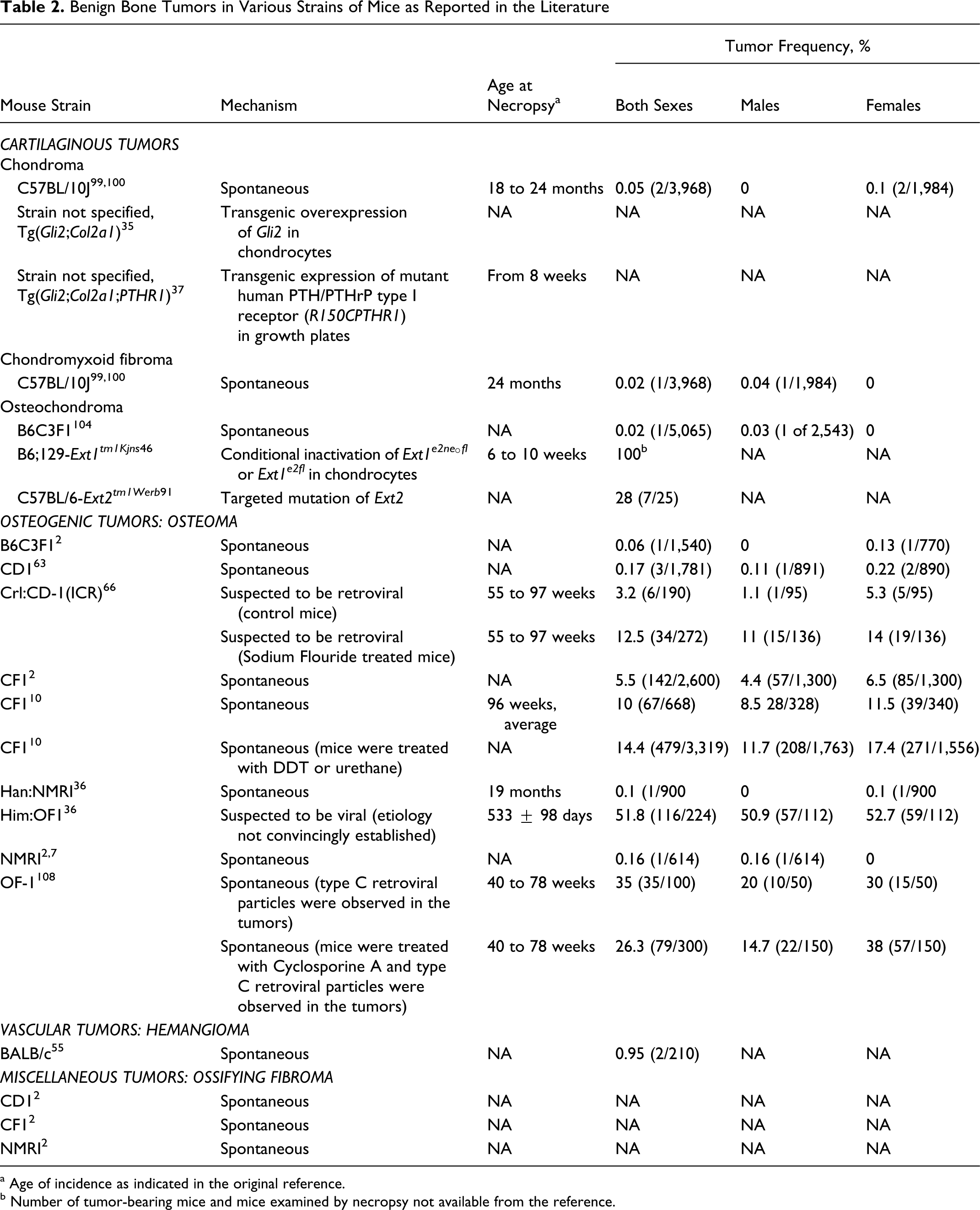
a Age of incidence as indicated in the original reference.
b Number of tumor-bearing mice and mice examined by necropsy not available from the reference.
The Mouse Tumor Biology Database (http://www.informatics.jax.org/, September 2010), 52 Mouse Genome (http://www.informatics.jax.org/, September 2010), 21 and JAX Mice Database (http://www.jaxmice.org/query/, September 2010) at The Jackson Laboratory were used to verify information on tumor types, tumor frequencies, genes, alleles, and mouse strains. Tumor frequencies or comments pertaining to tumor frequencies that reference the Mouse Tumor Biology Database are identified in subscript as (MTB).
Genes with a confirmed involvement in the pathogenesis of bone neoplasms in genetically engineered mouse models and genes known to significantly interact with the aforementioned genes were listed for gene network analysis. For performing the network analysis, each gene was assigned a hypothetical fold change score. If the loss or inactivation of a gene resulted in the development of a bone neoplasm in a mouse model, that gene was considered downregulated and assigned a hypothetical fold change score of –99 (eg, tumor suppressor genes). If the transgenic overexpression or dysregulated activation of a gene resulted in the development of a bone neoplasm in a mouse model, that gene was considered upregulated and assigned a hypothetical fold change score of 99 (eg, oncogenes). Ingenuity IPA Pathway Analysis Software (Redwood City, CA) was used for the gene network analysis. The list of genes is available in Supplementary Table 3(available at http://vet.sagepub.com/supplemental).
Malignant Bone Tumors in Various Strains of Mice as Reported in the Literature
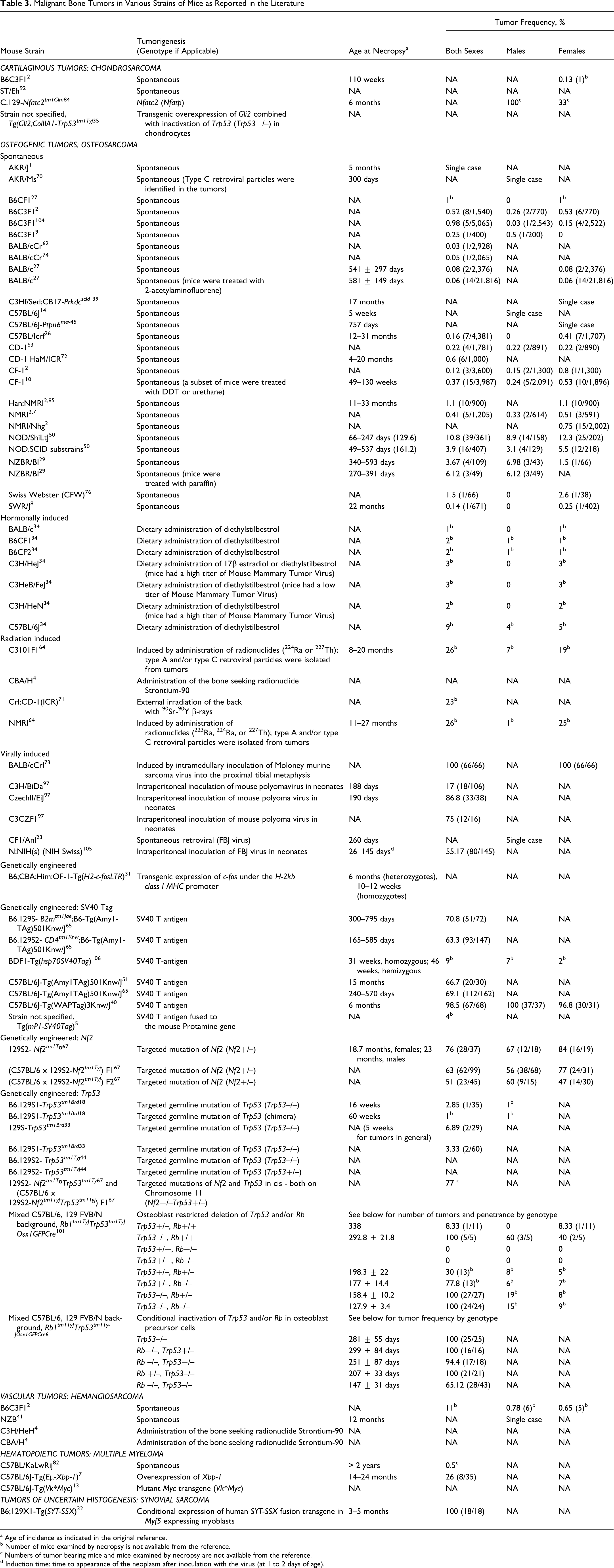
a Age of incidence as indicated in the original reference.
b Number of mice examined by necropsy is not available from the reference.
c Numbers of tumor bearing mice and mice examined by necropsy are not available from the reference.
d Induction time: time to appearance of the neoplasm after inoculation with the virus (at 1 to 2 days of age).
Results
Comparison of Classification Schemes
The WHO Classification of Bone Tumors in humans 25 broadly categorizes tumors as cartilage, osteogenic, giant cell, notochordal, vascular, smooth muscle, lipogenic, neural, miscellaneous, fibrogenic, fibrohistiocytic, primitive neuroectodermal, and hematopoietic, as well as miscellaneous lesions and joint lesions. Synovial sarcoma is not included under joint lesions or other categories in this scheme because of its ambiguous histogenesis. 25 The WHO Classification of Bone Tumors in humans is also used for the mouse pathology ontology (http://eulep.pdn.cam.ac.uk/Pathology_Ontology/index.php), a system for the histopathology of genetically engineered mice in the European mutant mouse pathology database, Pathbase (http://www.pathbase.net).
The Histological Classification of Bone and Joint Tumors of Domestic Animals 89 broadly categorizes them as benign tumors, malignant tumors, and tumorlike lesions. Malignant tumors are subcategorized as central tumors, peripheral tumors, joint tumors, miscellaneous tumors, and tumors of bone marrow. 89
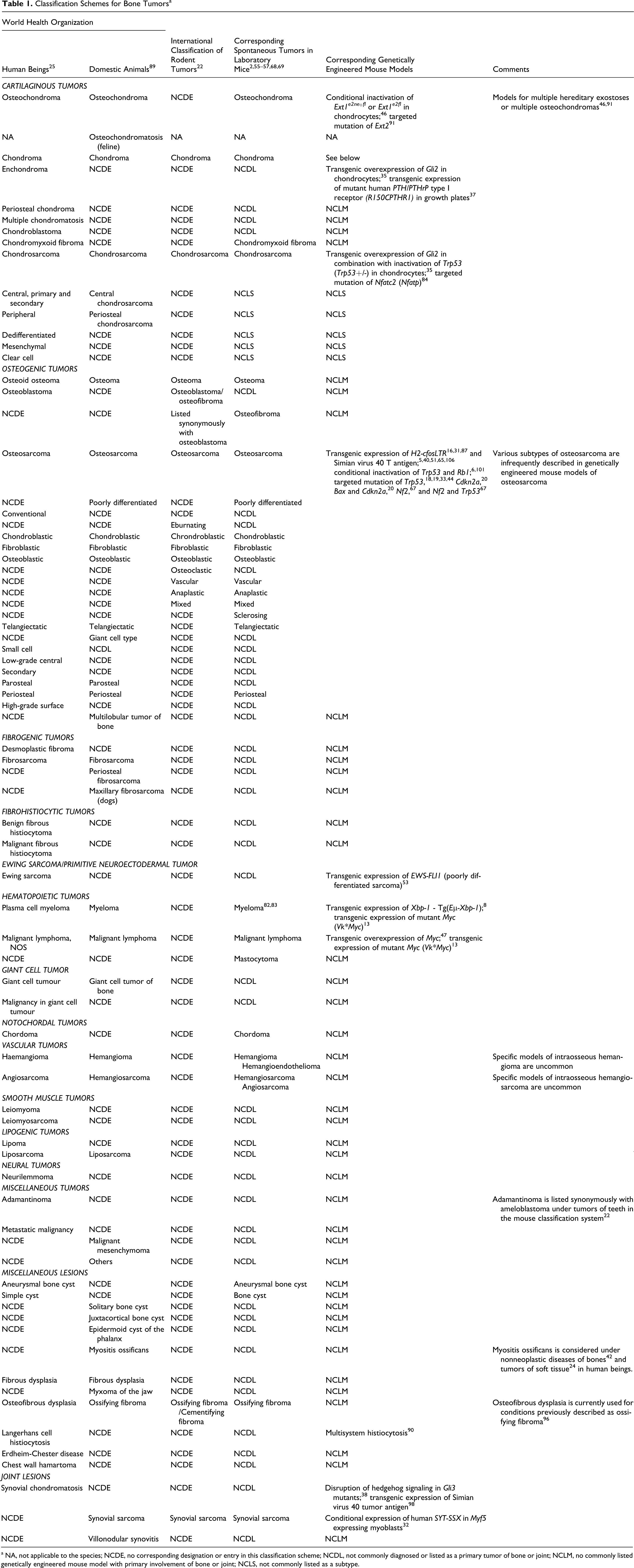
The most commonly used scheme for classification of mouse bone tumors is the one detailed in the International Classification of Rodent Tumors: The Mouse. 22 Tumors of the skeletal system listed in this scheme include osteoma, osteosarcoma, osteofibroma, ossifying fibroma, chondroma, chondrosarcoma, and synovial sarcoma. 22 This classification scheme is disparate with extant WHO classifications of bone tumors in humans 25 and domestic animals, 89 as well as with some of the descriptions of bone tumor types observed in laboratory mice.55 –61 The mouse classification system describes (1) osteofibroma as being synonymous with osteoblastoma and (2) ossifying fibroma as being synonymous with cementifying fibroma. 22 Ossifying fibroma and osteofibroma are not described in the human World Health Organization classification scheme. 25 In classifications and descriptions of human bone tumors, osteoblastoma is considered similar to osteoid osteoma,25,42,86,96 and the designation ossifying fibroma has been replaced by osteofibrous dysplasia.25,42,96 The International Classification of Rodent Tumors: The Mouse does not consider intraosseous vascular neoplasms under tumors of the skeletal system. 22 In some descriptions of mouse tumors, hemangioma and hemangioendothelioma are considered synonymous, 55 although the latter is considered synonymous with angiosarcoma in descriptions of human tumors.42,96Table 1 compares and summarizes classification schemes for tumors of bones and joints; Tables 2 to 4 summarize details of these tumors, as reported in the literature and observed at The Jackson Laboratory.
Cartilaginous Tumors
Osteochondroma
Osteochondromas are benign, cartilage-capped, partially ossified tumors that are also known as exostoses. They are typically reminiscent of an epiphyseal plate rotated 90° to the long axis of bone, given that they arise from the surface of an endochondral bone adjacent to a physis and are attached to the underlying skeleton by a bony stalk. Histologically, osteochondromas consist of chondrocytes arranged in a manner similar to that in epiphyses.42,86,89,95,96 Spontaneous osteochondromas are the most common benign bone tumor in humans42,86,96 and are reported in cats, dogs, and horses. They are extremely rare in laboratory mice 56 and were reported in B6C3F1 mice (0.03% frequency in males). 104 A single osteochondroma was found in a WBB6F1/J-KitW/KitW-v/J mouse at The Jackson Laboratory.
Chondroma
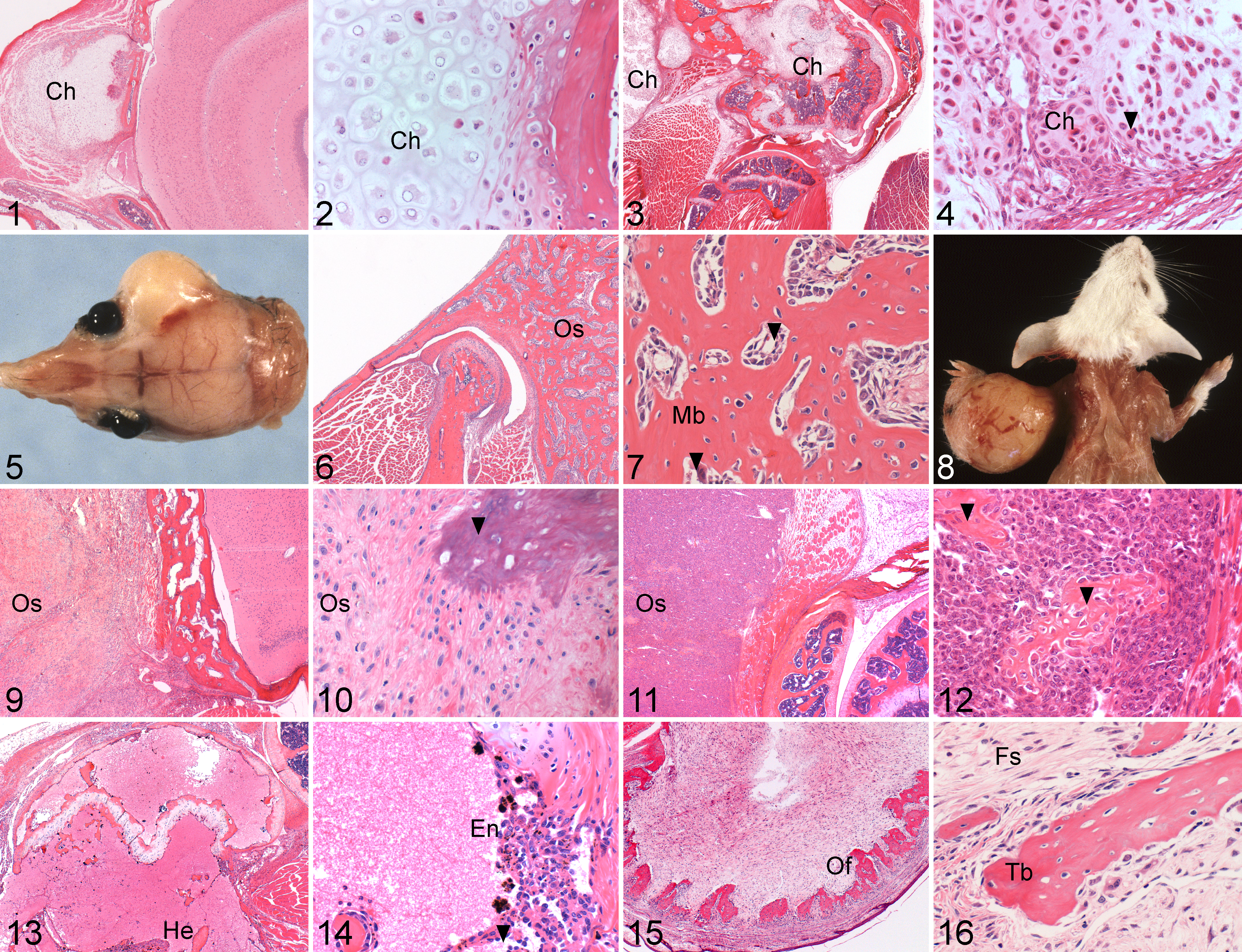
Chondromas are benign tumors of chondrocytes consisting mostly of mature hyaline cartilage. They are typically circumscribed, encapsulated, slow growing, expansile, and smooth to nodular, and they usually occur in bones of enchondral origin. They can arise within the medullary cavity, where they are known as enchondromas, or on the surface of bone, where they are known as periosteal or juxtacortical chondromas. Histologically, chondromas consist of nodular aggregates of well-differentiated chondrocytes with little to no cellular and nuclear pleomorphism (Figs. 1, 2).22,42,55–57,86,89,95,96 Spontaneous chondromas are extremely rare in laboratory mice.2,22,55–57,78–80 Chondromas were reported in C57BL/10J (0.1% frequency in females),99,100 and a single case was found in a NOD.129P2(B6)-B2mtm1Unc/J mouse (Figs. 1, 2) at The Jackson Laboratory.
Chondromyxoid fibroma
Chondromyxoid fibromas are benign cartilaginous tumors that typically involve the metaphyses of long bones. Histologically, chondromyxoid fibromas are usually lobulated, consisting of nodules of poorly formed hyaline cartilage mixed with variable amounts of myxoid matrix delinated by fibrous septae.42,86,96,99,100 Chondromyxoid fibromas are extremely rare in humans,42,86,96 domestic animals, 89 and mice and were reported in C57BL/10J mice (0.04% frequency in males).99,100
Chondrosarcoma
Chondrosarcomas are malignant cartilaginous neoplasms that arise de novo in bone or undergo malignant transformation within a preexisting benign cartilaginous tumor. They are typically expansile and sometimes lobulated, and they more commonly involve flat bones than long bones in most species. They have also been reported to arise in locations with extraskeletal cartilage, such as the larynx in humans and mice. Chondrosarcomas may arise from the medullary cavity (central chondrosarcoma) or periosteum (peripheral chondrosarcoma), with the former being more common. Histologically, chondrosarcomas are often lobulated, consisting of different proportions of hyaline or myxoid cartilage composed of chondrocytes that vary in cytologic atypia, mitotic activity, and degree of differentiation (Figs. 3, 4). They primarily produce chondroid and fibrillar matrix but rarely osteoid. However, occasional chondrosarcomas may contain reactive or metaplastic bone matrix.22,42,55–57,86,89,95,96 Chondrosarcoma is the third-most common malignancy of bone in humans, after myeloma and osteosarcoma,42,86,96 and it is reported in cats, dogs, and sheep89,95 but is extremely rare in mice.2,22,55–57 Spontaneous chondrosarcomas were reported in B6C3F1 mice (0.13% frequency in females), 2 in C57BL/10J mice (0.03% frequency in females),99,100 and in a ST/Eh mouse (single case) 92 and observed in a 325 day-old male C57BL/6J mouse (Figs. 3, 4) at The Jackson Laboratory.
Osteogenic Tumors
Osteoma
Osteomas are rare benign tumors of osteoblasts composed of mature well-differentiated bone with a predominantly woven arrangement. They are typically solitary, dense, and smooth, and they preferentially arise from the periosteal surface and merge with underlying cortical bone. Osteomas most commonly occur in bones of the skull in most species (Fig. 5). An osteoma may rarely be seen on the surface of a long bone. Histologically, osteomas are composed of dense coalescing spicules and trabeculae of bone lined by osteoblasts and osteoclasts. Fatty or hematopoietic marrow may be present within the trabeculae (Figs. 6, 7). The intertrabecular connective tissue is composed of spindle-shaped cells, and it becomes increasingly sparse as the tumor becomes more compact and sclerotic.22,42,55-57,61,86,89,95,96 Spontaneous osteomas are rare in most strains of mice and seem to be more common in females than in males, with the difference being attributed partly to a female hormonal influence.
Cases of spontaneous osteoma were documented in the following mice strains: AKR, 61 B6C3F1,2,9 C57BL/6NNia(MTB), CBA/H(MTB), CD-1,63,66,72 CF-1,2,10,61 CFW/CarWHanJena(MTB), NMRI2,7 NZO/Bl(MTB), Him:OF1, 36 and OF-1. 108 CF-1, Him:OF1, and OF-1, which are genetically related outbred strains of mice and have the highest documented frequency of osteomas among all mouse strains.2,61 The reported frequency of spontaneous osteomas in CF-1 mice was 5.5% in one study 2 (4.4% in males and 6.5% in females) and 10% in another (8.5% in males and 11.5% in females). 10 In the latter, the combined average age of occurrence for both sexes was 96 weeks. 10 Spontaneous osteomas are quite rare in other strains, with frequencies ranging from 0.02 to 0.20% in various reports. 2 In one study, a frequency of 0.13% was observed in female B6C3F1 mice (1 case in 770 mice examined). 2
Reports exist of higher frequencies of osteomas with concurrent identification and isolation of retroviral particles. 61 In a study on skeletal lesions of sodium fluoride toxicity in Crl:CD-1 (ICR) outbred mice, the reported frequency was 12.5% in sodium fluoride–treated mice (11% in males and 14% in females) and 3.2% in controls (1.1% in males and 5.3% in females) with a suspected retroviral etiology at 55 to 97 weeks of age. 66 In a study on the carcinogenic potential of cyclosporine A in OF-1 mice, a 26.3% frequency was reported in cyclosporine-treated mice (14.7% in males and 38% in females), with 35% in controls (20% in males and 30% in females), at 40 to 78 weeks of age. 108 Type C retroviral particles were identified in tumors from both groups, but an etiologic correlation was not conclusively established. In an aging study, Him:OF-1 mice were reported to have a frequency of 51.8% (50.9% in males and 52.7% in females) at 533 ± 98 days of age, and viruslike particles were seen on ultrastructural observation of tumor cells and necrotic tissues. 36 Osteomas induced by neonatal administration of murine polyomavirus were documented in C3H/BiDa mice. 17 At The Jackson Laboratory, osteomas were diagnosed in mice of strains A/J, C57BL/6J (Fig. 5), DBA/2J, B6C3F1/J, C3.SW-H2b/SnJ, and C57BL6/J-Tg(Amy1Tag)501Knw/J (Figs. 6, 7)—one case in each strain, all in females.
Osteosarcoma
Osteosarcomas are malignant mesenchymal neoplasms in which tumor cells typically produce osteoid or bony matrix.2,22,25,42,56,57,60,69,75,86,89,95,96,102They are typically invasive, and they frequently metastasize.2,22,42,55-57,60,75,86,89,95,96,102 Osteosarcomas most commonly arise within the medullary cavities of bones—particularly, the metaphyseal regions of long bones—and invade the adjacent cortex (Fig. 8); these are referred to as central osteosarcomas. Osteosarcomas less commonly arise from the periosteum; these are called peripheral osteosarcomas and are of two types—namely, periosteal or parosteal (juxtacortical) osteosarcomas. Histologically, osteosarcomas consist of malignant osteoblasts, osteoclasts, or less differentiated mesenchymal cells that often form osteoid matrix (Figs. 9, 10, 11, 12).42,86,89,95,96 Based on histologic and cytologic features, osteosarcomas are often subtyped as osteoblastic, osteoclastic, fibroblastic, chondroblastic, telangiectatic/vascular, mixed, or anaplastic.22,86,89 With the exception of myeloma and lymphoma in humans, osteosarcoma is the most common primary bone tumor in most species.2,22,42,55,57,86,89,95,96,102Osteosarcomas rarely arise in extraskeletal soft tissues independent of adjacent bones. 24
Osteosarcomas are the most commonly reported bone tumors in laboratory mice,2,22,55,57,68,69 but the average frequency of spontaneous osteosarcomas in most strains of mice is relatively low(MTB).2,56 There have been single-case reports of osteosarcoma in a 5-month-old AKR/J male mouse, 1 300-day-old AKR/Ms mouse, 70 5-week-old C57BL/6J male mouse, 14 224-day-old C57BL/6J female mouse(MTB),757-day-old C57BL/6J-Ptpn6mev/Ptpn6mevfemale mouse, 45 and 17 month-old C3Hf/Sed;CB17-Prkdcscid/Prkdcscid female mouse. 39 A recent study reported a relatively higher frequency of spontaneous osteosarcomas in NOD/ShiLtJ and NOD-derived mice at The Jackson Laboratory. 50 Spontaneous osteosarcomas were also observed in mice of strains C3Hf/He (0.4% to 1.0% frequency in females)(MTB), C57BL/6NNia (1.1% frequency)(MTB), RF/Un (0.7% frequency)(MTB), and SWR/J (0.6% frequency)(MTB). The age of occurrence of osteosarcomas, as reported in various mouse strains, varied from 35 days (5 weeks) in a C57BL/6J male mouse 14 to 31 months (more than 900 days) in C57BL/Icrf mice. 26
Virally induced osteosarcomas were periodically reported, and most notable among them are the ones induced by retroviruses (Finkel Biskis Jenkins murine osteosarcoma virus and Moloney murine sarcoma virus)23,73,77,105 and murine polyomavirus.17,97 Periosteal chondro-osseous tumors resembling human parosteal osteosarcomas were induced in National Institutes of Health Swiss mice by neonatal intraperitoneal inoculation of Finkel Biskis Jenkins virus. 105 Osteosarcomas with and without metastases in CzechII/EiJ and C3H/BiDa mice, respectively, were induced by neonatal intraperitoneal inoculation of the RA strain of murine polyomavirus. 97 Osteosarcomas in cranial bones are a relatively frequent observation in many SV40 Large T antigen transgenic mice regardless of the promoter, suggesting a direct effect (J.P.S., unpublished observation).
Osteosarcomas induced by intraperitoneal administration of bone-seeking radionuclides 224Ra, 223Ra, and 227Th were reported in C3H/HeEl.102, (BALB/c x CBA) F2, (C3Hx101) F1, and NMRI mice 64 and by external irradiation with 90Sr-90Y beta rays in ICR mice. 71 In other studies, administration of radiostrontium (90Sr) was reported to cause osteosarcomas and skeletal hemangiosarcomas in CBA and C3H mice.
Strains of mice varied widely with respect to metastases of osteosarcomas, and metastases were infrequent or nonexistent in some. Metastases of spontaneous osteosarcomas were noted in BALB/c, 27 C3Hf/Sed;CB17-Prkdcscid/Prkdcscid, 39 C57BL/6J, 14 C57BL/10J, 100 C57BL/Icrf, 26 CF-1, 10 NZB/Bl, 29 and RF/Un(MTB) mice. Metastases were quite infrequent in NOD/ShiLtJ and NOD-derived mice 50 and absent in AKR 1 and B6C3F1 9 mice. Among virally induced osteosarcomas, metastases were prominent in BALB/c and CD-1 mice inoculated with Moloney murine sarcoma virus 73 and CzechII/EiJ and CZC3F1 mice inoculated with Murine polyoma virus. 97 Metastases were not evident in CBA/J mice inoculated with the Finkel Biskis Jenkins virus. 77
At The Jackson Laboratory osteosarcomas were diagnosed in mice of strains 129Sv/J, A/J, AKR/J (Figs. 9, 10), B6;129P2-Nos2tm1Lau/J, B6129SF2/J, B6C3F1/J, BALB/cJ, BALB/cByJ, BKS.Cg-Dock7mLeprdb/++/J, C3D2F1/J, C3H/HeJ, C3.Smn.CB17-Prkdcscid/J, C57BL/6J, CAF1/J, CBA/J, CBXNO-7, CWDAKRF1/J, CXJ-8/SlkJ, NOD/ShiLtJ (Figs. 11, 12) and NOD derived (Fig. 8), NOR/LtJ, and WBB6F1/J-KitW/KitW-v/J. The age at necropsy varied from 49 days (NOD.CB17-Prkdcscid/J female) to 537 days (NOD.Cg-PrkdcscidI12rgtm1Wjl/SzJ female). Osteosarcomas were also found in mice transgenic for the Simian virus 40 tumor antigen (SV40 Tag)40,51,65,106 and in mice bearing targeted mutations of Trp53 (Trp53tm1TyJ),44 in agreement with previous reports in the literature.
Vascular Tumors
Hemangioma
Hemangiomas are benign vascular tumors composed of blood-filled, endothelium-lined spaces. They are typically soft tissue tumors but can rarely arise as primary bone tumors, occurring more frequently in the axial skeleton than elsewhere in humans.42,96 Histologically, hemangiomas consist of a partially circumscribed network of vascular spaces, usually lined by a single layer of well-differentiated endothelial cells embedded in a fibrous stroma. Intraosseous hemangiomas are extremely infrequent in laboratory mice2,55 as well as dogs and cats.89,95 They were reported in BALB/c mice (0.95% frequency). 55 An intraosseous hemangioma was observed in an HRS/J +/+ mouse at The Jackson Laboratory.
Hemangiosarcoma
Hemangiosarcomas (hemangioendothelial sarcomas) are malignant vascular tumors composed of endothelium-lined vascular channels supported by various proportions of stroma.30,42,89,95,96 They are typically soft tissue tumors but can occasionally arise as primary bone tumors.30,42,89,95,96 Histologically, hemangiosarcomas consist of malignant endothelial cells that differ in their degree of differentiation and cytologic atypia and form vascular channels and solid sheets embedded in a fibrovascular stroma (Figs. 13, 14).30,42,89,96 Intraosseous hemangiosarcomas are extremely infrequent in laboratory mice and domestic animals.2,55,56,89 The primary differential diagnosis for an intraosseous hemangiosarcoma is a telangiectatic osteosarcoma in which osteoblasts rather than neoplastic endothelial cells line the blood-filled spaces.42,89,95,96 A spontaneous intraosseous hemangiosarcoma (angiosarcoma) was reported in an NZB mouse. 41 Hemangiosarcomas of bone were found in mice at The Jackson Laboratory of strains CD-1, DBA/2J, HRS/J, FVB/NJ-Trp53tm1TyJ/J, B6;129-Oxttm1Wsy/J, B6.CAST-Gpi-1a/EiJ, and NOD.Cg-PrkdcscidI12rgtm1Wjl/SzJ (Figs. 13, 14).
Miscellaneous Tumors and Lesions
Ossifying fibroma, osteofibroma, and osteofibrous dysplasia
Ossifying fibromas are rare fibro-osseous tumors that usually involve the mandible and maxilla in most species. They are typically solitary, slow growing, expansile, and sharply demarcated, and they distort the normal contour of the affected bone. They are morphologically similar to a fibroma but contain metaplastic bone. Histologically, ossifying fibromas are well demarcated from surrounding tissue by bone, and they consist of spindle-shape fibroblasts. The fibroblasts appear to transform into osteoblasts forming irregular spicules of woven bone. In general, ossifying fibromas are of relatively low cellularity with a higher proportion of fibrous stroma than bone. Lamellar bone is extremely rare in an ossifying fibroma but may be formed where woven bone trabeculae are resorbed and replaced.2,22,57,58,89,95
A few authors consider ossifying fibromas as being synonymous with osteofibroma or fibrous osteoma, 89 while others describe them as being synonymous with cementifying fibroma when teeth are involved.22,57,58 The latter authors describe osteofibromas as being distinct from ossifying fibromas and more similar to osteoblastomas. Such osteofibromas are typified as benign, expansile, noninvasive bone-forming tumors with a predilection for the vertebral column. Histologically, osteofibromas are characterized by mature trabecular bone within a densely cellular spindle cell stroma.22,57,59 Human tumors previously described as ossifying fibroma are currently designated as osteofibrous dysplasia.25,42,96 Osteofibrous dysplasia is characterized by a hypocellular spindle cell proliferation with the production of immature woven bone. 42 Spontaneous ossifying fibromas (incidence not available) were reported in CD-1, CF-1, and NMRI mice.2,57 A single case was diagnosed in a NONcNZO10/LtJ mouse (Figs. 15, 16) at The Jackson Laboratory. The latter lesion had some microscopic features resembling those described in osteofibrous dysplasia of humans.42,96
Experimental and Genetically Engineered Mouse Models
Numerous genetically engineered mouse models have contributed to the elucidation of genetic mechanisms involved in skeletal tumors. 49 Selected models and their characteristics are summarized below.
Exostoses (multiple) 1 and 2 (Ext1, Ext2)
The genes exostoses (multiple) 1 (EXT1) and exostoses (multiple) 2 (EXT2) encode proteins that function in the biosynthesis of heparan sulfate on the cell surface and in the extracellular matrix.46,86,91 Germline loss-of-function mutations in EXT1 and EXT2 were associated with cartilaginous tumors and multiple hereditary exostoses in humans.42,46,86,91 Mice with conditional inactivation of Ext1 (Ext1e2ne°fl and Ext1e2fl) in chondrocytes were shown to develop osteochondromas by 6 to 10 weeks of age. 46 Mice with a targeted mutation of Ext2 (Ext2tm1Werb) were shown to develop foci of chondrocyte hypertrophy by 2 weeks of age that progress to osteochondromas. 91 Both are regarded as models of human multiple hereditary exostoses and are among the few currently available genetically engineered mouse models of benign bone tumors.
GLI-Kruppel family member oncogene (Gli2)
Enchondromas were shown to develop with high frequency in mice with transgenic overexpression of the GLI-Kruppel family member oncogene (Gli2) in chondrocytes and with expression of a mutant form of the human parathyroid hormone/parathyroid hormone–related protein receptor (R150CPTHR1) in growth plates, both regulated by the Collagen 2a1 (Co12a1) promoter. 37 Chondrosarcomas were found to develop on account of transgenic overexpression of the Gli2 gene in chondrocytes under regulation of the Co12a1 promoter in heterozygous Trp53tm1TyJ mice. 35 In the enchondroma model, chondrocyte proliferation mediated by Indian hedgehog (Ihh) signaling is thought to be the mechanism of tumorigenesis. 37 In the chondrosarcoma model, overexpression of Gli2 and deficiency of Trp53 additively downregulate insulin-like growth factor binding protein 3 (Igfbp3), which in turn inhibits chondrocyte apoptosis to facilitate proliferation and transformation. 35
Nuclear factor of activated T cells, cytoplasmic calcineurin dependent-2 gene (Nfatc2)
Mice with targeted mutations in the Nfatc2 gene (Nfatc2tm1Glm) were shown to develop cartilaginous proliferations resembling chondrosarcoma. In these mice, chondrosarcomas developed predominantly in the shoulder, knee, hip, and ankle joints. In this model (by an unclear mechanism), Nfatc2 functions as a repressor of chondrocyte differentiation and proliferation, and its loss facilitates excessive proliferation of chondrocytes. 84
Osteosarcoma-associated genes
The molecular mechanisms and genetic pathways implicated in osteosarcomagenesis include aberrations involving transformation-related protein 53 (Trp53),6,15,18,19 retinoblastoma 1 (Rb1),6,15,43,49,101,107 myelocytomatosis oncogene (Myc), 49 Finkel Biskis Jenkins osteosarcoma oncogene (Fos),16,31,87,103 hypermethylated in cancer 1 (Hic1),11,12 GLI-Kruppel family member (Gli1), 49 fibroblast growth factor receptor 2 (Fgfr2), 49 runt-related transcription factor 2 (Runx), 49 osterix or the Sp7 transcription factor 7 (Osx or Sp7), 49 neurofibromatosis 2 (Nf2), 67 and bone morphogenetic proteins and their receptors. 49 Genetically engineered mouse models that have contributed to understanding these mechanisms include mice with targeted mutations or conditional inactivation of BCL-2-associated x protein (Baxtm1SjK) and cyclin-dependent kinase inhibitor 2A (Cdkn2atm1Cjs), 20 Hic1 (Hic1tm1Sbb),11,12Nf2 (Nf2tm1TyJ), 67 Trp53 (Trp53tm1Brd/Trp53tm1TyJ/Trp53tm1Brn),18,19,33,44,48,54Trp53 (Trp53tm1TyJ) and Nf2 (Nf2tm1TyJ), 67 Trp53 (Trp53tm1TyJ) and Rb1 (Rb1tm1TyJ), 107 Trp53 (Trp53tm1TyJ), and cyclin-dependent kinase inhibitor 2C (Cdkn2ctm1Yxi) 15 and mice with transgenic expression of c-fos (under the human metallothionein promoter or H2-Kb class I MHC promoter)31,49,87,103 or the SV40 Tag.6,40,51,65,106
Mice with mutations of Trp53 in tandem with mutations of Rb1 specifically in osteoblast precursors are among the most successful models of osteosarcoma.6,101 In one study, mice with conditional alleles of Trp53 (Trp53tm1Brn) and Rb1 (Rb1tm3TyJ) developed tumors that ranged in histology from fibroblastic with little osteoid or mineral to osteoblastic with abundant osteoid and mineral. Such tumors occurred most commonly in the lower jaw and upper jaw, followed by the hip and ribs and less frequently in the dorsal cranium, forelimb, and sternum. 101 In another study employing concurrent loss of Trp53tm1TyJ and Rb1tm1TyJ, tumors arose in the femur, nasal bones, vertebral column, and skull. 6 Between both studies, the average age of tumor occurrence varied from 125 days in Trp53 and Rb1 double-null mice to 338 days in mice heterozygous for Trp53 (Trp53+/–, Rb1+/+). Transformation of an Osterix (Osx1) expressing preosteoblast cell due to concurrent loss of Trp53 and Rb1 is implicated in the pathogenesis of osteosarcoma in these models.6,101
Mice that bear the tumor antigen gene from SV40 Tag in their genome were also shown to develop osteosarcomas5,40,51,65,106 and lesions resembling synovial chondromatosis. 98 The lesions of chondromatosis reportedly involved the articular cartilages of the legs and feet in these mice. 98 In SV40 Tag–induced osteosarcomas, sequestration and inactivation of the P53 and RB1 proteins by the SV40 Tag (usually, large T antigen) resulted in tumorigenesis in a promoter-independent manner. Tumors arose predominantly in the axial skeleton with variable involvement of the skull, vertebral column, sternum, ribs, shoulder, and pelvic girdle, with less frequent involvement of the humerus and femur.5,40,51,65,106
Human synovial sarcomas are marked by a signature translocation-mediated fusion of the Synovial Sarcoma Translocation, chromosome 18 (SYT) gene on chromosome 18q11 to the Sarcoma, Synovial, X breakpoint (SSX1, SSX2, or SSX4) gene on chromosome Xp11. 32 The recently reported transgenic mouse model of synovial sarcoma based on conditional expression of the human SYT-SSX fusion gene is a unique genetically engineered model of synovial sarcoma, 32 an extremely rare spontaneous tumor in laboratory mice.22,55 In these mice, tumors were induced by conditional expression of the human SYT-SSX fusion gene in myoblast precursors expressing the myogenic regulatory factor Myf5. 32 Tumors occurred between 3 to 5 months of age in the skeletal muscle in intercostal areas and near joints of the limbs. These tumors recapitulated the histologic, immunohistochemical, and transcriptional profiles of human synovial sarcoma. This study implicates Myf5-specific myoblast lineage cells in the histogenesis of synovial sarcoma. 32 Reports also exist of synovial sarcomas induced by interscapular injections of cannabinoids in BALB/c and C57BL/6 mice. In this model, cannabinoids are thought to initiate tumorigenesis, and sex steroids are thought to facilitate progression. A clear mechanistic correlate is yet to be elucidated. 93
Gene Network Analysis
A list of candidate genes was created from genes with a confirmed involvement in the pathogenesis of neoplasms of bone and additional genes known to significantly influence the former. Hypothetical fold change scores were assigned to each listed gene, and gene network analysis was performed to deduce network interactions between genes.
Results of the analysis (Supplementary Table 4 and Supplementary Figs. 1–4, available at http://vet.sagepub.com/supplemental) suggested that transformation related protein 53 (Trp53), retinoblastoma 1 (Rb1), neurofibromatosis 2 (Nf2), Werner syndrome homolog, human (Wrn), nuclear factor of activated T cells, cytoplasmic calcineurin dependent-2 (Nfatc2), and hypermethylated in cancer 1 (Hic1) could be most significant among those genes whose loss or inhibition led to the development of bone neoplasms in mouse models. Results of the analysis also suggested that bone morphogenic protein receptor type 2 (Bmpr2), cadherin 11 (Cdh11), collagen type 2 alpha 1 subunit (C012a1), catenin beta 1 (Ctnnb1), fibroblast growth factor receptor 2 (Fgfr2), Finkel Biskis Jenkins osteosarcoma oncogene (Fos), GLI-Kruppel family member GLI1 (Gli1), and GLI-Kruppel family member GLI2 (Gli2) could be most significant among those genes whose overexpression or activation led to the development of bone neoplasms in mouse models. In addition to the aforementioned genes, the analysis suggested that constitutive photomorphogenic homolog subunit 3, Arabidopsis thaliana (Cops3), endothelin 1 (Edn1), twist homolog 1, Drosophila (Twist1), sequestosome 1 (Sqstm1), RecQ protein-like 4 (Recq14), and insulin-like growth factor binding protein (Igfbp3) could be most significant among the network of genes involved in the pathogenesis of bone neoplasms.
Benign and Malignant Bone Tumors in Strains of Mice From The Jackson Laboratory Diagnostic Necropsy Archives a
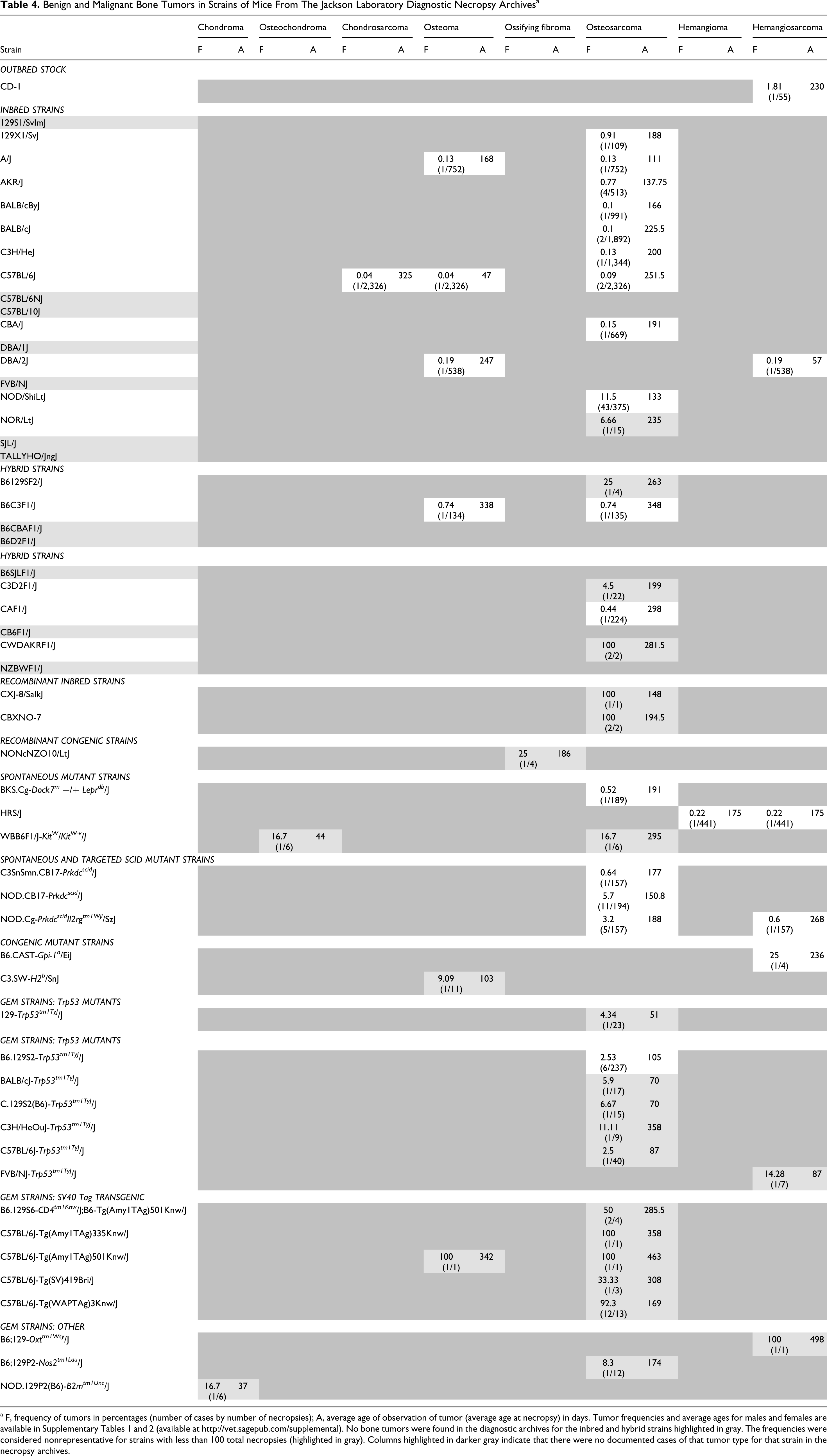
a F, frequency of tumors in percentages (number of cases by number of necropsies); A, average age of observation of tumor (average age at necropsy) in days. Tumor frequencies and average ages for males and females are available in Supplementary Tables 1 and 2 (available at http://vet.sagepub.com/supplemental). No bone tumors were found in the diagnostic archives for the inbred and hybrid strains highlighted in gray. The frequencies were considered nonrepresentative for strains with less than 100 total necropsies (highlighted in gray). Columns highlighted in darker gray indicate that there were no documented cases of that tumor type for that strain in the necropsy archives.
Discussion
Spontaneous bone tumors were infrequent in most strains of mice with the exceptions of osteosarcomas in NZBR/BI, NOD, and NOD-derived mice and osteomas in CF-1 and OF-1 mice. The most popular inbred strains of mice currently available through The Jackson Laboratory are 129S1/SvImJ, 129X1/SvJ, A/J, AKR/J, BALB/cByJ, BALB/cJ, C3H/HeJ, C57BL/6J, C57BL/6NJ, C57BL/10J, CBA/J, DBA/1J, DBA/2J, FVB/NJ, NOD/ShiLtJ, and SJL/J (JAX Mice, http://www.jaxmice.org/, September 2010). Among these, 129S1/SvImJ, C57BL/10J, DBA/1J, FVB/NJ, and SJL/J mice had no documented bone neoplasms in the diagnostic necropsy archives. This is noteworthy even though the colony sizes of these strains have varied through the years, many of the submitted mice were 8 months or younger, and focus of the diagnostic necropsy program was evaluation of sick mice rather than a systematic study of cancer with equal numbers of age- and sex-matched mice. Systematic aging studies 109 have been designed to better address such questions. In those mice with documented neoplasms, spontaneous osteogenic neoplasms occurred more frequently than cartilaginous, vascular, and hematopoietic ones, with osteoma being the most common benign bone tumor and with osteosarcoma being the most common malignancy of bone. Spontaneous cartilaginous neoplasms were the least frequent tumors of bone in mice, in direct contrast to chondrogenic neoplasms, the second-most common bone tumor type, after hematolymphoid malignancies in humans. In this context it is worth noting that chondrogenic tumors were produced by targeted mutations of Nfatc2 in mice, 84 but no correlative mutations of NFAT1 were identified in a set of human chondrosarcomas and enchondromas. 3 This disparity needs further investigation.
Another noteworthy aspect is that strains of mice with spontaneous neoplasms of bone varied in their propensity to develop metastases. Metastases were infrequent or nonexistent in many studies. At The Jackson Laboratory, the age at necropsy for mice with spontaneous or genetically engineered malignant neoplasms of bone ranged from 41 to 537 days (M = 215.55) and 35 to 990 days (M = 393.17) in the literature, and metastases were relatively rare. Most metastases that occurred were of osteosarcomas, and lung was the favored site. In some of these cases with relatively younger ages of occurrence, mice not living long enough to develop metastases could be a contributory factor. A more important determinant of strain-specific differences in metastasis is their inherent diversity with respect to genes that influence cell–cell and cell–matrix adhesion and cell migration. In a recently published study, the difference between C3H/BiDa and CzechII/Ei mice in metastasis of Murine polyoma virus–induced osteosarcomas was attributed to variability in the levels of the metalloproteinase MMP-2 and the transcription factor NFAT. 97 CZC3F1 mice in this study exhibited metastases, suggesting dominance of the metastatic phenotype. 97 In studies on spontaneous osteosarcomas, metastases were evident in “black” strains of mice, such as C57BL/Icrf, 26 C57BL/6J, 14 and C57BL/10J, 100 and absent in B6C3F1 mice. 9 Such an apparent contrast between 2 F1 hybrid strains of mice involving the C3H strain warrants further investigation of bone tumor metastases in inbred and hybrid strains.
Conspicuous disparities exist between classification systems for human, veterinary, and mouse bone and joint tumors. Unified systems of classification and characterization for human, veterinary, mouse, and genetically engineered bone tumors that reconcile differences in designation for similar entities would enhance comparative studies of tumors of the skeleton across species. The Mouse Pathology Ontology (Pathbase; http://eulep.pdn.cam.ac.uk/Pathology_Ontology/index.php) developed by a consortium of veterinary and physician pathologists has reconciled these differences.
Footnotes
Acknowledgements
We gratefully acknowledge D. Boggess for retrieval of archived necropsy records and materials; J. Miller, E. Taylor, and M. McKluskey for retrieval of archived glass slides; the Department of Histopathology and Microscopy Sciences for preparation of histologic sections from archived paraffin blocks; J. Hammer for assistance with preparation of figure panels; T. Stearns for assistance with the Ingenuity IPA Pathway Analysis Software and preparation of gene networks; N. Buckley for assistance with formatting some references; Dr. C. Bult for advice on interpretation of the gene network analyses; and Drs E. Leiter, L. Shultz, and W. Beamer for critical comments and suggestions.
The authors declared that they received no commercial financial support for research or authorship of this article.
This work was supported in part by grants from the National Institutes of Health (CA34196, JPS), the Ellison Medical Foundation (JPS), and institutional shared services at The Jackson Laboratory (AK, OF).
References
Supplementary Material
Please find the following supplemental material available below.
For Open Access articles published under a Creative Commons License, all supplemental material carries the same license as the article it is associated with.
For non-Open Access articles published, all supplemental material carries a non-exclusive license, and permission requests for re-use of supplemental material or any part of supplemental material shall be sent directly to the copyright owner as specified in the copyright notice associated with the article.