
Abstract
A 7-year-old male miniature schnauzer dog with unilateral cryptorchidism was presented for elective orchiectomy. Surgery to remove the cryptorchid testis revealed a fully formed uterus with horns attached to both testis and the body and cervix terminating at the prostate gland. The gross and microscopic diagnosis for the genital tract was persistent Müllerian duct syndrome with unilateral cryptorchidism. Additional associated lesions included cystic endometrial hyperplasia and a solitary, intratubular seminoma within the undescended testis. Persistent Müllerian duct syndrome is rare among domestic animals but is more common in miniature schnauzer dogs because of inheritance as an autosomal recessive trait.
Keywords
History, Gross Findings, and Lab Results
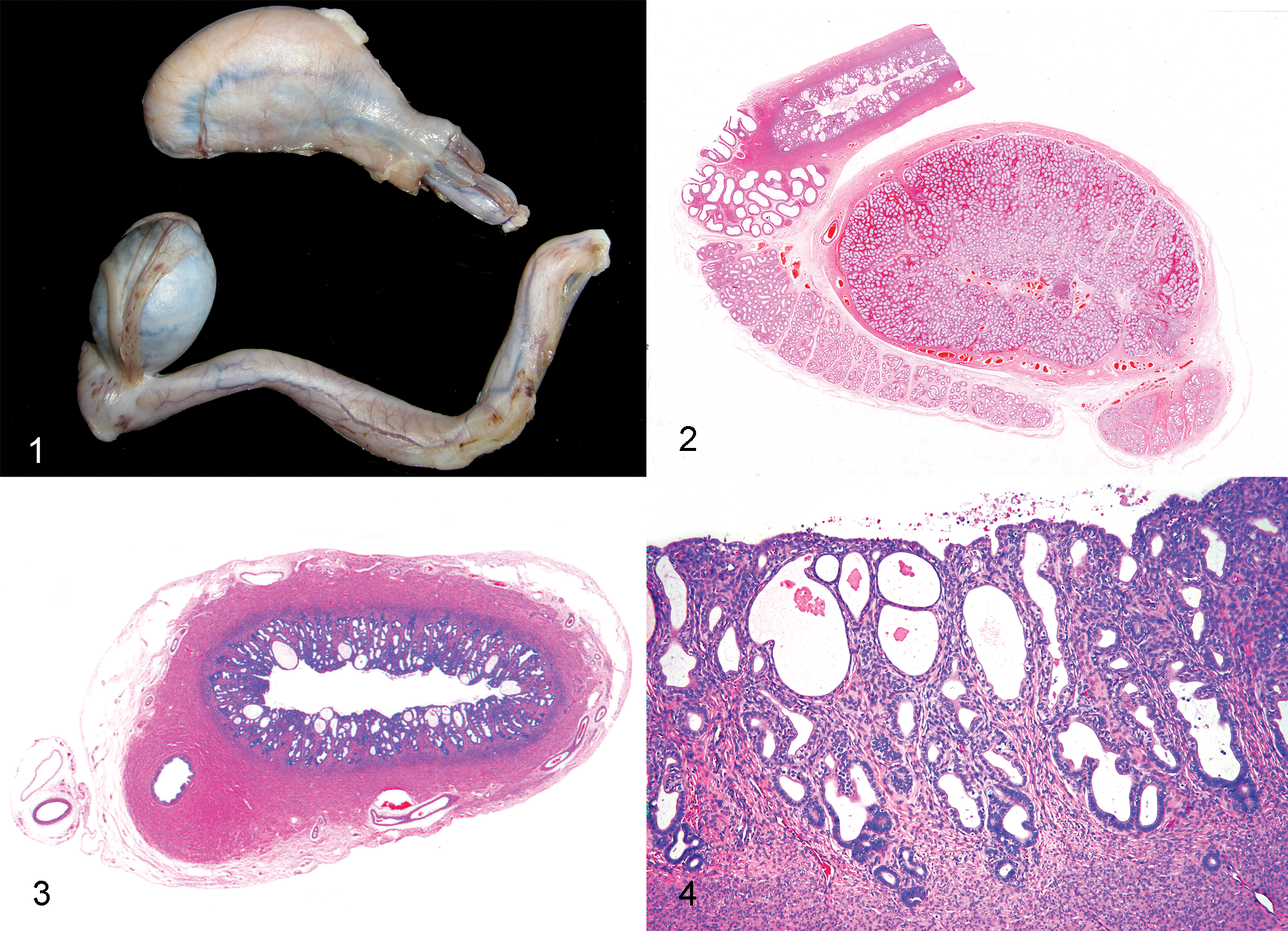
A 7-year-old intact male miniature schnauzer dog with a history of previously siring 2 litters and having offspring of 11 puppies in all was presented for elective orchidectomy. Only 1 testis, the left, was present within the scrotum; the right testis was not palpable in the scrotum or the inguinal area. Surgical exposure of the cryptorchid testis, in the region of the right internal inguinal ring, revealed a fully formed uterus, with the cranial aspect of the right uterine horn attaching to the tail of the epididymis. The uterine body and cervix terminated blindly on the dorsal aspect of the prostate gland, and the left uterine horn extended through the left inguinal ring, where it attached to the scrotal testis. Gonadohysterectomy was performed, ligating and transecting the uterine body near the cervix and transecting the right uterine horn as it passed through the inguinal ring. The scrotal testis and portion of the attached uterine horn was also surgically removed. The gonads and tubular genital tract were submitted for histopathological examination (Fig. 1).
Differential Diagnoses
Diagnoses to be considered for the presence of a feminized tubular genital tract accompanied by external male genitalia and gonads that grossly resemble testes include a true hermaphrodite, in which the gonads contain both ovarian and testicular components, and a male pseudohermaphrodite, in which the gonads are composed of only testicular tissue but are accompanied by a feminized or ambiguous tubular tract or external genitalia. Sex reversal, in which a genetic female with an XX karyotype develops into a phenotypic male or true hermaphrodite, could have been a consideration, had the dog not been a successful sire.
Microscopic Findings
The tubular portion of the genital tract was composed of fully formed uterine horns with development of all layers. The cranial portion of the uterine horn terminated near the tail of the epididymis (Fig. 2). The smooth muscle of the myometrium merged with the thick tunica muscularis of the ductus deferens (Fig. 3), the lumen of which was lined by typical pseudostratified and often ciliated columnar epithelium that occasionally demonstrated epithelial hyperplasia, forming intraepithelial lumens similar to that described in both epididymides. In the uterine horn attached to the undescended testis, the endometrium was approximately 1 mm thick, and numerous glands, many of which were moderately dilated, were lined by flattened epithelium and contained lightly eosinophilic amorphous material (Fig. 4). The lumen of this uterine horn contained similar eosinophilic material admixed with a small amount of cellular debris. The uterine horn associated with the scrotal testis had a much thinner endometrial layer (0.3 mm) with fewer glands in general, although several of the glands present were dilated, similar to those seen in the contralateral horn.
The cryptorchid testis was hypoplastic and had seminiferous tubules that were devoid of germinal epithelium and lined solely by morphologically normal Sertoli cells. This testis also contained a single discrete and unencapsulated focus in which 4 or 5 tubular lumens were filled with and obscured by sheets of large germinal cells accompanied by a few columnar or spindle-shaped cells presumed to be remaining Sertoli cells. In the epididymis, ducts were lined by moderately hyperplastic epithelium with the occasional formation of intraepithelial lumens. No sperm were seen within duct lumens. The scrotal testis was histologically unremarkable and had normally maturing germinal epithelium within tubules and abundant, mature spermatozoa within the lumen of the epididymis. Epithelial hyperplasia with intraepithelial lumen development, similar to that described in the contralateral gonad, was also seen in the ductus epididymis of the scrotal testis.
Diagnosis
The diagnosis was persistent Müllerian duct syndrome accompanied by cystic endometrial hyperplasia and unilateral cryptorchid testis containing a solitary, intratubular seminoma.
Discussion
Abnormalities in sexual development leading to intersex conditions can occur at 3 different levels, which include the chromosomal composition, gonadal structures, and phenotypic sexual development of the animal. 10 Pseudohermaphroditism, in which chromosomal and gonadal sex are the same but the development of internal or external genitalia are ambiguous or opposed, is the result of abnormal sexual development at the phenotypic level. 10 Persistent Müllerian duct syndrome (PMDS) is an example of male pseudohermaphroditism, and although rare among domestic animals, PMDS has been shown to be inherited as an autosomal recessive trait in miniature schnauzer dogs. 5 Affected dogs are typically normally masculinized males with an XY karyotype and testes accompanied by complete internal male genitalia as well as a female tubular genital tract composed of bilateral oviducts, bicornuate uterus, uterine body, cervix, and cranial vagina. 5 As in this case, many affected dogs (approximately 50%) are either unilaterally or bilaterally cryptorchid. 5 Bilaterally cryptorchid dogs are sterile, whereas dogs with PMDS and bilateral scrotal testes are usually fertile, despite lower than expected sperm counts. 13 Unilaterally cryptorchid affected males typically have lower than normal sperm counts 13 ; despite unilateral cryptorchidism, the dog described in this report was able to breed successfully.
In addition to cryptorchidism, other abnormalities that may be associated with PMDS in dogs include endometritis, pyometra, or hydrometra; cystitis; prostatitis; epididymitis; epididymal malformations; and testicular neoplasia.3,5,7,11,12 A small-diameter connection between the cranial vagina and the prostatic urethra has been described in 1 dog with PMDS accompanied by cystitis and pyometra, and this connection was the likely route of ascending infection. 5 Testicular neoplasms, especially Sertoli cell tumors, have been reported in several dogs with PMDS and are most frequently seen in the cryptorchid testis.1,3,11 The association between testicular tumors, namely, Sertoli cell tumors, seminomas, and mixed germ cell–stromal tumors, and failure of testicular descent, is well recognized.2,9 Abnormalities related to PMDS present in the dog described here included endometrial hyperplasia (without significant inflammation) in the uterine horn associated with the undescended testis and a single intratubular seminoma in the cryptorchid testis
Male pseudohermaphroditism may occur either because of failure of androgen-dependent masculinization or failure of Müllerian duct regression. In PMDS, androgen-dependent masculinization is normal; however, Müllerian ducts fail to regress. In normal males, Müllerian ducts regress as a result of Müllerian inhibiting substance (MIS) secretion by Sertoli cells. 6 In miniature schnauzers with PMDS, it has been shown that a normal amount of biologically functional MIS is secreted by Sertoli cells during the critical stage of gonadal development4–6; however, the Müllerian ducts fail to regress as a result of mutation in the MIS type II receptor gene (MISRII), such that the encoded receptor protein is truncated and either rapidly degraded or nonfunctional. 13 A molecular test for PMDS in miniature schnauzers has been developed based on the identical nature of the MISRII mutation in affected dogs of this breed. 8 Since approximately 50% of PMDS–affected miniature schnauzers have completely normal external genitalia and are fertile, this test could prove to be essential in decreasing and ultimately eliminating this inherited anomaly. 8
Footnotes
The authors declared that they had no conflicts of interest with respect to their authorship or the publication of this article.
The authors declared that they received no financial support for their research and/or authorship of this article.