
Abstract
A 14- × 12- × 6-cm (approximately 600 g) cryptorchid testis was surgically removed with the omentum from the posterior portion of the abdominal cavity of a 2-year-old male American Shorthair cat. At the time of a previous orchiectomy, a unilateral testis had been discovered in the scrotum. The cut surface of the cryptorchid testis consisted of solid areas with many cysts containing fluid and spongy soft areas. Histologically, the cryptorchid testis was composed of neuroectodermal components and diffuse immature glial tissues, cystic, tubular, and papillary epithelial tissues, and immature cartilaginous tissues. A metastatic mass in the omentum had histologic structures similar to those of the cryptorchid testis. The present case was diagnosed as teratoma in a feline unilateral cryptorchid testis.
Keywords
Teratomas are complex tumors with recognizable elements of multiple germ layers in various stages of maturation and are thus composed of multiple tissues foreign to the part of the body in which they arise. Testicular teratoma has been most commonly reported in the horse and is extremely rare in other animals, in which teratomas generally develop in the ovary. 1,2,4 Testicular tumors are rare in cats, and only two cases have been reported; both were Sertoli cell tumors. 3 Here, we describe a spontaneous teratoma in the unilateral cryptorchid testis of a cat.
A 2-year-old male American Shorthair cat was referred to the Animal Hospital with distention of the abdomen. In an exploratory laparotomy, a 14- × 12- × 6-cm mass (approximately 600 g) was found in the posterior portion of the abdominal cavity, and it was surgically removed together with the omentum. Because a unilateral testis had been found in the scrotum at the time of a previous orchiectomy, the mass in the abdominal cavity was suspected to be a cryptorchid testis.
The mass and the omentum were submitted to the laboratory for histopathologic examination. The cut surface of the mass consisted of solid areas with many cysts containing fluid and spongy soft areas. Gross examination of the omentum revealed a metastatic mass that was approximately 1 × 0.5 × 0.5 cm. Ultrasonography was performed approximately 1 month later and demonstrated ascites and many intra-abdominal masses that were approximately 2 cm in diameter. The cat died shortly thereafter, and a postmortem examination was not conducted.
The tissue samples were fixed in 10% phosphate-buffered formalin and processed routinely for light microscopic examination. Replicate sections of the tissue samples were used for immunohistochemical staining by the avidin–biotin–peroxidase complex method (Vectastain Elite kit, Vector Laboratories, Burlingame, CA) using primary antibodies against keratin, S-100 protein, lysozyme, carcinoembryonic antigen (CEA), glial fibrillary acidic protein (GFAP) (Dako Corp., Carpinteria, CA), neuron-specific enolase (NSE) (Nichirei Corp., Tokyo, Japan), neurofilament (monoclonal, Nichirei Corp.) vimentin, desmin, sarcomeric actin, and alpha-smooth muscle actin (monoclonal, Dako Corp.).
Histologically, the neoplasm was composed of primitive neuroectodermal, entodermal, and mesodermal tissues. Neuroectoderm-derived tumor components consisted of either dense clusters or multilayered rosettes of dark tumor cells surrounded by immature glial tissues (Fig. 1). Entodermal components consisted of single or multilayered epithelium-lined cysts, ranging in shape from cuboidal to columnar, with complex papillary invaginations into the cyst cavity (Fig. 2). Mesodermal components consisted of immature cartilaginous tissues containing hyaline cartilage, poorly differentiated chondrocytes, and fibrous connective tissues (Fig. 2). Numerous atypical mitotic figures were present in all the tumor cell types observed. Large areas of necrosis, edema, and hemorrhage were present. The metastatic mass in the omentum consisted of unorganized immature tumor cells, entodermal cysts, and immature cartilaginous tissues, but neuroectodermal tubular structures and multilayered rosettes were not present.
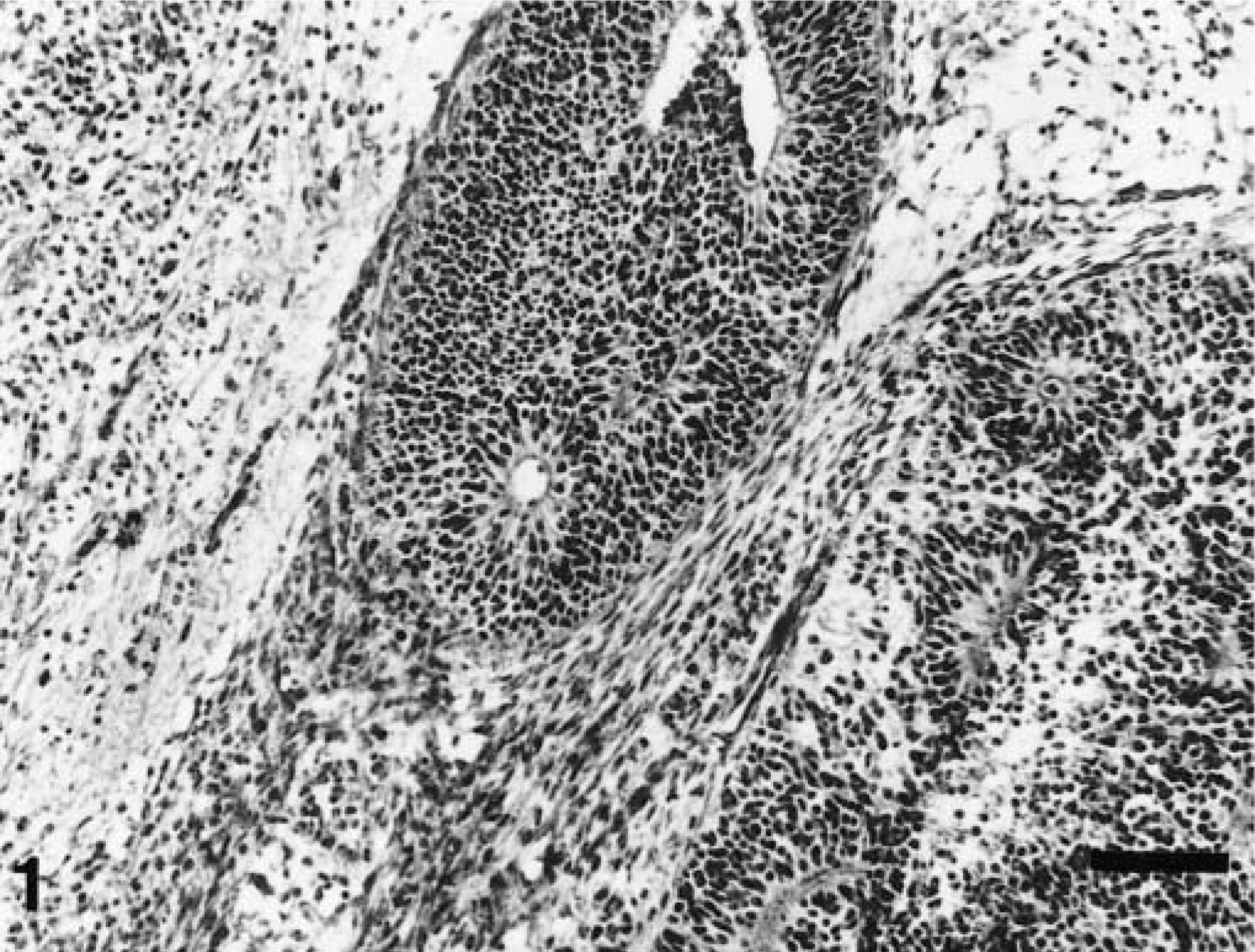
Cryptorchid testis; cat. Neuroectodermal tumor components consist of multilayered rosettes surrounded by immature glial tissues. HE. Bar = 60 µm.
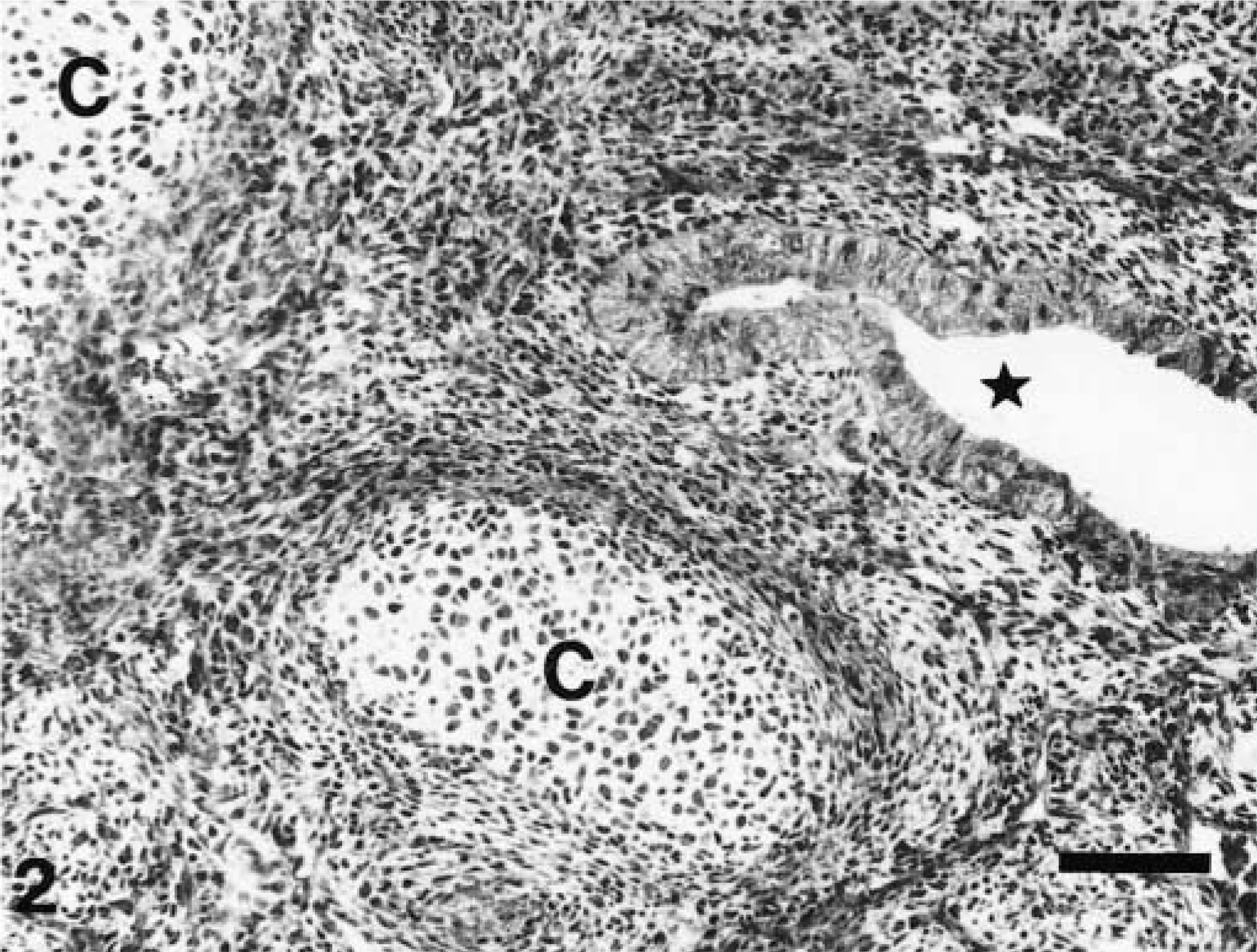
Cryptorchid testis; cat. Tumor mass contains a multilayered columnar epithelium–lined cyst (asterisk) and nests of hyaline cartilage surrounded by immature chondrocytes (C). HE. Bar = 60 µm.
Immunohistochemically, neuroectodermal dark tumor cells were positive for NSE, and immature glial cells were positive for GFAP. Entodermal components were positive for keratin and CEA. Mesodermal components were positive for vimentin.
Teratoma of the human testis is histologically classified as either mature teratoma or immature teratoma. 5 Mature teratoma is a neoplasm composed of stratified flattened epithelium, cartilage, bone, smooth muscle, or mature glandular tissue. Immature teratoma contains of primitive neuroectoderm, entoderm, or mesoderm that resembles normal embryonic tissues. Pure immature teratoma is extremely rare. In the present case, based on the clinical history of the cat and the results of histopathologic and immunohistochemical examination, the tumor was diagnosed as teratoma in the unilateral cryptorchid testis. The abdominal masses observed on ultrasonography were presumed to be metastases to the peritoneum. In humans, teratomas most often metastasize through the lymphatic vessels, 5 but in the absence of a postmortem examination we were unable to determine whether metastasis was via lymphatics or implantation.