
Abstract
White lions (Panthera leo krugeri) have never been common in the wild, and at present, the greatest population is kept in zoos where they are bred for biological and biodiversity conservation. During the years 2003 to 2008 in a zoological garden in northern Italy, 19 white lions were born to the same parents, who were in turn paternally consanguineous. Out of the 19 lions, 4 (21%) were stillborn, 13 (69%) died within 1 month, and 1 (5%) was euthanatized after 6 months because of difficulty with prehension of food. Six lions (32%) showed malformations involving the head (jaw, tongue, throat, teeth, and cranial bones). One lion (5%) still alive at 30 months revealed an Arnold–Chiari malformation upon submission for neurological evaluation of postural and gait abnormalities. Paternal consanguinity of the parents, along with inbreeding among white lions in general, could account for the high incidence of congenital malformations of the head in this pride of white lions.
White lions (Panthera leo krugeri) were first recorded in 1928 and in the early 1940s in the Timbavati and Kruger National Park regions of South Africa. They came to public attention in the 1970s in Chris McBride’s books 5,6 and following the activity of the Global White Lion Protection Trust. In 2003, the first reintroduction of white lions to their natural endemic range in South Africa was successfully initiated. 3,14 White lions are considered a special part of the biodiversity of the region because they are revered by the local communities, which hold them sacred.
White lions could be considered a separate subspecies (http://www.whitelions.org/); they have never been common in the wild. At present, the greatest population of white lions is in zoos, where they are bred for biological and biodiversity conservation.
In this article, we report congenital cranial malformations in a pride of white lions. A few reports of skull and brain malformations in captive wild felids exist in the literature, and these are mostly regarding Arnold-Chiari malformation in lions. 1,7,8,11,12
Materials and Methods
During the years 2003 to 2008 in a zoological garden in northern Italy, 19 white lions were born to the same parents, who were paternally blood related (ie, from parents born to a common father). Out of the 19 lions, 4 were stillborn (case Nos. 12, 13, 17, and 18); 13 died within 1 month (case Nos. 1–4, 6–11, 14–16); and 1 was artificially fed until 6 months and then euthanatized because of difficulty with prehension of food (case No. 5) (see Table 1 ). Necropsies were performed on all animals. Radiographs were taken in 4 cases (case Nos. 5, 12–14), and a computerized axial tomography (CAT) scan was carried out in 1 (case No. 17). The only lion still alive, at 30 months of age, was submitted for neurological evaluation because of posture and gait abnormalities (case No. 19).
Nineteen White Lions Bred in a Zoological Garden in Northern Italy, 2003–2008
a Still alive.
Results
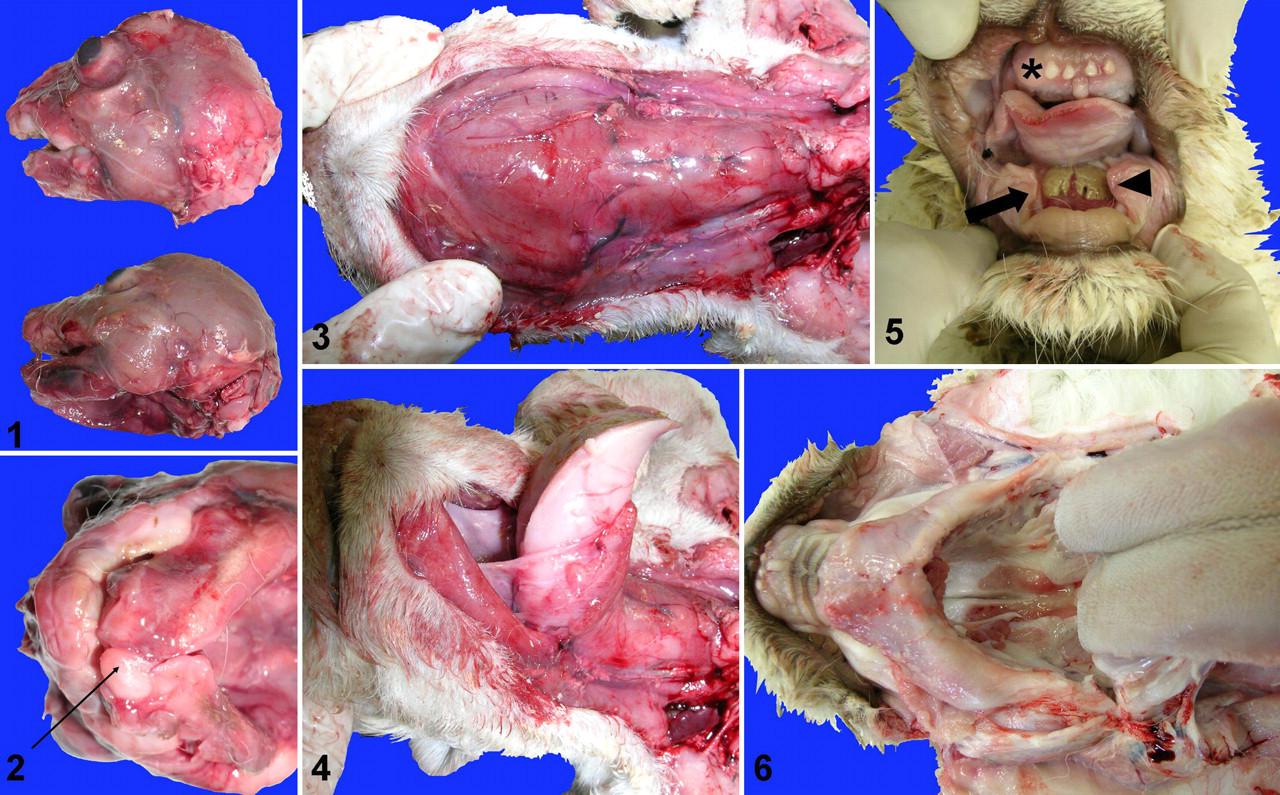
At necropsy, 1 stillborn, 3 newborn, and 1 juvenile lion showed micrognathia (case Nos. 5, 6, 10, 12, and 13; Fig. 1). In 1 lion, there was a left atrial cardiac cyst 3 mm in diameter (case No. 10). One lion (case No. 13) also showed gnathoschisis (Fig. 2), increased volume of soft tissues of the throat and epiglottis (Fig. 3), and macroglossia (Fig. 4).
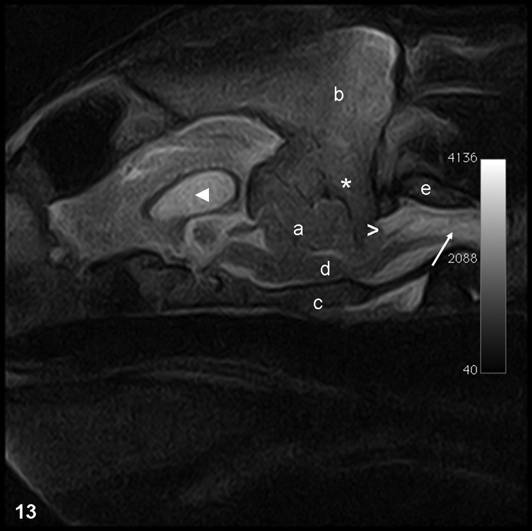
White lion, case No. 19. Magnetic resonance image, fast spin echo T2-weighted image (TR, 4000 ms; TE, 114 ms) of the brain on sagittal plane showing thickening of the occipital bone and crowding of the caudal fossa (asterisk), mild hydrocephalus (arrowhead), cerebellum compression with herniation through the foramen magnum (chevron) into the vertebral canal, and syringohydromyelia (arrow). Performed with 0.23 T MRV (Paramed, Genoa, Italy). a, cerebellum; b, occipital bone; c, basioccipital bone; d, medulla oblongata; e, atlas.
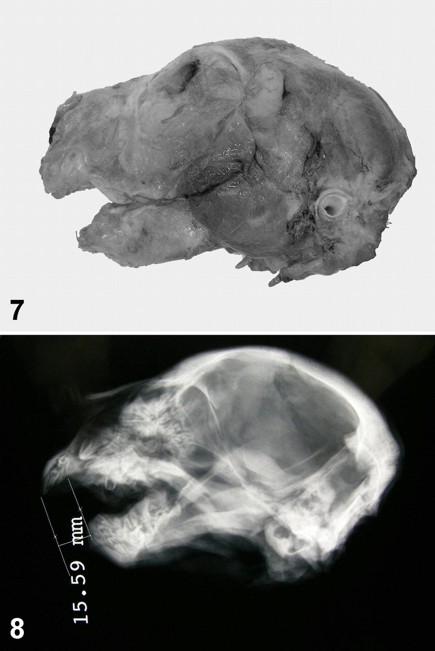
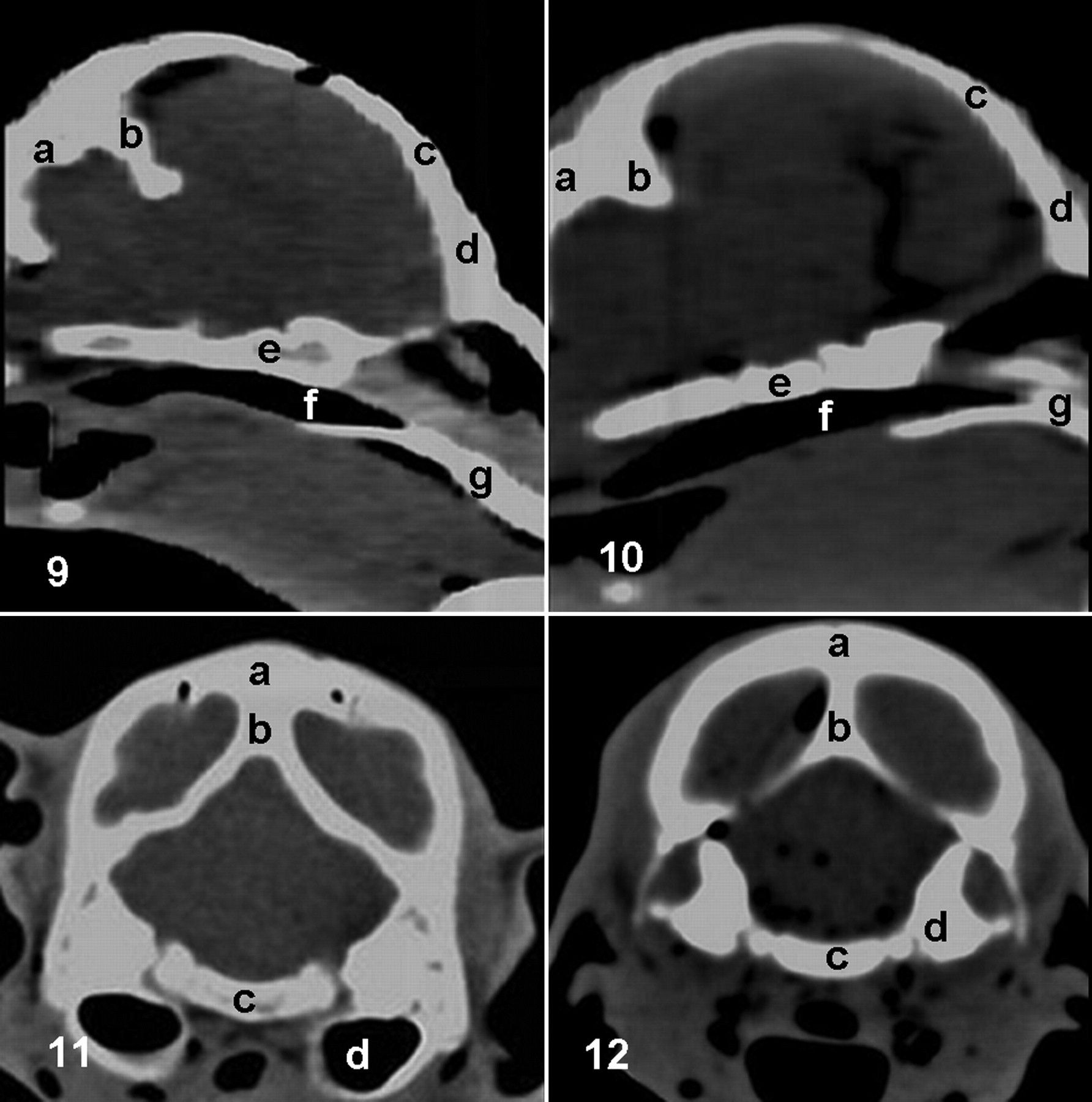
The juvenile, euthanatized at 6 months (case No. 5), also showed maxillary oligodontia, mandibular anodontia, gingival ulcers of the lower jaw (Fig. 5), increased thickness of the mandibular symphysis (Fig. 6), micrognathia (Figs. 7, 8), and small renal cysts. In 3 lions, a gross morphologic diagnosis of suppurative bronchopneumonia was made (case Nos. 3, 4, and 16), whereas in 3 animals, milk was found within the airways, allowing a diagnosis of aspiration pneumonia (case Nos. 1, 2, and 15). In 1 stillborn lion (case No. 17), CAT scan revealed a diffuse thickness of the cranial bones and sagittal craniosynostosis (Figs. 9–12).
No other gross skull abnormalities (or any other abnormalities in general) were detected in the lions.
The only lion that survived (case No. 19) showed moderate ataxia, slight horizontal nystagmus with fast phase to the right, absence of menace response, and slight mydriasis. Magnetic resonance imaging revealed thickening of the occipital bone, crowding of the caudal fossa, resultant mild hydrocephalus, cerebellar compression, and herniation of the cerebellar vermis through the foramen magnum. A pathological hyperintensity in the cervical spinal cord was observed, consistent with syringohydromyelia (Fig. 13). Following these investigations, a diagnosis of Arnold–Chiari malformation was made.
Discussion
Inbreeding depression is a decline in the value of a trait as a direct consequence of inbreeding. 13,15 The most common estimates of inbreeding depression involve traits closely related to fitness, such as reproductive traits (eg, number of eggs laid, number of young surviving), or metric traits indirectly associated with fitness (eg, ejaculate volume, body mass). 2
Most of the literature concerning lesions associated with inbreeding in animals has concentrated on domestic or captive-bred wild species 4,9 because of the obvious difficulties in estimating their incidence in free-living animals. 2
Congenital anomalies and malformations described in white lions here involved the head (jaw, tongue, throat, teeth, and cranial bones), with resulting nervous symptoms and clinical signs. The Arnold–Chiari malformation is a well-known syndrome in humans and dogs (mostly found in Cavalier King Charles Spaniels) that is also reported in lions. 1,7,8,11,12 For this condition (one considered a developmental abnormality), an overly small skull and the resultant pressure on the spinal cord are responsible for an increase in pressure of cerebral spinal fluid flow, resulting in compression of the cerebellum and, eventually, syringomyelia. There is strong evidence that the condition is heritable. 10
The present high incidence of cranial malformations detected in white lions derived from paternally consanguineous parents further attests to inbreeding depression in white lions, as already suggested by the limited number of these animals in zoos.
Even if the aim is to reestablish white lions within their natural distribution range, care must be taken to avoid excessive inbreeding, which can limit the gene pool in the existing population.
Footnotes
Acknowledgements
We thank Dr Federica Sammartano and Dr Anna Tomba for their assistance in computerized axial tomography scan and magnetic resonance imaging interpretation.
The authors declared that they had no conflicts of interest with respect to their authorship or the publication of this article.
The authors declared that they received no financial support for their research and/or authorship of this article.