
Abstract
Canine renal cell carcinomas (RCCs) are uncommon aggressive tumors that occur mainly in middle-aged male dogs. Their histologic classification bears no relationship with prognosis, and little information is available concerning their immunohistochemical properties. In this retrospective study, formalin-fixed, paraffin-embedded tissues from 13 canine RCCs were retrieved from the archives, classified histologically, and evaluated immunohistochemically. The dogs were 7 males and 6 females (1 spayed) of 10 different breeds, averaging 8 years in age. The tumors were classified as papillary, tubulopapillary, papillary–cystic, solid, or sarcomatoid. All 13 tumors were immunohistochemically positive for uromodulin, 12 for c-KIT, 11 for vimentin, 9 for wide-spectrum-screening cytokeratins, 7 for cytokeratins AE1/AE3 and carcinoembryonic antigen, 4 for cytokeratins CAM 5.2, and 3 for CD10. All 3 solid RCCs expressed vimentin, c-KIT, and carcinoembryonic antigen and were negative for cytokeratins. All 7 papillary and tubulopapillary tumors expressed vimentin; 6 (86%), cytokeratins; and 6 (86%), c-KIT. Both papillary–cystic RCCs were positive for cytokeratins and c-KIT and negative for vimentin. These results indicate that the different histologic types of RCC have characteristic immunohistochemical profiles and that c-KIT may be involved in the pathogenesis of canine RCC.
Renal cell carcinomas (RCCs)—also known as renal carcinomas, malignant nephromas, clear cell carcinomas, or Grawitz tumors—are uncommon in domestic animals. Even so, they are the most common canine primary renal tumor, with reported prevalence from 1.5 per 100 to 1.5 per 100,000 dogs. 3,13 Other canine renal epithelial neoplasms include renal adenoma, oncocytoma, transitional cell carcinoma, papilloma, and squamous cell carcinoma. 11 RCC is more common in male middle-aged dogs (8 to 9 years old), without breed predilection. 8 Metastatic disease is detected by thoracic radiography in 50% of dogs at presentation. 8 RCCs are classified on the basis of predominant histologic pattern, as papillary, tubular, or solid. These histologic types can be further classified as chromophobic, eosinophilic, or clear cell variants; all 3 cell types are usually present. 12 Unfortunately, classification by histologic or cytologic criteria is not predictive of biological behavior in domestic animals. 11 The immunohistochemical characterization of human RCC has proved useful in refining tumor classification and notions about its biological behavior. 1 The purpose of this study was to correlate the immunohistochemical profile of 13 canine RCCs with histologic classification.
Materials and Methods
Thirteen paraffin blocks representing 13 canine RCCs diagnosed between 2000 and 2007 were retrieved from among 13,037 canine neoplasms in the archives of three laboratories (Instituto de Ciências Biomédicas Abel Salazar, University of Porto; University of Trás-os-Montes e Alto-Douro; and Gram Laboratório de Anatomia Patológica Veterinária). The specimens, which had been fixed in formalin for 24 to 72 hours, were checked for strong vimentin immunoreactivity in stromal cells to ensure suitability for immunohistochemistry.
Table 1 presents the signalment of the 13 dogs. All tumors had been obtained by nephrectomy. Because no dog had other masses detected by physical examination, radiography, or laparotomy or any history of malignant tumors, the renal tumors were considered primary. Regional lymph nodes were not evaluated histologically.
Signalment of 13 Dogs with Renal Cell Carcinoma
a M, male; F, female; FS, spayed female.
b NR, not recorded.
Serial sections from each tumor, 2 μm in thickness, were used for hematoxylin and eosin staining and immunohistochemistry following a standard avidin–biotin–peroxidase complex method. Table 2 presents the source, clone, dilution, and antigen retrieval method for each antibody. Antibodies against cytokeratins (CKs) included AE1/AE3 (high molecular weight CKs 1, 2, 3, 4, 5, 6, 10, 14, 15, and 16 and low molecular weight CKs 7, 8, and 19), CAM 5.2 (CKs 7/8), and wide-spectrum screening representing a range of bovine muzzle epidermal keratins. Each tumor was also tested with antibodies against uromodulin, vimentin, c-KIT, CD10, and carcinoembryonic antigen (CEA).
Antibody Source, Dilution, and Antigen Retrieval for Immunohistochemistry a
a CK, cytokeratin; WSS, wide-spectrum screening; CEA, carcinoembryonic antigen.
b Citrate buffer, pH 6.9.
Tumors were classified by predominant histologic pattern according to World Health Organization criteria. 12 The presence or absence of necrosis and nuclear pleomorphism (+ slight, ++ moderate, +++ marked) were also noted. Mitotic index was assessed as the number of mitotic figures per 10 high-power fields (400×) in areas with the predominant histologic pattern near the tumor’s margins. The immunoreactivity for each marker was assessed semiquantitatively (– negative, + weakly positive, ++ moderately positive, +++ strongly positive).
Results
All 13 tumors were unilateral solitary masses. The tumors were classified as papillary (n = 4), tubulopapillary (n = 3), papillary–cystic (n = 2), solid (n = 3), and sarcomatoid RCCs (n = 1; Table 3 ).
Histologic and Immunohistochemical Features of 13 Canine Renal Cell Carcinomas
a Scoring: +, slight; ++, moderate; +++, marked.
b Mitotic index: No. mitotic figures per 10 high-power fields.
c Scoring: –, negative; +, weak; ++, moderate; +++, strong. See Table 2 for details on immunohistochemical antibodies.
Papillary and tubulopapillary tumors consisted of a dense, cuboidal to columnar cell population, forming papillae alone or mixed with tubules and supported by a delicate fibrovascular stroma. The neoplastic cells had a round, central, or basal nucleus, slight to moderate nuclear pleomorphism, and low to moderate amounts of pale eosinophilic cytoplasm.
Papillary–cystic tumors had cysts filled with mucin and cellular debris, surrounded by a sparser, cuboidal to columnar cell population forming small intracystic papillae supported by a dense fibrous stroma with moderate multifocal infiltration by macrophages with hemosiderin and ceroid accumulation. Cytologically, the neoplastic cells of papillary–cystic RCCs resembled those of papillary tumors, but the cells had more abundant, mostly vacuolated cytoplasm.
The single sarcomatoid RCC was composed of a dense spindle-cell population arranged in whorls in a moderately dense, poorly vascular fibrous stroma. Multifocally, neoplastic cells were polygonal with limited or abortive tubule formation. Nuclei were markedly pleomorphic; the cytoplasm was pale eosinophilic with scattered vacuolation and distinct cell borders.
Solid tumors were densely cellular. Sheets of neoplastic cells were supported by scanty, inconspicuous fibrous stroma. Neoplastic cells were polygonal to round with a high nucleus:cytoplasm ratio.
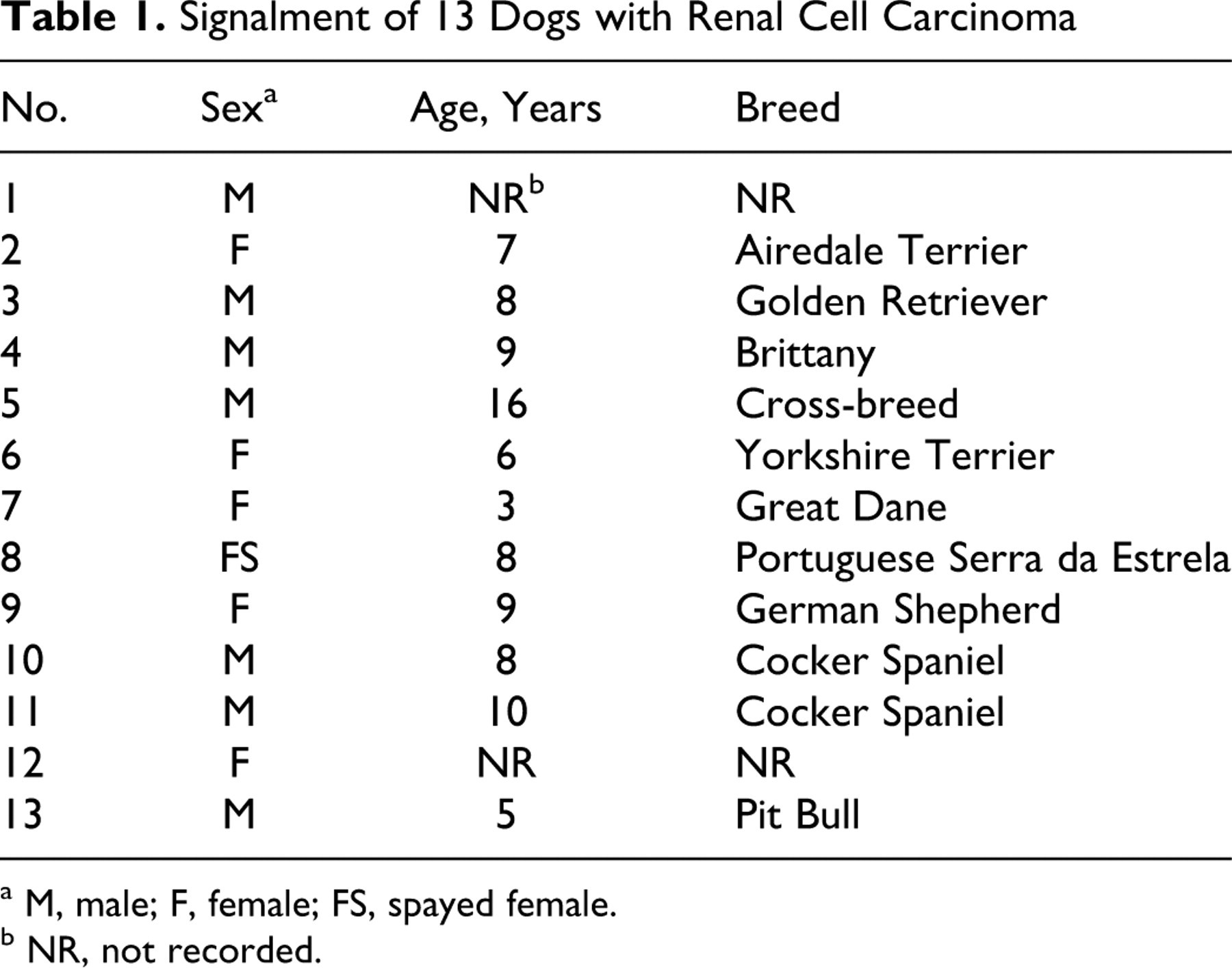
Among the 13 RCCs, mitotic index varied from 4 to 62 mitotic figures per 10 high-power fields (Fig. 1 ). All but case No. 5 had multifocal to coalescing necrosis. Renal tissue at the periphery of the tumor was compressed, with glomerular atrophy and interstitial fibrosis. In one tumor (No. 12), neoplastic cells invaded vessels. In another case (No. 2), the tumor invaded the adjacent adrenal gland.
Table 3 presents immunohistochemistry results. All tumors had weak to strong, diffuse cytoplasmic expression of uromodulin (Fig. 2 ). The three different antibodies to CKs—wide-spectrum-screening CKs, AE1/AE3, and CAM 5.2—were reactive with 9 tumors (Nos. 1, 2, and 4–10), 7 (Nos. 2, 4, and 6–10), and 4 (Nos. 2 and 7–9), respectively. CK immunoreactivity was concentrated in a few cell clusters in many cases and was membranous, especially apical, but extending to the basolateral cell membrane or into the cytoplasm (Fig.3 ). Moderate to strong, diffuse cytoplasmic vimentin expression (Fig. 4 ) was detected in 11 tumors (Nos. 1–7 and 10–13). Cytoplasmic, granular, generally moderate c-KIT immunoreactivity (Fig. 5 ) was observed in 12 tumors (Nos. 1–6 and 8–13). CD10 immunoreactivity was detected in 3 tumors (Nos. 2, 8, and 11), mostly in the apical cell membrane or, less commonly, as a focal cytoplasmic reactivity (Fig. 6 ). Weak to moderate CEA expression was detected in 7 tumors (Nos. 2, 4, 8, and 10–13), either in the apical cell membrane or diffusely in the cytoplasm. In all cases with adjacent nonneoplastic renal tissue, only distal tubules had intense, diffuse cytoplasmic immunoreactivity for uromodulin. Renal tubular and ductal epithelium and the urothelium had strong, multifocal to diffuse cytoplasmic labeling with antibodies to CKs. Stromal cells had strong, diffuse immunoreactivity for vimentin. Tubular epithelium had weak, granular cytoplasmic immunoreactivity for c-KIT. Renal tubules also had strong, diffuse immunoreactivity for CD10 and moderate reactivity for CEA (for both antibodies) with the apical cell membrane. Strong, diffuse cytoplasmic CEA expression was also detected in the urothelium.
Discussion
The number of tumors retrieved from the archives constituted approximately 10 RCCs per 10,000 canine neoplasms. Because there is no accurate count of the Portuguese canine population and because this population is served by laboratories besides those contributing to this study, these data cannot be readily compared with reported prevalence. 3,13 The average age of the dogs in the present study was 8 years. No sex or breed predilection was identified, although male dogs are reportedly at increased risk of developing RCCs. 11
Human RCCs are classified into three types with distinct prognostic implications: clear cell (conventional), papillary, and chromophobe RCCs. 4 The histologic patterns and cytologic features of canine RCCs do not match those of human RCC. 10 Instead, canine RCCs often have a mixture of cell types (clear cell, chromophobe, eosinophilic) 12 that renders the human classification inapplicable. Tubular carcinoma is considered the most common histologic pattern in RCCs of domestic animals. 11 However, in this series, papillary carcinoma was the most common pattern; 9 tumors had papillary differentiation, whereas only 3 had predominantly tubulopapillary differentiation. In accordance with the literature, 11 all tumors had a mixture of histologic patterns, and necrosis was identified in all but one case. The average mitotic index was 30.8. Interestingly, the 2 papillary–cystic tumors had lower mitotic indices (only 4 or 5 mitotic figures per 10 high-power fields). Remarkably, despite the high mitotic indices in some tumors, only one case had histologic evidence of vascular invasion, and the mitotic index in that case was 18. Follow-up data would be needed to correlate mitotic index with case outcome in canine renal carcinomas.
A hereditary form of renal cystadenocarcinoma and nodular dermatofibrosis has been described in German Shepherds with germline mutations of the BHD gene. 9 Dog No. 9 was a female German Shepherd with a papillary–cystic RCC, but neither nodular dermatofibrosis nor BHD gene mutation was reported in this dog.
To our knowledge, this is the first immunohistochemical characterization of canine RCCs. As in a bovine study, 7 uromodulin immunoexpression was detected in all renal cell tumors. This finding supports the hypothesis that RCCs originate from the distal convoluted tubules. The sarcomatoid RCC (a histologic type seldom recognized in veterinary pathology 19 ) was also positive for uromodulin and for vimentin and CK coexpression, which helped confirm the diagnosis. CKs were detected immunohistochemically in 9 of 13 tumors. Wide-spectrum-screening anti-CKs labeled a few more tumors than did antibodies to AE1/AE3 or CAM 5.2. Interestingly, 8 of 9 RCCs with papillary differentiation reacted with at least one of these anti-CKs, whereas all solid RCCs (a less differentiated pattern) were negative for CKs. Vimentin, a cytoskeleton protein commonly found in mesenchymal cells, was moderately to strongly expressed by all RCCs, except the 2 papillary–cystic tumors. In mammals, the renal epithelium develops from the mesenchymal blastema, losing vimentin and gaining CK expression. 6 The expression of vimentin and CKs by epithelial cells (as in the epithelial component of porcine nephroblastomas) has been proposed to reflect origin from remnants of embryonic mesenchyme. 6 It might also reflect an origin from partly undifferentiated renal stem cells or an acquired epithelial–mesenchymal transition associated with the development of an invasive phenotype. 16
C-KIT is a transmembrane tyrosine kinase growth factor receptor that—when bound by its ligand, stem cell factor—triggers signaling pathways that lead to cell proliferation and resistance to apoptosis. Mutations of the c-kit proto-oncogene that produce constitutively active forms of c-KIT have been associated with several canine and human malignancies. 5 Cytoplasmic c-KIT immunoexpression was detected in all but one tumor, regardless of histologic type, in contrast to human RCCs, in which c-KIT expression is characteristic of the chromophobe type. 14,18 In canine mast cell tumors, changes in c-KIT expression, from the normal membrane-associated pattern to a cytoplasmic pattern, are related to c-kit exon 11 activating mutations 17 and have important prognostic implications. The cytoplasmic pattern of c-KIT expression in canine RCCs raises the possibility that c-kit mutations might also have a role in the pathogenesis of these tumors. CD10 (also known as common acute lymphocytic leukemia antigen) is a cell surface metalloendopeptidase called neprilysin, expressed on the brush border of renal tubules and by human clear cell and papillary RCCs. 2 CD10 immunoexpression was present in only 3 of 13 cases and did not seem to be associated with any specific histologic pattern. In contrast, all 3 solid RCCs had moderate CEA immunoreactivity, compared with the only occasional positivity in other histologic types. Human RCCs are usually negative for CEA, whereas human urothelium and urothelial carcinomas are mostly positive. 15 However, the polyclonal antibodies recognize a number of CEA family and other related proteins, 15 so these results should be corroborated with studies using monoclonal antibodies.
In summary, all 3 solid tumors were negative for CKs and positive for vimentin, CEA, and c-KIT. All 7 papillary or tubulopapillary tumors expressed vimentin; 6 of 7 expressed CKs; and 6 of 7 expressed c-KIT. In contrast, both papillary–cystic RCCs were negative for vimentin but positive for CKs and c-KIT and had lower than average mitotic index.
The immunohistochemical profile of these canine RCCs differs from that of human RCC. 1 For example, CD10 expression was detected in only 23% of the canine RCCs but is present in up to 90% of all clear cell RCCs and 67% of all papillary human RCCs. 1 C-KIT immunopositivity was detected in 8 of 9 and in 3 of 3 canine tumors with papillary or solid differentiation, respectively, whereas this marker is characteristically absent in human papillary or clear cell RCCs but is found in 55% of human chromophobe RCCs. 1,14,18 The expression of CEA in 7 of 13 canine RCCs highlights another difference from human RCC, which is generally negative for this marker. 15 However, both species have vimentin expression in certain RCC types. 10,13,17
A weakness of this study is the lack of information regarding postsurgical survival or case outcome. Invasion of adjacent adrenal gland was observed in one dog; another had evidence of local vascular invasion. The lack of evidence of metastatic disease at diagnosis might simply reflect an earlier stage of disease in these dogs that were selected for nephrectomy. However, regional lymph nodes were not evaluated at surgery; thoracic or abdominal imaging studies were not available; and necropsy was not performed on any dog. Thus, in this retrospective study, we could not correlate histologic pattern or immunohistochemical profile of the RCCs with their biological behavior.
Footnotes
Acknowledgements
Rui M. Gil da Costa is supported by FCT research grant SFRH/BD/37565/2007, financed by the Portuguese Ministry of Science and Technology and the Social European Fund.
The authors declared that they had no conflicts of interest with respect to their authorship or the publication of this article.
The authors declared that they received no financial support for their research and/or authorship of this article.