
Abstract
An approximately one-and-a-half-year-old, neutered male, mixed-breed dog was presented for a chronic history of vomiting. Profuse diarrhea was also noted during examination. An exploratory laparotomy was performed, bone chips were removed from the stomach, and a raised, circular area of gastric mucosa was biopsied. Histologically, there was severe gastric cryptosporidiosis as well as numerous spiral bacteria, consistent with Helicobacter spp. Polymerase chain reaction revealed visible bands for the 18S ribosomal RNA gene for Cryptosporidium spp. The polymerase chain reaction product was sequenced and was found to be most similar to Cryptosporidium muris. Both the gastric location and the species of Cryptosporidium are unusual in a dog.
Keywords
A one-and-a-half year old Chihuahua mix dog was presented to the referring veterinarian with a 2-month history of vomiting. During examination, the dog also had multiple episodes of profuse diarrhea. Hematocrit was 22%. Abdominal radiographs revealed radio-opaque material in the stomach. Based on radiographic findings, the dog was scheduled for an exploratory laparotomy. During surgery, numerous bone fragments were removed from the stomach and distal esophagus. A slightly raised, 0.75 × 1.0 cm, reddened, circular area was noted on the mucosal surface of the stomach. An incisional, full-thickness biopsy of this area was obtained, placed in 10% formalin, and submitted to the Athens Veterinary Diagnostic Laboratory for histopathologic evaluation. Following surgery, the dog was treated with one fourth of a 250-mg metronidazole tablet twice daily for 6 days, 100 mg of ferrous sulfate once daily for 30 days, and 5 mg of omeprazole once daily for 14 days. At a recheck 3 weeks post surgery, hematocrit had improved to 35%. Six weeks post surgery, the client reported that the dog had returned to normal, and the dog was subsequently lost to follow-up.
Differential Diagnoses
There are numerous differential diagnoses for chronic vomiting and diarrhea in dogs. Based on laparoscopic findings, the presence of foreign bodies (bone chips) with focal gastric irritation was the primary rule out. However, other causes of vomiting and diarrhea in dogs include Campylobacter spp, Helicobacter spp, Pythium insidiosum, coccidia, and inflammatory bowel disease. Causes of diarrhea without vomiting include Giardia lamblia, metazoan parasites such as roundworms or whipworms, Histoplasma capsulatum, and Prototheca spp. Causes of vomiting without diarrhea include hypersensitivity reactions, neoplasia, and gastric ulcers. Because the dog had a 2-month history of illness and appeared bright and alert, systemic diseases such as parvoviral enteritis and distemper were not considered.
Microscopic Findings
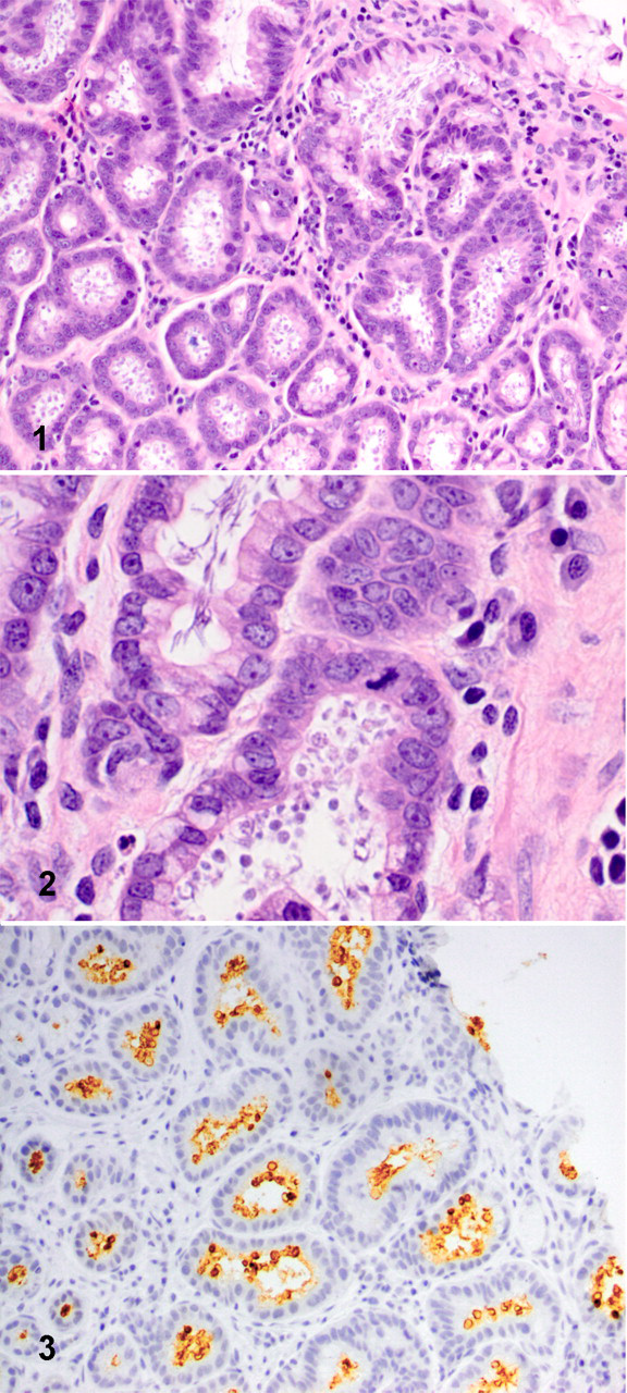
Gastric glands were filled with round, 2 to 4 μm, basophilic organisms (Fig. 1). Numerous corkscrew shaped bacteria 4 to 10 μm in length, consistent with Helicobacter spp, were also noted along the mucosal surface, within gastric glands, and less frequently within the cytoplasm of parietal cells (Fig. 2 ). Few lymphocytes and plasma cells and rare eosinophils were noted in the lamina propria between glands. Immunohistochemistry for Cryptosporidium spp was performed on formalin-fixed paraffin-embedded tissue using a peroxidase labeling system with diaminobenzidine as the chromagen. The primary antibody was an anti-Cryptosporidium spp mouse monoclonal from VMRD Inc. (Pullman, WA) and was used at a 1:100 dilution. Immunohistochemistry for Cryptosporidium spp was strongly positive (Fig. 3 ).
Polymerase Chain Reaction
Polymerase chain reaction (PCR) was performed as previously described using Cryptosporidium spp 18S ribosomal RNA (rRNA) as the target. 1 Amplification of DNA extracted from formalin-fixed, paraffin embedded sections of stomach and subsequent sequencing confirmed the organism to be C muris. Additional PCR for Cryptosporidium spp using additional gene targets to further confirm the phylogenetic placement of the isolate and PCR for Helicobacter spp were not performed because of a lack of any additional sample on which to perform these tests.
Discussion
The exact causes of clinical disease in this dog are unknown because only a gastric biopsy was submitted. The bone chips in the stomach may have played a role through interference with gastric emptying and alterations in acid and enzyme secretion, resulting in an abnormal microenvironment that allowed proliferation of C muris and Helicobacter spp. However, the profuse diarrhea during the physical examination suggests that there may have been an enteric pathogen as well. Although delivery of maldigested ingesta to the intestine is a possible cause for diarrhea, primary enteritis cannot be excluded.
Both Cryptosporidium spp and Helicobacter spp are considered rare or uncommon causes of gastroenteritis in dogs. Helicobacter spp are commonly present in both normal dogs and those with gastrointestinal disease. One study found that 82.3% of dogs tested by PCR performed on nucleic acid extracted from gastric biopsies were positive for Helicobacter spp. 4 A second study of 30 dogs using a combination of urease activity, histology, PCR, and enzyme-linked immunoassay found a similar infection rate with Helicobacter spp of 76.7%. 11 In this study, there was no significant association between Helicobacter spp infection and proinflammatory cytokine expression or severity of gastritis. However, in a study of Helicobacter felis and C muris in mice, inoculation of H felis acted similarly to stress to activate C muris, resulting in gastric inflammation as determined by histologic examination of gastric tissue. 9
Clinical signs of cryptosporidiosis, which may include diarrhea, decreased appetite, and weight loss, occur only sporadically in dogs, although prevalence rates of 2 to 80% have been reported using either an auramine–rhodamine fluorescent staining procedure on feces or indirect immunofluorescence assay on serum. 3,10 There are at least 10 distinct species in the genus Cryptosporidium. In dogs, Cryptosporidium canis is by far the most common, as determined by fecal PCR. 1,7 However, a study of a group of dogs in Texas identified C muris as the only isolate using a fecal enzyme immunoassay and subsequent PCR. 5 Cryptosporidial organisms are typically found in the intestines of dogs, and there is only one other report, associated with C canis, of gastric cryptosporidiosis in a dog. 6 Identification of the isolate in our case as C muris helps to explain its location in the stomach, because C muris typically is found in the stomach, most commonly in mice but rarely in primates and dogs. 2,5,8 However, because only a gastric biopsy was submitted, the possibility of both gastric and intestinal colonization cannot be ruled out. Significant lesions were not associated with presence of the parasite in either mice or primates. 2,8 No pathologic examination was performed on the Texas dogs, so the location of the organisms or presence of inflammatory changes could not be confirmed. 5 In summary, this is the first report of natural gastric cryptosporidiosis in a dog associated with C muris infection.
Footnotes
Acknowledgement
The authors thank Lisa Whittington for assistance with molecular testing.
The authors declared that they had no conflicts of interest with respect to their authorship or the publication of this article.
The authors declared that they received no financial support for their research and/or authorship of this article.