
Abstract
A 6-year-old castrated male ferret presented with multiple black and tan proliferative skin lesions. Histologically, the lesions were characterized by multifocal plaques of irregular epidermal hyperplasia and full-thickness dysplasia, with loss of normal epithelial stratification, loss of nuclear polarity, and rare eosinophilic intranuclear inclusion bodies in the superficial layers of the epidermis. Immunohistochemical staining with a monoclonal antibody against papillomaviruses was strongly immunoreactive. Ultrastructurally, large numbers of hexagonal viral particles approximately 50 nm were observed within the nuclei of dysplastic superficial keratinocytes. To the authors' knowledge, this is the first report of a ferret multicentric squamous cell carcinoma in situ associated with papillomavirus.
Papillomaviruses are small, circular, double-stranded DNA viruses with mucosal and skin tropism associated with neoplastic transformation of infected cells. 9,18 It is well established that papillomaviruses can infect humans, 4 dogs, 1 cats, 6,9,15,17,18,30 horses, 8 and bovines. 2 Immunosuppressed humans, 4 dogs, 11 and cats 3,5,19,28 are especially predisposed to the development of cutaneous neoplastic lesions associated with papillomaviruses. Other species reported with neoplastic cutaneous lesions associated with papillomaviruses include bats, 13,25 rabbits, 14 Western barred bandicoots, 31 and cetaceans. 29
Squamous cell carcinoma in situ in humans, also known as Bowen disease, is typically characterized by slowly progressive well-demarcated erythematous patches or plaques with a scaling or crusted surface, which can be pigmented or verrucous. The lesions in Bowen disease are often solitary, but in a low percentage of cases (10 to 20%), they occur at multiple sites. 21 Similar lesions to those described in humans are observed in cats, and the term bowenoid in situ carcinoma (BISC) has been proposed to describe the feline condition. 7 In this species, the lesions are often multifocal crusted plaques histologically characterized by irregular epidermal hyperplasia and dysplasia, which can progress to invasive squamous cell carcinoma. 6,17 Papillomavirus has been identified in these BISCs. 6,9,15,17,18,30 The purpose of this report is to describe the features of a case of multicentric squamous cell carcinoma in situ in a ferret associated with papillomavirus.
A 6-year-old castrated male ferret was presented to the referring veterinarian in June 2008 with multiple black and tan proliferative skin lesions that resembled keratin plaques. Five skin punch biopsies were collected and submitted to the Dermatopathology Specialty Service at Texas A&M University. Two months earlier, the ferret was presented to a different veterinarian, with similar lesions. Biopsies collected at that time were diagnosed as papillated epidermal hyperplasia and dysplasia with secondary bacterial and mycotic infection, suggestive of papillomavirus infection or a preneoplastic lesion. Immunohistochemical staining was negative for papillomavirus at that time. The ferret had a history of insulinoma and adrenal disease, that was never confirmed histologically. In August 2008, the ferret died, and a necropsy was performed by the referring veterinarian. The cause of death was inconclusive but suggestive of cardiomyopathy.
The skin biopsies submitted to Texas A&M University were fixed in 10% buffered formalin, embedded in paraffin, and routinely processed for hematoxylin and eosin. Histologically, the epidermis was multifocally thickened into plaques of moderate irregular hyperplasia, with sharply demarcated margins and occasional involvement of superficial follicular infundibula (Fig. 1). Within these areas of hyperplasia there was full-thickness dysplasia with loss of normal epithelial stratification and loss of nuclear polarity. Several keratinocytes exhibited pleomorphic round to oval nuclei, finely stippled chromatin with 1 to 2 prominent nucleoli, and moderate to plentiful amphophilic to basophilic homogeneous cytoplasm. At the base of the mass, keratinocytes were elongated (Fig. 2). Occasional cells in the stratum corneum had abundant, clear, basophilic homogeneous to vesicular cytoplasm and vesicular to clumped chromatin. Rarely, keratinocytes in the superficial stratum granulosum or deep stratum corneum contained 5- to 7-µm amphophilic to eosinophilic intranuclear inclusion bodies (Fig. 3). Anisocytosis and anisokaryosis were moderate. Mitotic activity was high (average, 4 per 400× field). There were numerous scattered apoptotic cells and occasional multinucleated keratinocytes. The overlying stratum corneum was markedly thickened and parakeratotic. Histologic findings were characteristic of multicentric squamous cell carcinoma in situ.
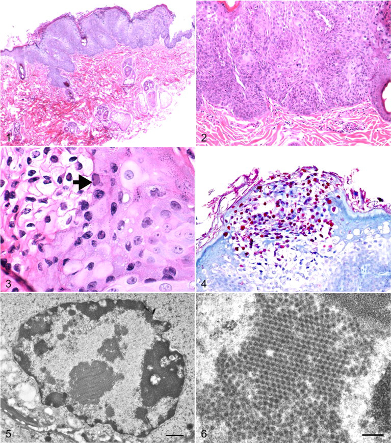
The presence of intranuclear inclusion bodies strongly suggested a viral-induced lesion. Immunohistochemical staining was performed with a monoclonal antibody against human papillomaviruses 1, 6, 11, 16, 18, and 31 (Chemicon International Inc, Temecula, CA). 1 Briefly, immunohistochemical staining was performed with an automated staining system (Benchmark, Ventana Medical Systems Inc, Tucson, AZ) that incorporated a commercial detection system (Enhanced V-Red detection system, Ventana Medical Systems Inc). Antigen retrieval was achieved by incubating slides in a high-pH antigen retrieval solution for 60 minutes. The primary antibody was applied at a concentration of 1:100 for 30 minutes. Sections were counterstained with hematoxylin. Positive control specimens included tissues known to be infected with canine papillomavirus. For negative control specimens, the primary antibody was replaced with homologous nonimmune serum. Strong nuclear immunorectivity was present primarily within the superficial dysplastic cells (Fig. 4).
For the polymerase chain reaction (PCR) assay, DNA was extracted from paraffin-embedded specimens with a commercial kit and tested with a PCR assay previously reported to be capable of detecting a broad range of human papillomaviruses. 12 The assay targeted a 450–base pair region of the papillomavirus L1 gene and incorporated primers MY11 (5′-CMCAGGGWCATAAYAATGG-3′) and MY09 (5′-CGTCCMARRGGAWACTGATC-3′). Assay specifications were similar to those previously described, 2 with the reaction carried out in a multiplex format. PCR failed to amplify a segment of the viral genome from the extracted samples.
Ultrastructural examination was performed on skin samples fixed in formalin and embedded in paraffin. Tissue segments were excised from the paraffin block, cleared with xylene, and rehydrated with water. The hydrated tissue was stained with 1% osmium tetroxide with 0.5% potassium ferrocyanide for 1.5 hours, then dehydrated with alcohol and embedded in epoxy resin. Thin sections were cut with an ultramicrotome (MTX, RMC Products, Boeckeler Instruments, Inc, Tucson, AZ), collected on copper grids, and poststained with uranyl acetate and lead citrate. The samples were imaged with a Hitachi H-7000 transmission electron microscope, operating at an accelerating voltage of 75 kV. Ultrastructurally, large numbers of aggregates of electron-dense, approximately 50-nm-diameter hexagonal viral particles compatible with papillomavirus were observed within the nuclei of dysplastic superficial keratinocytes (Figs. 5, 6).
Cutaneous neoplasms are common in ferrets, and the skin appears to be the third-most commonly reported organ affected by neoplasms. 23 The most frequently reported neoplasm is the mast cell tumor, followed by basal cell tumors and sebaceous gland neoplasms. 22,23,27,32 Only one report described multifocal cutaneous invasive squamous cell carcinoma in a ferret, affecting the digits of the left hindfoot, the left tarsus, and right front footpad; however, histologic features of a viral infection were not observed. 22 To our knowledge, the lesions observed in the ferret of this report have not been described, and they resemble those of BISC as described in cats, 6,7,9,16,17,20,30 humans (Bowen disease), 21 and a dog. 7
In cats, BISC has been associated with papillomavirus. 6,9,10,17,18,20 Papillomavirus is also implicated in the development of other feline skin neoplasms, such as cutaneous fibropapillomas (also called feline sarcoid), 26 oral papillomas, 28 cutaneous viral papillomas 15,28,30 and invasive squamous cell carcinoma. 16,20,28 The lesions of BISC in cats are multicentric, similar to the distribution of the lesions observed in the present ferret. 6,7,30 Histologically, the lesions are similar and characterized by plaques of irregular epidermal hyperplasia and dysplasia. However, viral inclusion bodies within keratinocytes, as described in this ferret, have not been identified in the BISC lesions in cats. 6,7,20 Given the general species specificity of papillomaviruses, 24,28 this difference suggests that a different papillomavirus could be associated with the lesions in this ferret. Some reports described intracytoplasmic pseudoinclusions in BISC in cats, 28,30 which consist of finely granular to fibrillar electron-dense material of aberrant intermediate filament assembly. 28 These intracytoplasmic pseudoinclusions were not observed in the ferret lesions.
Strong nuclear immunoreactivity for papillomavirus and the presence of approximately 50-nm viral particles forming crystalline arrays within the nuclei of superficial keratinocytes confirm that the multicentric squamous cell carcinomas in situ in the present ferret were associated with papillomavirus. Unfortunately, multiple trials of PCR failed to amplify viral DNA, and we were unable to classify this papillomavirus. Because this is likely a different papillomavirus, one not characterized in ferrets, the performance of this assay is not known. The PCR assay is designed for formalin-fixed tissues; however, degenerate primers are only moderately sensitive and can fail to amplify viral genome in some cases. The morphologic difference of the lesions observed in this ferret—such as the presence of intranuclear inclusion bodies, which are absent in cats—also suggests that this is a different papillomavirus.
In conclusion, ferrets can develop multicentric squamous carcinoma in situ associated with papillomavirus, similar to the lesions observed in felines with BISC.
Footnotes
Acknowledgments
We thank Dr John Roths for his invaluable assistance with photographic images, Sarah Jones and Kristen Sledge for preparation of sections for microscopic examination, and Dr Robert Droleskey at the Food and Feed Safety Research Unit, Southern Plains Area Research Center, Agricultural Research Service, US Department of Agriculture, for his assistance with the transmission electron microscope.
The authors declared that they had no conflicts of interest with respect to their authorship or the publication of this article.
The authors declared that they received no financial support for their research and/or authorship of this article.