
Abstract
This report documents an unusual case of congenital foregut cyst with dysphagia and stridor in a Thoroughbred foal. Histologically, the bilocular cyst, near the junction of larynx and trachea, had an epithelial lining of bronchogenic and esophageal origin. Concomitant malformation of the laryngeal muscles and cartilage resulted in a combination of anomalies that have not been reported in the human or veterinary literature.
Bronchogenic and esophageal cysts are rare anomalies that are attributed to abnormal budding or division of the primitive foregut. 1,17,19,21 These malformations are classified by the components of the cyst wall: the lining epithelium, glands, muscular layer and cartilage. 1,17,19 Esophageal cysts have 2 muscular layers, whereas bronchogenic cysts have cartilage rings. 17 Esophageal cysts have been reported in several horses 14,18,20 and a dog. 5 Bronchogenic cysts have been reported in 2 horses, 2,15 1 of which also had an esophageal component. 15 We describe a foregut cyst with bronchogenic and esophageal portions and concurrent malformation of laryngeal musculature and cartilage in a Thoroughbred foal.
Clinical and Pathologic Findings
A neonatal male Thoroughbred foal was presented with abnormal respiratory sounds, dysphagia, and suspected aspiration pneumonia. Rostral displacement of the palatopharyngeal arch, dorsal displacement of the soft palate, and left displacement of the arytenoid process were detected endoscopically. Owing to poor prognosis, the foal was euthanized 2 days after birth. The mare had previously had 8 foals with no problems.
At necropsy, a cyst about 20 × 3 × 3 cm was found just caudal to the larynx along the left side of the trachea (Fig. 1 ). The cyst had C-shaped cartilage rings and a dorsal membranous wall; both ends were tapered and closed (Fig. 2). A more pliable cyst about 9 × 4 × 4 cm was found dorsal to the larynx and adhered to the esophagus (Fig. 1). This cyst had a muscular wall and white membranous lining. Both cysts were filled with viscous white fluid. A 1-cm-diameter hole connected the lumen of each cyst, but no communication was detected between the cyst and the trachea, esophagus, or larynx. The tracheal cyst was, however, attached to the left cricoid cartilage with left rotation of the cartilage. The left cricoarytenoideus dorsalis muscle appeared hypoplastic; the right muscle appeared disorganized (Fig. 3). The left cricothyroideus muscle was not found; instead, supernumerary muscles connected the cricoid cartilage and the left thyroid cartilage to the tracheal cyst (Fig. 2). The corniculate processes of the artenoid cartilage were displaced to the left (Fig. 3). The left caudal cornu of the thyroid cartilage was shorter than the right. No other anomalies were detected in the respiratory or digestive system.
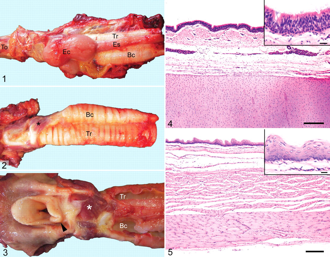
Histologically, the tracheal cyst was lined by pseudostratified ciliated columnar epithelium; its wall was composed of submucosa with serous glands and hyaline cartilage (Fig. 4). The more pliable cyst was lined by stratified squamous to pseudostratified ciliated columnar epithelium (Fig. 5). Mucous glands were in the submucosa beneath the pseudostratified columnar epithelium. The cyst wall had 2 muscular layers, with fibers of the outer layer oriented perpendicular to those of the inner layer (Fig. 5). Based on these findings, the bilocular structure was classified as a communicating foregut cyst of bronchogenic and esophageal types. Histologic findings in the lungs were attributed to aspiration pneumonia.
Discussion
A similar congenital foregut cyst with cranial esophageal and caudal bronchogenic portions was reported in an Arabian filly; the bronchogenic portion had only 2 semicircles of cartilage, 15 in contrast to our case with more mature tracheal structures. No laryngeal malformations were reported in the Arabian filly. 15 In horses, congenital laryngeal defects are frequently associated with dorsal displacement of the soft palate, 7,13 rostral displacement of the palatopharyngeal arch, 6,22 or epiglottic entrapment by arytenoepiglottic folds. 3 Congenital foregut cysts generally have not been associated with these conditions, although a case of paralaryngeal bronchogenic cyst with concomitant cricoid cartilage malformation was reported in a Thoroughbred gelding. 2 In humans, several cases of mixed foregut cysts of bronchogenic and esophageal type have been described, with the cysts located in the mediastinum or pleural cavity and frequently accompanied by extralobar pulmonary sequestration. 4,8,9,11,23 To our knowledge, the combined bronchogenic and esophageal cysts with concurrent laryngeal malformation, as seen in the horse of this report, have not been reported in the human or veterinary literature.
During development, the trachea arises from the ventral wall of the foregut, whereas laryngeal cartilage and muscles originate from mesenchyme of the fourth and sixth pharyngeal arches. 12,16,17 The foregut, separated from the respiratory diverticulum, then elongates to form the esophagus. 12,16 Bronchogenic or esophageal cysts, especially those in the cervical region, can cause dysphagia and respiratory distress owing to compression, rupture and subsequent inflammation, or (rarely) malignant transformation. 1,2,4,9,10,14,15,18-21 Therefore, most congenital foregut cysts are excised in veterinary and human medicine, almost always with favorable outcome. 1,2,5,8,9,15,17-19,21 However, potential surgical candidates should be thoroughly evaluated for the presence of concurrent laryngeal malformations, which might worsen the prognosis.
Footnotes
The authors declared no potential conflicts of interest with respect to the authorship and/or publication of this article.
The authors received no financial support for the research and/or authorship of this article.