
Abstract
A cranial cervical mass was surgically removed from a dog. On histologic examination, the mass was consistent with an esophageal duplication cyst, a condition rare in humans and not reported in the dog.
Esophageal duplication cysts are congenital anomalies of the foregut, typically found adjacent to the esophagus. 1 In humans, they result from developmental errors between the fifth to eighth weeks of embryologic development. In all species, lesions in the cranial third of the esophagus are typically asymptomatic after presentation; however, they can cause dysphagia and respiratory distress, because of compression of adjacent structures, and occasional gastrointestinal bleeding. Esophageal duplication cysts are rare in both humans and domestic animals, accounting for less than 2.5% of esophageal tumors in humans. 14 In the veterinary literature, this condition has been reported in 4 horses and a Cynomolgus monkey. 9,10,12
Esophageal cysts are classified as duplications when they satisfy 3 criteria: the cyst 1) must be within the esophageal wall; 2) usually contains 2 muscle layers; 3) must contain squamous, columnar, cuboidal, pseudostratified, or ciliated epithelium. 1 This is the first description of an esophageal duplication cyst in the dog.
A 1-year-old castrated male Border Collie was presented to the Veterinary Teaching Hospital, Atlantic Veterinary College. A slowly growing, fluctuant mass had been noted at the cranioventral aspect of the neck since birth. At 6 months of age, the mass was aspirated by the referring veterinarian and was found to contain a mucoid fluid. The mass rapidly refilled. At the time of referral, there were no clinical signs associated with the mass. The mass was located on the left side of the neck, extending from the laryngeal region to the thoracic inlet. It was surgically excised and found to lie between the sternocephalicus, sternohyoideus, and sternothyroideus muscles. An esophageal tube was placed, and the mass was found to be within the esophageal wall deep to the muscle layers. On gross examination, the mass was fluctuant and well circumscribed, was easily dissected from the esophagus, and did not communicate with the esophagus. The mass was submitted for histologic evaluation. On macroscopic examination, the submitted tissue was a well-circumscribed hollow mass with a thin wall, filled with a turbid serosanguineous fluid. On microscopic examination, the mass was composed of a central cavity lined with a partially keratinized stratified squamous and pseudostratified epithelium. Within the submucosa, abundant submucosal mucous glands were present (Fig. 1). A broad circumferential striated muscularis externa was present, composed of variably defined longitudinal and circular layers. The surrounding adventitia was diffusely infiltrated with mixed inflammatory cells, including abundant neutrophils, lymphocytes, and plasma cells. Based on the gross and histologic findings, the mass was diagnosed as an esophageal duplication cyst.
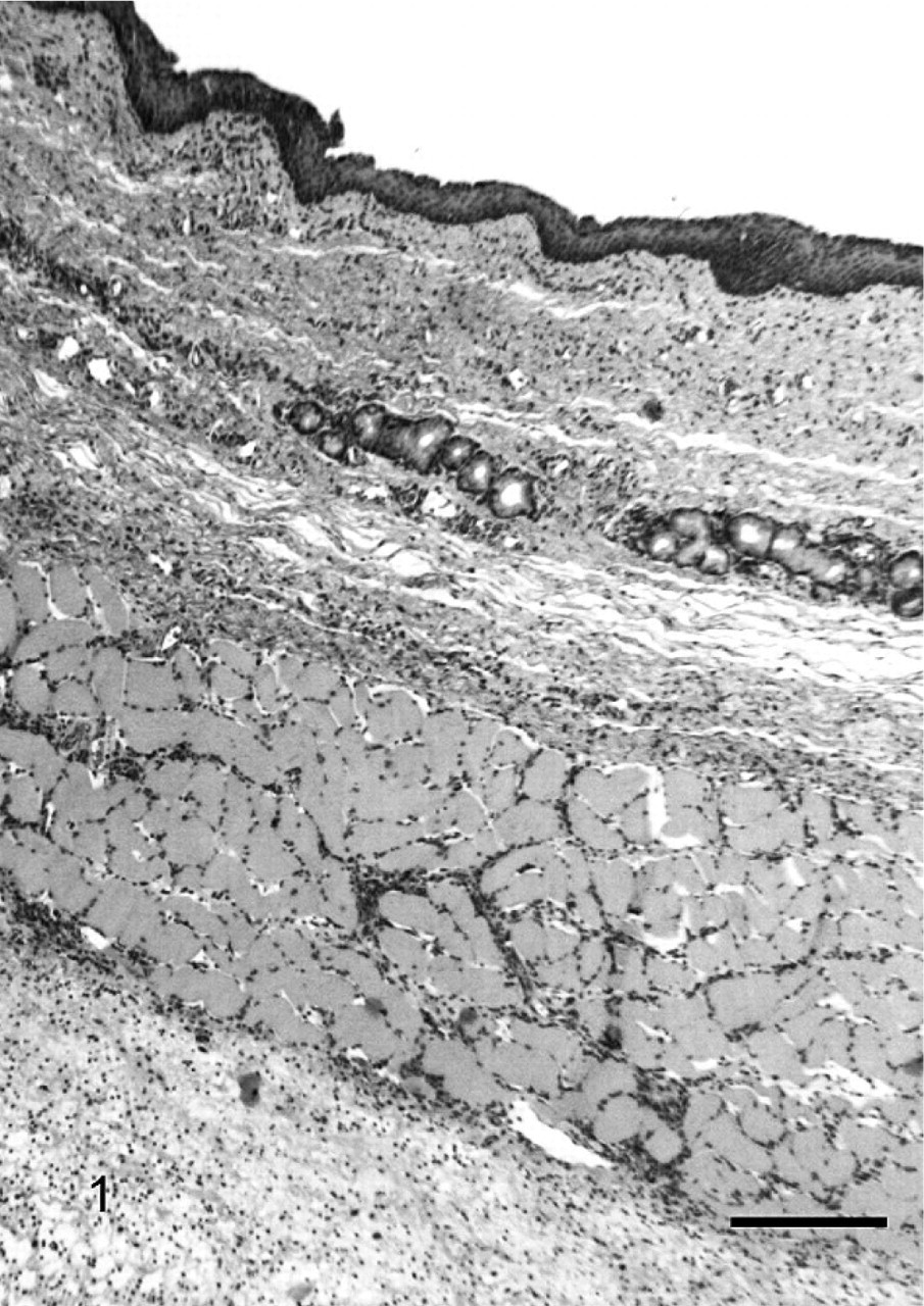
Cross section, esophageal duplication cyst; dog. There is a stratified to pseudostratified epithelium, with underlying mucus glands and a distinct (striated) muscularis externa. The adventitia is edematous and diffusely infiltrated with mixed inflammatory cells. HE. Bar 250 μm.
Cervical swellings in veterinary medicine are relatively common. In the dog, these could include a wide range of conditions, such as abscesses, mucoceles, lymphadenopathies, non-neoplastic or neoplastic enlargements of the thyroid gland, thyroglossal duct cysts and neoplasms, or branchial pouch anomalies. The distinct, fluctuant mass described was highly suggestive on presentation of a cystic structure. Because of its location close to the thyroid, clinically, the main differential diagnosis was a thyroid-associated mass. However, on histologic examination, the presence of pseudostratified epithelium, submucosal glands, and a muscularis externa recapitulated the esophagus, and, therefore, supported the classification of esophageal duplication cyst. 4 In the current case, the presence of skeletal muscle in the muscularis externa layer is consistent with an origin of the proximal esophagus, since embryologically, the proximal one third of the esophageal musculature is a continuation of the striated muscle of the pharynx. 13
Congenital duplication cysts are unusual findings in humans and rare in veterinary medicine. This is the first reported case of an esophageal duplication cyst in a dog and has only previously been reported in 4 horses and a single nonhuman primate. A number of studies indicate its rarity in human medicine 1 ; however, because of the potential complications associated with rupture or compression, the condition is well described. Clinical signs in humans associated with compression of adjacent structures are caused by a combination of vascular compression, partial esophageal obstruction, dyspnea, and commonly, for cysts that are located in the mediastinum, mediastinitis because of rupture. 2–5,7,8,11
Duplication cysts respond well to complete excision. The current recommendation in human medicine is for complete surgical excision immediately after diagnosis, before serious complications. 1 Most complications are caused by compression; however, there is 1 reported case of malignant transformation. 6 In the case presented, complete surgical excision was performed, and the patient has made an uneventful recovery, with no indication of recurrence.
Footnotes
Acknowledgements
We thank Ramona Taylor, Dianne O'Connell, and Len Doucette for their technical assistance.