
Abstract
Congenital vascular tumors of the skin have been described in people and a few animals, but unlike infantile hemangiomas in children, spontaneous regression has not been described in animals. A 2-day-old male Belgian Blue cross calf was presented for multiple congenital cutaneous masses that were soft, alopecic, and hyperemic; the calf had no other apparent abnormalities. Two weeks later, one mass had regressed. Surgical excision of one of the remaining masses was performed; histopathologic and immunohistochemical findings were considered diagnostic for epithelioid hemangioma. Eight months following initial presentation, all the masses had regressed spontaneously. This constitutes the first account in the veterinary literature of spontaneous regression in a congenital vascular tumor.
Hemangiomas of infancy are the most common benign tumor of childhood. 1 Several variants have been identified; the infantile hemangiomas (IH) are most common. IH are marked by rapid postnatal proliferation and slow spontaneous involution. 2 Congenital or neonatal vascular tumors of the skin have been described in a few animals, often calves, 5 but spontaneous regression, as occurs routinely in IH in children, has not been described.
In humans, a remarkable diversity of benign endothelial tumors is recognized, including lobular capillary hemangioma, tufted hemangioma, glomeruloid hemangioma, and epithelioid hemangioma. 3 Epithelioid variants of vascular endothelial tumors, characterized by the plump epithelial-like appearance of the neoplastic endothelial cells, are well-recognized entities. 4 In veterinary medicine, cases of epithelioid hemangiomas have been recently described. 7 This report describes a calf with multiple congenital epithelioid hemangiomas that spontaneously regressed.
History
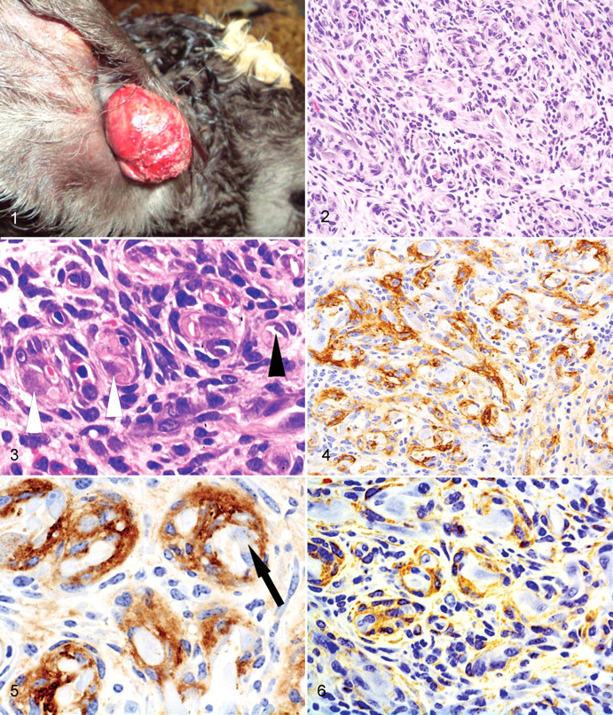
A 2-day-old male Belgian Blue cross calf was presented for the evaluation of congenital cutaneous masses (Fig. 1 ). No signs of systemic illness were reported, and physical examination was otherwise unremarkable. Five soft, pedunculated, alopecic, and hyperemic masses were in the skin of the thorax, both flanks, muzzle, and right auditory meatus. The masses ranged from 2 to 7 cm in diameter and were superficially ulcerated. Two weeks later, the muzzle lesion had regressed, leaving a small ulcerated area, whereas the other four masses were still present and covered by hemorrhagic crusts. All masses were grossly similar. The thoracic mass was excised under local anesthesia, fixed in 10% neutral-buffered formalin, and submitted for histopathologic examination.
The tissue was embedded in paraffin, sectioned at 4 μm, and stained with hematoxylin and eosin. The tissue was also evaluated immunohistochemically for the expression of alpha smooth muscle actin (α-SMA) and von Willebrand factor (vWF). Four-micrometer-thick sections were mounted on charged microscope slides (Menzel-Glaser, Brunswick, Germany). Immunohistochemistry was performed using the EnVision system (Dakocytomation, Glostrup, Denmark) and the Shandon Sequenza Coverplate system (Thermo Fisher Scientific, Waltham, MA). Briefly, endogenous peroxidases were blocked with peroxidase-blocking solution (Dakocytomation) for 15 minutes at room temperature. Antigen retrieval was performed using 0.05% protease XIV (Sigma-Aldrich, St. Louis, MO) at 37°C for 15 minutes (vWF) or citrate antigen unmasking solution (Vector Labs, Burlingame, CA) in a steamer for 15 minutes (α-SMA). Slides were incubated with primary antibody for 30 minutes at the following dilutions: α-SMA (Novocastra Laboratories, Newcastle upon Tyne, UK) at 1:50 or vWF (Dakocytomation) at 1:50. 3,3-Diaminobenzidine-tetrahydrochloride (DAB; Dakocytomation) was used as chromogen and counterstained with hematoxylin. As a negative control, a duplicate of each section was incubated with normal rabbit immunoglobulin G (IgG; vWF) and normal mouse IgG (α-SMA) for the same time as the primary antibody. Staining of normal blood vessels within the dermis was used as internal controls for vWF and α-SMA.
Pathologic Findings
Microscopic examination revealed a nonencapsulated, partially ulcerated, exophytic neoplasm that effaced the epidermis, infiltrated and replaced the dermis, and multifocally involved the subcutis (Fig. 2). The mass formed sheets of whorled structures, some with erythrocytes, but in many, a central lumen suggested a vasoformative pattern of growth. The vascular structures contained two distinct cell populations (Fig. 3): One consisted of nests of central large polygonal cells, each with a moderate amount of amphophilic cytoplasm, a single large vesicular nucleus, and often a large intracytoplasmic vacuole. These were encircled by smaller spindloid cells with sparse cytoplasm. Lymphocytes and a few eosinophils infiltrated the interstitium. Immunohistochemistry for vWF (Figs. 4, 5) revealed strong granular cytoplasmic reactivity in the innermost whorled cells (consistent with endothelium) and, more weakly and variably, in the larger polygonal cells. The smaller, spindloid cells were weakly positive for cytoplasmic α-SMA (Fig. 6) consistent with pericytes. In some vascular spaces, there was more diffuse brown immunoreactivity for vWF, which might represent binding to free vWF in plasma. Based on macroscopic and microscopic appearance, a vasoformative tumor was diagnosed; the plump luminal cells and the mixed infiltrate were consistent with epithelioid hemangioma. 7
Five months later, only two masses remained; by 8 months of age, all cutaneous masses had regressed. The calf was growing normally at the time of this writing (at 9 months of age), and no other abnormalities were reported.
Discussion
Classifications of vascular masses are confusing; conventionally, hamartomas are seen early in life and are nonneoplastic, whereas hemangiomas are considered to be clonal neoplasms presenting from birth to maturity. Congenital skin neoplasia is rare in calves. 9 Cases of multicentric cutaneous fetal or congenital hemangioma have been reported in 10 cattle, 5,8,9 but features consistent with the epithelioid variant were not described. One case of epithelioid hemangioma was reported in a 7-month-old calf; 7 however, it is not known if this was present at birth. Two cases of congenital dermal capillary hemangioendothelioma have been reported in foals; however, the histological description of vascular structures lined by flattened endothelial cells would not be consistent with an epithelioid variant. 6
In humans, hemangiomas are the most benign of endothelial tumors and, in some cases, such as IH, regress spontaneously. The biology of this human tumor has been studied extensively in an attempt to understand how spontaneous regression is mediated. Endothelial cells of IH have some unique features, including an immature arrested phenotype 2 and the expression of a glucose transporter protein normally found in the placenta. 1 During the involution phase, specific genes, including an angiogenesis inhibitor and interferon inducer, become activated and may participate in tumor regression. 2 The case in this calf had similar clinical features, although regression was much faster than that in IH in children. To the best of our knowledge, this is the first report in the veterinary literature of congenital epithelioid hemangioma with spontaneous regression.
Footnotes
The authors declared that they had no conflicts of interest with respect to their authorship or the publication of this article.
The authors declared that they received no financial support for their research and/or authorship of this article.