
Abstract
Of 1146 caprine necropsy or biopsy specimens submitted from 1987 through 2011 to the Veterinary Diagnostic Laboratory at Oregon State University, 100 goats (8.7%) had 102 tumors. Detailed records were available for 89 cases. Fifty-five goats were female, 17 were castrated males, and 12 were intact males. Breeds included 21 Nubian, 16 Pygmy, 10 Pygora, 8 Alpine, 4 Angora, 4 Saanen, 2 Toggenburg, and 9 crossbred goats. Dwarf, Nubian, and Saanen goats were overrepresented and Alpine and Boer goats underrepresented among cases with neoplastic disease in comparison to submissions overall. Age ranged from 7 months to 19 years (median, 7 years). Histopathology was performed on 97 tumors. Lymphoma (n = 17) was the most common tumor, followed by cutaneous squamous cell carcinoma (n = 10) and thymoma (n = 9). Most lymphomas were multicentric. All 7 mammary neoplasms were adenocarcinomas. Five of 7 vascular proliferations were hemangiosarcomas. All 4 melanocytic tumors were classified as (malignant) melanoma. Rarely reported caprine tumors included a choroid plexus carcinoma, 2 rhabdomyosarcomas, and 3 pheochromocytomas. Cutaneous round cell tumors were provisionally diagnosed as 2 histiocytomas and 5 mast cell tumors. Single cases of previously unreported caprine tumors included amyloid-producing odontogenic tumor, myxosarcoma, sebaceous carcinoma, apocrine sweat gland adenoma, and thyroid carcinoma. Nonneoplastic entities included 2 cases of mammary fibroadenomatous hyperplasia and single cases of vascular hamartoma, cervical adenomatous hyperplasia, and cervical leiomyofibromatosis. The results of this 25-year retrospective study indicate that lymphoma in particular and tumors in general are common in goats.
The agricultural importance of goats in the United States is largely driven by an increasing demand for alternative meat and milk sources, a trend that is projected to continue. 1 Retrospective studies of caprine tumors have been published in the past 40 years, 4,6,10,12,14,19,34 but some had a narrow focus or included few new cases. For example, a 1996 study of cutaneous neoplasms was focused on melanomas and squamous cell carcinomas. 14 Another report primarily on nonneoplastic external masses. 12 Three studies published 30 or more years ago 4,6,10 and a more recent abstract 19 cover a wide spectrum of conditions in a larger number of goats. The paucity of comprehensive and recent reports of caprine tumors prompted the current retrospective study of 102 tumors among the caprine biopsy and necropsy submissions to the Veterinary Diagnostic Laboratory at Oregon State University from 1987 to 2011.
Materials and Methods
Cases
Cases were retrieved from the archives of the Veterinary Diagnostic Laboratory (VDL) at Oregon State University (OSU). Entries in 2 different database systems, Data General (1987–2001) and Visualab (2001–2011), were analyzed. All diagnostic codes, including the term neoplasm, were used to identify cases. Additional information, including signalment, history, and gross necropsy descriptions, was retrieved from the submission sheets and archived reports. The total number of caprine necropsy and biopsy submissions for the period was used to calculate the prevalence of neoplastic disease and specific neoplasms.
Histopathology
Histopathology was performed on all cases for which either archived slides or paraffin blocks were available (n = 97). Histologic sections stained with hematoxylin and eosin (HE) were evaluated without knowledge of the original diagnosis. In selected cases, serial sections were stained with Giemsa, toluidine blue, Churukian-Schenk, Fontana Masson, and Alcian blue at pH 2.5 and Masson’s trichrome stain. Histopathologic findings were recorded for all available sections of tumor tissue; tumors were categorized as mesenchymal, epithelial, thymic, melanocytic, endocrine, odontogenic, nervous system, or nonneoplastic. Specific tumors were classified according to published references or the pertinent fascicle of the World Health Organization’s “International Histological Classification of Tumors of Domestic Animals,” jointly published by the Armed Forces Institute of Pathology, C. L. Davis DVM Foundation, American Registry of Pathology, and the World Health Organization Collaborating Center for Worldwide Reference on Comparative Oncology.
Immunohistochemistry
Immunohistochemistry (IHC) was performed on an autostainer (Dako Autostainer Universal Staining System; Dako, Carpinteria, CA) according to standard operating procedures. Paraffin sections were high-temperature antigen retrieved with BDTM Retrieval A solution (Dako). Endogenous peroxidase activity was blocked by immersing slides in methanol containing 3% hydrogen peroxide for 10 minutes. The following primary antihuman antibodies (DakoCytomation, Carpinteria, CA) were applied for 30 minutes at room temperature: wide-spectrum cytokeratin antiserum (WSS; 1:500), cytokeratin antibody cocktail (AE1/AE3; 1:200), vimentin (clone Vim 3B4; 1:100), muscle actin (clone HHF35; 1:50), sarcomeric actin (clone Alpha-Sr-1; 1:20), and von Willebrand factor (vWF aka factor VIII–related antigen, clone F8/86; 1:400). Antibodies against the canine or feline β2 subunit of β2 integrins (Peter Moore, University of California, Davis; clone CA16.3C10 at 1:20 and FE3.9F2 at 1:320) were used in an attempt to detect CD18. MaxPoly-One Polymer HRP Rabbit or Mouse Detection solution (MaxVision Biosciences, Bothell, WA) for 7 minutes at room temperature and Nova Red (SK-4800; Vector Labs, Burlingame, CA) as chromagen were used with Dako hematoxylin (S3302) as counterstain. Positive control tissues for histochemistry and IHC included skin of a goat with eosinophilic dermatitis, a grade III canine cutaneous mast cell tumor, normal caprine skin, caprine lymph node, and a composite block containing canine skeletal muscle, smooth muscle, and a rhabdomyosarcoma. Serial sections of neoplastic tissue incubated with Dako Universal negative serum served as negative controls.
Statistical Analysis
Prevalence of breeds among goats with tumors was compared with prevalence of breeds among all caprine accessions during the sample period by the χ2 test using GraphPad Prism (GraphPad Software, La Jolla, CA; free online version at http://graphpad.com/quickcalcs/chisquared1/).
Results
Animals/Cases
From 1987 to 2011, 1146 caprine necropsy or biopsy cases were received. Breed distribution is summarized in Table 1. In 705 cases, sex was recorded as 489 female, 129 male, and 87 castrated male goats. Age ranged from fetal to 17 years (median, 3.1 years). The 393 cases submitted for pathology from 2001 through 2011 included 124 biopsy specimens and 269 necropsies (65 gross examination only; 204 with histopathology).
Prevalence of Breeds and Goats With Tumors in Caprine Necropsy or Biopsy Accessions
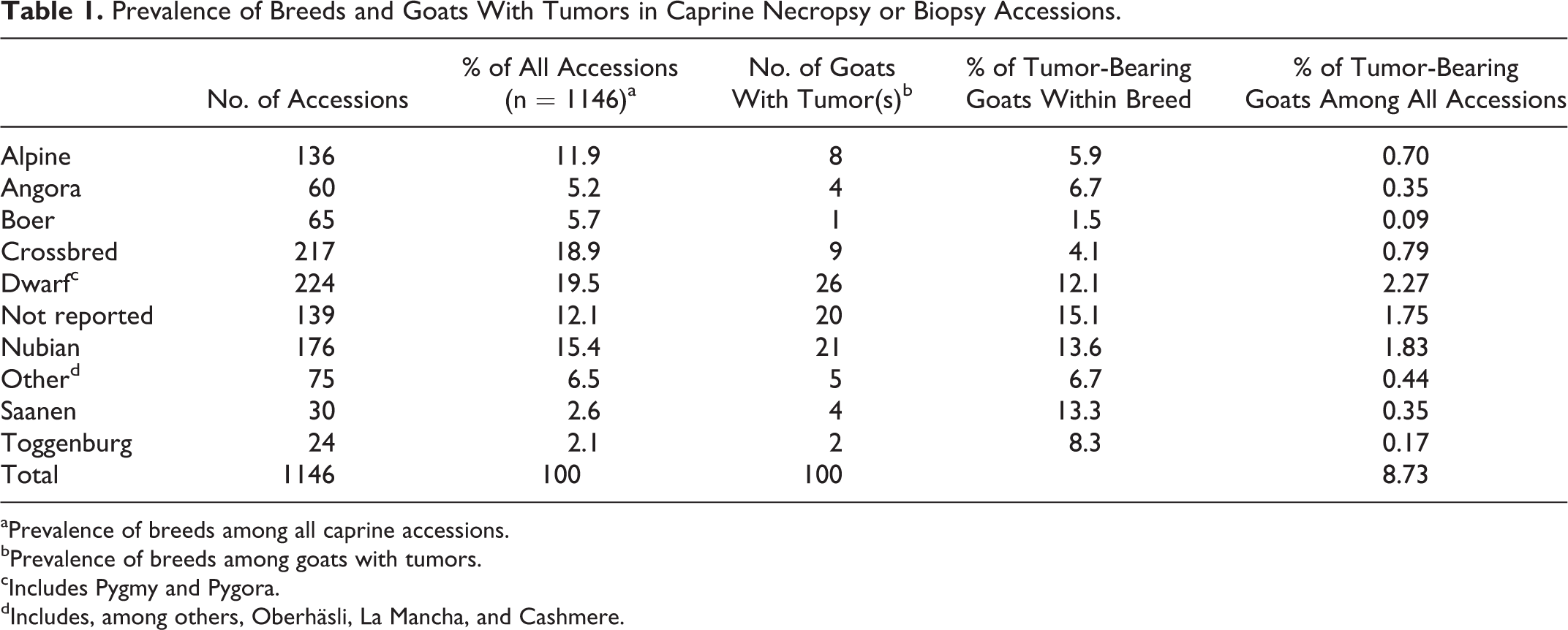
aPrevalence of breeds among all caprine accessions.
bPrevalence of breeds among goats with tumors.
cIncludes Pygmy and Pygora.
dIncludes, among others, Oberhäsli, La Mancha, and Cashmere.
A total of 102 distinct tumors had been diagnosed in 104 submissions from 100 goats. Two goats had both a biopsy specimen and a postmortem examination. Another goat had 3 distinct tumors. Detailed records from 57 biopsy and 33 necropsy submissions (27 with histopathology) were available for 89 of the 100 goats. Breeds included Nubian, Pygmy, Pygora, Alpine, Angora, Saanen, Toggenburg, “other,” crossbred, and single cases of Oberhäsli, Cashmere, and Boer (Table 1). The breed was not recorded for 10 of the 89 goats. Sex distribution was 55 female (61%), 17 castrated male (19%), 12 male (13%), and 5 unrecorded. Age in 84 of the 89 goats ranged from 7 months to 19 years (median, 7 years). For 11 goats, no information beyond species and diagnosis was on file, but histologic slides or paraffin blocks were available.
Tumors
Histologic slides or paraffin blocks of formalin-fixed tissue were available for 96 of the 100 goats with tumors. The original diagnosis was revised upon review of HE-stained slides or, in selected cases, upon histochemistry or IHC. The diagnosis, signalment, sample type, and site of the tumor are listed for each case in Supplemental Table S1. The 102 tumors included 42 mesenchymal neoplasms, 35 epithelial neoplasms, 9 thymomas, 4 melanomas, 4 endocrine neoplasms, a choroid plexus carcinoma, and an amyloid-producing odontogenic tumor. One goat with a pheochromocytoma also had a ruminal papilloma (not available for histopathology) and a uterine leiomyoma. Six nonneoplastic proliferative lesions are included based on the sensu lato definition of tumor.
Mesenchymal Tumors
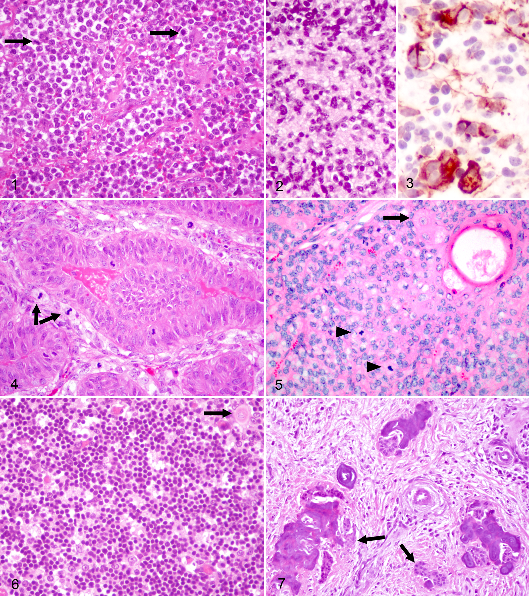
Lymphoma was diagnosed in 17 goats. Age ranged from 1 to 9 years (median, 3 years). Most lymphomas were diagnosed at necropsy and were multicentric with involvement of lymph nodes, liver, kidneys, and lungs or, less commonly, the nasal mucosa/submucosa, central nervous system (spinal cord or olfactory lobes of brain), eyelids, and gingiva. Histologically, lymphomas were either dominated by small or large neoplastic lymphocytes or were a mixture of many small and fewer large cells. Mitotic index varied but was generally low in small-cell lymphomas and high in large-cell lymphomas (Fig. 1).
Cutaneous round cell tumors, diagnosed as mast cell tumor or histiocytoma, comprised a homogeneous population of large cells with abundant cytoplasm. Only 1 of the 5 mast cell tumors had metachromatic cytoplasmic granules in Giemsa- or toluidine blue–stained sections. Anisocytosis and anisokaryosis were marked in another case. Cutaneous histiocytoma was diagnosed in a 7-month-old and a 14-month-old goat; immunohistochemistry for CD18 did not label cells in normal caprine lymph node or in either tumor.
Vascular neoplasms were diagnosed in 6 goats. Four cases had been reported as part of a previous study. 5 One case of cutaneous hemangioma and 5 cutaneous hemangiosarcomas were diagnosed. One of the 5 hemangiosarcomas had been reported as hemangioma but was reclassified as hemangiosarcoma based on histologic examination of a newly prepared HE-stained section. The tumor consisted of densely packed, small irregular vascular spaces lined by large endothelial cells with an average of 1 mitotic figure per 400× field. In another case, the affected goat had proliferative vascular lesions of the skin of the distal aspects of both hind limbs that ranged from hamartoma or hemangioma to hemangiosarcoma.
Neoplasms of muscular differentiation included 4 leiomyomas (2 uterine, 1 vaginal, and 1 rectal) and 2 rhabdomyosarcomas. One pleomorphic rhabdomyosarcoma was subjacent to nonhaired, nonpigmented, stratified squamous epithelium with prominent keratohyalin granules. Histologically, the tumor was composed of a mixture of streaming spindled cells, groups of large stellate cells, and rare strap cells. The cytoplasm of most neoplastic cells expressed muscle actin, whereas expression of sarcomeric actin was patchy and limited to slender spindle cells. The second rhabdomyosarcoma was in the hard palate/maxilla and was classified as embryonal subtype. Small cells were arranged in dense sheets and poorly defined bundles (Fig. 2). Scattered clustering of nuclei suggested multinucleation; there were 11 mitotic figures in ten 400× fields. Fewer large round neoplastic cells (rhabdomyoblasts) had the most intense immunoreactivity for muscle actin and sarcomeric actin, especially in multinucleated cells (Fig. 3).
Tumors of fibroblastic differentiation included 1 fibroma and 2 fibrosarcomas. The interdigital fibroma had prominent vascular stroma. The fibrosarcoma from the claw was well differentiated with 4 mitotic figures per ten 400× fields. The fibrosarcoma that presented clinically as a draining lesion in an unspecified location was poorly differentiated with numerous and atypical mitotic figures.
The sole myxosarcoma consisted of large anaplastic, spindled to stellate cells that exfoliated well on impression smears. Histologically, neoplastic cells expanded the omentum in abundant mucinous matrix that stained brightly blue with Alcian blue stain, pH 2.5. Neoplastic cells were not detected in representative sections from other submitted organs (uterus, ovary, liver, kidney, and rumen).
Unclassified sarcomas
Two gingival sarcomas could not be classified. Both had large anaplastic to spindled cells, with few to moderate numbers of multinucleated giant cells, in interlacing bundles with numerous and atypical mitotic figures (≥10 per 400× field).
Epithelial Tumors
Squamous cell carcinoma (SCC) was diagnosed in 10 goats. Six of the 7 SCCs for which site was recorded originated in sparsely haired skin: udder (n = 3), perianal region (n = 3), horn base (n = 1), vulva (n = 1), and eyelid (n = 1). All goats with SCC of the udder had multiple SCCs, either on the udder (n = 1) or in other topographic locations (1 each, eyelid or vulva). The seventh SCC was in soft tissue of the neck and interpreted as a nodal metastasis.
The 7 goats with mammary adenocarcinoma were older than 4 years at diagnosis. All 6 goats with recorded sex were female. Breeds included 2 Alpine, 3 Nubian, and a crossbred goat. Mammary adenocarcinomas had the following patterns: simple tubular (n = 1), simple tubular to solid with cribriform areas (n = 3), and complex (n = 3). Mitotic figures were common in epithelial cells of all tumors but were common in myoepithelial cells in only 1 complex tubular adenocarcinoma (Fig. 4). Mineralization was present in a simple cribriform to solid and a complex tubular adenocarcinoma.
Intestinal adenocarcinomas were diagnosed in 3 middle-aged Pygora goats from different premises from 2002 to 2006. In 2 goats, the ileocecal junction was the primary site with metastasis to mesenteric lymph nodes. In the third case, a primary tumor was not identified, but the visceral pleura and the splenic capsule contained signet ring cells in a tubular to acinar arrangement; mesenteric lymph nodes were not examined histologically.
Epithelial neoplasms of the reproductive tract occurred in 3 female goats. Two adenocarcinomas were in the cervix or cervix and uterus, and 1 was in the vagina. One goat with cervical adenocarcinoma had histologic evidence of metastasis to lymph nodes and lungs.
Two nasal adenocarcinomas had a papillary to tubular pattern. One also contained ciliated and large atypical neoplastic cells.
Three papillomas occurred in the skin and 1, not available for histologic examination, in the rumen. The cutaneous papillomas included a fibropapilloma from the shoulder and perianal squamous papilloma. Cutaneous papillomas had neither koilocytes nor inclusion bodies.
Sebaceous gland tumors were classified as 2 epitheliomas and 1 carcinoma. Both sebaceous epitheliomas were from the perianal region. The sebaceous carcinoma came from the base of a horn and had continuity with the epidermis. Neoplastic cells had open, round to oval nuclei; an abundant finely vacuolated cytoplasm; and 21 mitotic figures in ten 400× fields (Fig. 5). This mass had been diagnosed originally as liposarcoma.
The sole apocrine sweat gland adenoma presented as an exophytic mass removed from the hind limb of a Nubian goat.
The primary site of 2 metastatic adenocarcinomas was undetermined, although uterus, mammary gland, and intestine were considered likely organs of origin. Histologically, both adenocarcinomas had large, anaplastic cells in tubular to cribriform arrangements and numerous and atypical mitotic figures.
Thymic Tumors
Nine goats with thymoma were of various breeds, ranged in age from 2 to 12 years (mean, 9.4 years), and included equal numbers of female and male goats. Slides or paraffin blocks were available for 7 cases. In 4 cases, epithelial cells predominated; in 2 cases, lymphocytes (Fig. 6); and in 1 case, both populations were present in approximately equal proportions. Hassall’s corpuscles were not readily identified. One thymoma was considered malignant based on invasion of mediastinal lymphatic vessels, but distant metastases were not detected. This goat also had hypoproteinemia, anasarca, and serous effusion in the thorax and abdominal cavity.
Melanocytic Tumors
All 4 melanocytic tumors were cutaneous, classified as (malignant) melanoma, and had areas with an epithelioid pattern. Neuroid or spindle cell arrangements were present in single melanomas. Two were from the coronary band of the hoof, and 1 was from the base of a horn. Metastasis was not detected in the sole case presented for necropsy.
Tumors of the Endocrine System
Two of the 3 pheochromocytomas involved both adrenal glands; all 3 invaded the vena cava. Two goats had metastases to liver, lung, spleen, and serosal surfaces; in 1 goat, the heart and cerebrum were also involved. The 2 cases evaluated histologically had variable staining of cytoplasmic granules with Churukian-Schenk histochemistry.
The sole thyroid carcinoma was composed of solid packets of anaplastic cells with rare follicle formation. Mitotic index was 2 per ten 400× fields.
Tumors of the Nervous System
Choroid plexus carcinoma was the only primary tumor of the central nervous system. A polypoid mass protruded into the lateral ventricles and compressed and multifocally invaded the periventricular neuropil. Although most of the tumor comprised uniform, bland cells without mitotic figures, areas of invasion had prominent cellular atypia and 2 mitotic figures per 400× field.
Odontogenic Tumors
The sole odontogenic tumor presented as an ulcerated gingival mass of well-differentiated dentinal tissues and was classified as an amyloid-producing odontogenic tumor (APOT). Odontogenic epithelium in bands and islands had peripheral palisading and prominent intercellular bridges (Fig. 7). The dense collagenous stroma contained globules of hyaline congophilic material, irregular concentrically layered globular to botryoid aggregates of mineralized acellular matrix (Liesegang rings), and scanty acellular brightly eosinophilic material with fine striations interpreted as dentin.
Nonneoplastic Mass Lesions
Six of the mass lesions were considered nonneoplastic. Fibroadenomatous hyperplasia of the mammary gland, diagnosed in 2 young female goats, was histologically indistinguishable from the condition in cats. One goat had adenomatous hyperplasia of the uterine cervical glands. Histologically, irregularly shaped, dilated tubules were lined by crowded, tall columnar to pseudostratified epithelium. Tubules were entrapped in dense collagenous tissue and protruded into the superficial smooth muscle bundles of the tunica muscularis. Leiomyofibromatosis of the uterine cervix multifocally expanded the tunica muscularis by poorly defined streams of spindled cells arranged in interlacing bundles with variable amount of collagenous stroma and poor demarcation. A nasal polyp had multifocal suppurative to ulcerative inflammation.
A vascular hamartoma was located in the skin of a juvenile Saanen goat.
Statistical Analysis
The χ2 value for breed prevalence of 21.082 with 9 degrees of freedom had a 2-tailed P value of .0123. This difference in prevalence of breeds among goats with tumors vs goats overall (Table 1) is considered statistically significant by conventional criteria, even though one of the expected values was less than 5 and thus violated a basic assumption of the test. Dairy breeds accounted for 32.2% and dwarf breeds for 19.5% of all caprine accessions and 38% and 26%, respectively, of goats with tumors. The prevalence of tumor cases among all caprine accessions was 8.7%. Prevalences of tumor cases within each goat breed are listed in Table 1.
Discussion
The goats in this study were of diverse breeding. The overrepresentation of dairy breeds (38%) in the goats with neoplastic disease reflects their prevalence among submissions to the VDL (32.2%) as well as in the Oregon state population. 2 The 8.7% prevalence of neoplastic disease for OSU VDL submissions is similar to that reported from the Veterinary Diagnostic Laboratory at the University of Georgia (USA) 19 but much higher than in older retrospective studies. 10,27 Breed prevalence among goats with tumors was significantly different from that of submissions overall, with dwarf, Nubian, and Saanen goats overrepresented and Alpine, Boer, and crossbred goats underrepresented. The high prevalence of tumors in dwarf, Nubian, and Saanen goats could indicate a predisposition of these breeds to neoplastic disease or simply reflect the higher median age of pet (dwarf) and dairy goats compared with goats of other agricultural uses or caprine cases in general.
In the current study, lymphoma was the most common neoplasm, and mesenchymal tumors outnumbered epithelial tumors in contrast to most previous reports, in which squamous cell carcinoma was the most common neoplasm. 4,10,19,25,27 In this study, SCC was the second most common neoplasm, followed by thymoma and mammary carcinoma.
Most caprine lymphomas were multicentric at diagnosis, typical of previously published cases. 9,23,31 The spinal cord/vertebral column or cranial structures, including conjunctiva, have been reported as major sites of lymphoma, but these structures were seldom involved in the current study. A retroviral cause for caprine lymphoma has been proposed because some goats experimentally infected with bovine leukemia virus (BLV) develop multicentric lymphoma. 23 Unfortunately, only 2 cases had been tested serologically for exposure to BLV and did not have titers. 31 The other presumptive leukocytic tumors in this study included cutaneous round cell tumors, which were provisionally classified as histiocytomas and mast cell tumors.
Squamous cell carcinoma was the most common epithelial tumor. As in previous studies, 4,14 cutaneous SCCs occurred exclusively in glabrous skin, suggesting ultraviolet light as a likely pathogenetic factor. Most SCCs were on the udder or in perianal skin. The latter is one of the most frequently reported topographic locations for caprine SCC. 4,14,19,34 None of the SCCs in this study occurred on the face and eyelids or in internal organs.
Thymoma is one of the more common neoplasms in goats, 19 especially in dairy breeds. 16 However, overrepresentation of dairy breeds among goats with thymoma was not identified in the current study, in which dwarf breeds (Pygmy and Pygora) were most frequently affected. Malignant caprine thymoma has been reported, 6,22 and lymphatic invasion warranted diagnosis of a malignancy in 1 thymoma in this study. The concurrent hypoproteinemia with anasarca was interpreted as a paraneoplastic syndrome in the absence of other plausible causes. Neither congestive heart failure nor megaesophagus was diagnosed in this study in contrast to reported cases of caprine thymoma. 24,26
Mammary gland tumors were more common in this study than in previous reports. 28,32 None occurred in male goats. Although 2 goats had fibroadenomatous hyperplasia, all 7 mammary neoplasms were malignant, and metastatic disease was detected in both cases in which necropsy was performed. Three adenocarcinomas were complex tumors, not previously reported in goats, with prominent epithelial and myoepithelial components.
Vasoproliferative lesions, including hamartoma, hemangioma, and hemangiosarcoma, have been recognized as common caprine tumors. 4,5,19 In contrast, melanocytic tumors were less common in the current study than in previous studies; 4,6,14,19 the relatively few Angora goats in the current study might explain this difference. 30 The perianal region and ears are the most commonly reported topographic sites of caprine melanoma 4,14,30 but were not involved in any of the current cases. All melanocytic tumors were malignant, consistent with reported cases. 4,6,14,19
Pheochromocytoma has been reported in goats 11,19,21 but is more common in cattle and dogs. 7 All 3 cases in this cohort were malignant, but invasion of the vena cava was more common than distant metastasis. The bilateral involvement of the adrenal glands is unusual in domestic animals. 7
Only 1 report of caprine rhabdomyosarcoma was found in the veterinary literature. 18 The head is a common site of embryonal rhabdomyosarcoma in dogs 8 and was affected in 1 of the 2 cases in this study. Choroid plexus carcinoma has been reported in a goat. 20 Metastatic disease along the central canal of the spinal cord is a criterion of malignancy; 29 however, invasion of periventricular parenchyma and marked cellular atypia prompted the diagnosis of a malignancy, even though the spinal cord/canal was not examined. Leiomyomas were not restricted to the reproductive tract; 1 tumor was in the rectum. Leiomyofibromatosis of the cervix is most commonly encountered in dogs, cats, and goats. 8 Tumors of fibroblastic differentiation appear to be uncommon in goats; 6,17,19 although gingiva has been the most commonly reported site, all cases in this cohort were cutaneous masses.
Intestinal adenocarcinomas are relatively common in sheep, mainly in the small intestine. 6,17 Interestingly, all intestinal adenocarcinomas in the current study occurred in Pygora goats and preferentially at the ileocecal junction.
Neoplasms in goats that, to the author’s knowledge, have not been reported, were thyroid carcinoma, amyloid-producing odontogenic tumor, sebaceous carcinoma, apocrine sweat gland adenoma, and myxosarcoma. Thyroid carcinoma is uncommon in domestic species other than dogs, 7 and prevalence increases with age. 33 Reported caprine sebaceous tumors include a sebaceous epithelioma 19 and a basosquamous carcinoma with sebaceous differentiation. 34 Misdiagnosis might contribute to this apparent rarity, as 2 of 3 cases in this study were originally diagnosed as squamous cell carcinoma or liposarcoma. Odontogenic neoplasms are rare in any domestic animal species. 17 Amyloid-producing odontogenic tumor was diagnosed in this study by the presence of congophilic material, odontogenic epithelium, and calcifying botryoid globules, even though dental epithelial caps cradling mesenchymal aggregates were not identified. Complex odontoma 13,17 was included in the differential diagnoses.
Six nonneoplastic tumors were identified in this cohort. Fibroadenomatous hyperplasia of the mammary gland is rarely reported in animals other than cats; 15 in goats, it has been described only in Nubians. 3,32 Hormonal dysregulation, such as the progesterone imbalances implicated in the feline condition, was not confirmed in either of the current or previously reported cases. 3,32
Lymphoma, squamous cell carcinoma, thymoma, and mammary adenocarcinoma were the most common neoplasms in goats in this 25-year retrospective study. With the paucity of published case reports, comprehensive retrospective studies, or tumor registries, it is difficult to assess the true prevalence of specific neoplasms. However, thyroid carcinoma, amyloid-producing odontogenic tumor, sebaceous carcinoma, and apocrine sweat gland adenoma have not been previously reported in goats. Results of this study and review of the literature suggest that these neoplasms and the seldom reported pheochromocytoma, rhabdomyosarcoma, choroid plexus carcinoma, cutaneous histiocytoma, and mast cell tumor are rare caprine neoplasms.
Footnotes
Acknowledgements
I am most grateful to past and current pathologists from the Veterinary Diagnostic Laboratory at Oregon State University, especially Drs. Rob Bildfell, Barry Cooper, Howard Gelberg, Amir Hamir, Ole Hedstrom, Jerry Heidel, Stan Snyder, and Beth Valentine. Their diagnoses played a critical role in retrieving cases for this retrospective study. The dedicated work of past and current histotechnology staff is much appreciated. A summary of the study was presented at the 37th Annual Conference of the New Zealand Society for Veterinary Pathology, Palmerston North, New Zealand (February 18–19, 2012).
Declaration of Conflicting Interests
The author(s) declared no potential conflicts of interest with respect to the research, authorship, and/or publication of this article.
Funding
The author(s) received no financial support for the research, authorship, and/or publication of this article.
References
Supplementary Material
Please find the following supplemental material available below.
For Open Access articles published under a Creative Commons License, all supplemental material carries the same license as the article it is associated with.
For non-Open Access articles published, all supplemental material carries a non-exclusive license, and permission requests for re-use of supplemental material or any part of supplemental material shall be sent directly to the copyright owner as specified in the copyright notice associated with the article.