
Abstract
At necropsy, an 11-year-old Japanese Black cow with anemia, leukocytopenia, and progressive hind limb ataxia had marked diffuse splenomegaly and multiple masses in the thoracic vertebrae. Histologically, neoplastic erythrophagocytic histiocytes were in the splenic red pulp, vertebral masses, and blood vessels of the liver and lungs. The spinal cord was compressed by the vertebral masses. Clinicopathological, macroscopic, and histologic findings were consistent with hemophagocytic histiocytic sarcoma. Vertebral involvement with spinal cord compression and resultant hind limb ataxia is an unusual presentation for this tumor, which has been described mainly in dogs and cats.
Histiocytic proliferative disorders are rare in cattle. 3,9 Dogs are more commonly affected with reactive or neoplastic histiocytic disorders, most of which probably arise from Langerhans cells or interstitial dendritic cells. 1,2,5,10 Recently, a neoplasm of macrophage lineage—hemophagocytic histiocytic sarcoma (HS)—was described in dogs and cats. 4-6,10 Affected animals were anemic and thrombocytopenic had diffuse splenomegaly without distinct masses and had a proliferation of neoplastic histiocytes in the spleen and bone marrow. 4-6 Many of the neoplastic cells had engulfed erythrocytes and, less often, leukocytes; immunohistochemical profiles were consistent with those of macrophages in the splenic red pulp and bone marrow. 4-6,10 We report a case of hemophagocytic HS in a cow with ataxia and paralysis secondary to neoplastic involvement of thoracic vertebrae.
History
An 11-year-old Japanese Black cow had decreased feed intake, decreased activity, and an abnormal gait that progressed to staggering and hind limb paralysis. The cow was anemic—erythrocyte count, 2.97 × 10⁶/µl (reference range, 5–10 × 106/µl); hematocrit, 19% (reference range, 24–46%)—and leukocytopenic: leukocyte count, 2,100/µl (reference range, 4,000–12,000/µl). 8 Serum biochemistry abnormalities included mild increases in glucose (83 mg/dl; reference range, 33–66 mg/dl), aspartate aminotransferase (134 IU/L; reference range, 43–127 IU/L), γ-glutamyltransferase (47 IU/L; reference range, 15–39 IU/L), total bilirubin (0.6 mg/dl; reference range, 0.01–0.47 mg/dl), and a slight decrease in total cholesterol (66 mg/dl; reference range, 80–120 mg/dl). 8 Seventeen days after the onset of clinical signs, the cow was euthanized.
Necropsy Findings
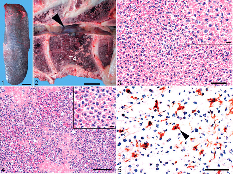
At necropsy, the cow weighed 456 kg. The spleen was diffusely enlarged to 102 × 33 × 8 cm and 11.9 kg (normal weight, 0.665–1.155 kg 7 ; Fig. 1 ). The splenic parenchyma was firm with indistinct white pulp and no discrete masses. The liver was 50 × 33 × 11 cm and weighed 7.3 kg (normal weight, 3.4–9.2 kg). 7 Dark red foci of telangiectasis were scattered through hepatic parenchyma. Thick-walled bile ducts contained thickened bile. The dorsal aspect of the fourth thoracic vertebral body protruded focally into the spinal canal (Fig. 2) because of dark-red nodular expansion of the subperiosteal marrow with irregular discontinuity of the associated cortex. Similar but milder lesions were in the second, sixth, and seventh thoracic vertebrae. Pertinent lesions were not detected in other organs, including the brain.
Histologic Findings
Histologically, the splenic red pulp was congested and expanded by numerous histiocytes (Fig. 3) and fewer erythroid precursors. The white pulp was compressed and atrophied. The neoplastic histiocytes had a pleomorphic nucleus with pale eosinophilic cytoplasm. Mitotic rate ranged from 0 to 2 per high-power field. Many had phagocytized one to several erythrocytes, which resulted in peripheral displacement of the nucleus.
The larger intrahepatic bile ducts had prominent periductal fibrosis; adjacent hepatic parenchyma had periportal to bridging fibrosis. Hepatocytes had mild diffuse hydropic degeneration. Erythrophagocytic histiocytes were detected in vascular lumina but not in the extravascular space. A similar distribution of erythrophagocytic histiocytes was observed in the lungs.
Histiocytic proliferation also comprised the vertebral masses and adjacent marrow infiltration (Fig. 4). The histiocytes in the vertebrae resembled those in the spleen but had higher nuclear atypia and less erythrophagocytosis. The neoplastic histiocytes had only rarely engulfed granulocytes. Focally, histiocytic proliferation penetrated the lateral cortical bone into the perivertebral adipose tissue. Axonal swelling was most prominent in the ventral funiculi of the fourth thoracic spinal cord segment, where the largest vertebral mass was. Central chromatolysis was observed in ventral horn neuronal soma from the third to fourth thoracic segments; macrophages infiltrated dilated myelin sheaths in the ventral funiculi of the cord from the third thoracic to third lumbar segments.
Immunohistochemistry
Paraffin-embedded sections of the vertebral mass and normal bovine spleen were evaluated immunohistochemically, using monoclonal antibodies against rat CD68 (clone ED1; Serotec, Oxford, UK), MHC-II (HLA-DR clone TAL.1B5; DakoCytomation, Glostrup, Denmark), CD18 (clone BAT75A; VMRD, Pullman, WA), human myeloid/histiocyte antigen (clone MAC387; DakoCytomation), human CD79a (clone HM57; DakoCytomation), polyclonal antibodies against human lysozyme (LabVision, Fremont, CA), human α-1-antitrypsin (Zymed, South San Francisco, CA), human CD3 (DakoCytomation), human CD20 (LabVision), and human lambda light chains (DakoCytomation). Approximately half the histiocytes in the vertebral mass (including erythrophagocytic histiocytes) expressed CD68 (Fig. 5), MHC-II, CD18, and lysozyme. In normal bovine spleen, cells positive for CD68 and lysozyme were confined to the red pulp, whereas reactivity for MHC-II and CD18 was detected in both red pulp and white pulp. The neoplastic histiocytes were negative for CD3, CD79a, CD20, and lambda light chains. Antibodies to myeloid/histiocyte antigen and α-1-antitrypsin did not bind to cells in our sections.
Discussion
This cow had characteristic lesions of hemophagocytic HS: diffuse splenomegaly without distinct mass formation and a proliferation of histiocytes with marked phagocytic activity in the spleen and bone marrow. In the liver and lungs, erythrophagocytic histiocytes were detected only in the blood vessels. Because the leukocytic count was diminished and no erythrophagocytic histiocytes were observed in the vessels of other organs, we considered the condition in this animal to be metastatic rather than leukemic. These findings are consistent with those reported in hemophagocytic HS of dogs and cats. 4-6,10
Hemophagocytic HS is a histiocytic proliferation of macrophage origin and is considered a variant of HS or malignant histiocytosis. 4-6,10 Neoplastic histiocytes primarily proliferate in the spleen and bone marrow, with subsequent intravascular spread to liver, lungs, and lymph nodes; the neoplastic histiocytes have avid phagocytic activity in all sites. 5,6,10
Histiocytic proliferative disorders are derived from two major lineages: monocyte–macrophage and dendritic or Langerhans cells. Cells of macrophage origin are characteristically CD11b+, CD14+, and CD68+, whereas dendritic cells are CD1+ and CD11c+. 5 In dogs, neoplastic histiocytes of hemophagocytic HS were CD11d+/CD18+/MHC-II+, whereas those in the nonhemophagocytic HS were CD1c+/CD11c+/CD18+/MHC-II+. 2,6 Furthermore, CD11d was expressed exclusively by macrophages of the splenic red pulp and bone marrow in normal dogs. 6 In a feline hemophagocytic HS, the immunophenotype was CD1c–/CD11b+/CD18+/MHC-II+. 4 From these phenotypic profiles, the neoplastic cells of hemophagocytic HS were considered to originate from macrophages residing in the splenic red pulp or bone marrow. 4,6 Antibodies to bovine CD1c, CD11b, CD11c, or CD11d were not available. In normal bovine spleen, however, cells immunohistochemically positive for CD68 and lysozyme were confined to the red pulp. This may be useful to distinguish macrophages from dendritic cells, which reside in the white pulp. In the present case, a proportion of the histiocytes with and without erythrophagocytosis in the vertebral mass were immunohistochemically positive for CD68, CD18, lysozyme, and MHC-II. These results are consistent with origin from macrophages rather than dendritic cells. Poor antigen preservation may explain the detection of immunoreactivity in only a proportion of the neoplastic histiocytes.
Although erythrophagocytic plasmacytoma has been reported, 11 plasmacytic origin was ruled out in this case by the lack of expression of CD79a or lambda light chains. Additionally, lack of expression of B-cell or T-cell markers helped to eliminate the possibility of lymphocytic origin.
Four cases of bovine malignant histiocytosis have been reported, with one animal having a distribution of neoplastic cells like that in this and canine cases. However, the phagocytic activity was inconspicuous in those cases, in contrast to that with hemophagocytic HS. 3,5,6 Therefore, the present case was diagnosed as hemophagocytic HS and is, to our knowledge, the first reported bovine case.
Dogs and cats with hemophagocytic HS have nonspecific clinical signs (eg, lethargy, inappetance, and weight loss) and hematologic and biochemical changes. 4-6 Anemia, hypocholesterolemia, and hyperbilirubinemia were detected in the cow of this report. Hind limb paralysis in this case was attributed to spinal cord compression by the vertebral masses. Formation of discrete masses is unusual with hemophagocytic HS.
Footnotes
The authors declared that they had no conflicts of interests with respect to their authorship or the publication of this article.
The authors declared that they received no financial support for their research and/or authorship of this article.