
Abstract
Ewing sarcoma (ES) is a highly aggressive bone and soft tissue tumor that occurs mainly in young children and adolescents and is associated with primary and metastatic disease. Intramedullary ES (either primary or secondary) is rare, and the ideal management remains inconclusive. We herein report intramedullary and extramedullary metastatic ES in a single patient. A 46-year-old woman was referred to our outpatient clinic from the oncology clinic with progressive paraparesis and paresthesia for 1 week prior to presentation. She had developed left clavicular ES 2 years earlier for which surgery and chemoradiotherapy had been performed. At the present evaluation, she was diagnosed with intramedullary thoracic and lumbar extradural masses. Thoracic surgery was performed, and a biopsy of the lesion was obtained. The diagnosis of ES was confirmed histopathologically, and she underwent adjuvant chemotherapy. Her neurological status did not improve after surgery, and she underwent rehabilitation and physical therapy. The lumbar lesion resolved with chemotherapy. Metastasis of ES to the spinal cord, especially intramedullary lesions, is extremely rare, and there is no standard management guideline. However, surgical decompression and adjuvant chemotherapy are the main treatments in these cases.
Keywords
Introduction
Ewing sarcoma is a highly aggressive bone and soft tissue tumor that occurs mainly in children, with a survival rate of 70% to 80% in localized disease and 25% to 30% in metastatic disease. 1 Ewing sarcoma is the second most common malignancy in childhood and adolescence, and treatment comprises surgical resection, radiotherapy, chemotherapy, and re-do surgeries. 2 Most patients present with locoregional disease, and a small number present with overt metastasis. Although the most common site of Ewing sarcoma is the diaphysis of the long bones, tumors can arise in any bone and soft tissue, and the symptoms vary accordingly. 2
Although metastasis of Ewing sarcoma to the central nervous system (CNS) as well primary CNS disease have been reported previously, involvement of the spinal cord is extremely rare and requires specific management and considerations. 3 To date, only 36 cases of intramedullary Ewing sarcoma have been reported (summarized in Table 1),4–36 among which there were 31 primary cases4–11,13,15–17,19–32,34,36 and 5 recurrent and metastatic cases.12,14,18,33,35 Because the number of reported cases of intramedullary Ewing sarcoma is limited, disease management and patient survival data, as well as patient outcomes, remain inconclusive. Previously, intradural extramedullary cases of Ewing sarcoma were reviewed extensively; 37 however, intradural intramedullary cases of Ewing sarcoma have not been reviewed. In the current article, we report metastatic intramedullary thoracic Ewing sarcoma in an adult following complete treatment of a previously detected Ewing sarcoma.
Summary of cases of metastatic intramedullary Ewing sarcoma in the literature.

Note: Age is listed in years unless otherwise stated.
m, months; N/A, not available.
Case presentation
A 46-year-old woman was referred to our outpatient clinic from the oncology clinic with progressive paraparesis and paresthesia for 1 week prior to presentation (August 2021). She was diagnosed with Ewing sarcoma in December 2019, and again, with left clavicular Ewing sarcoma approximately 2 years prior to presentation. She presented with a progressively enlarging neck mass on the left side that had developed within the previous 2 months. Spiral computed tomography (CT) of the chest and mediastinum revealed a large mildly enhanced mass measuring approximately 60 × 80 mm in the apex of the left lung with small extension to the base of the neck on the left side, just touching the tracheal wall. No signs of bone destruction or bone erosion were seen. The patient underwent surgery, and the tumor was totally excised and histopathologically proven to be Ewing sarcoma of the clavicle. She then underwent chemotherapy with vincristine sulfate, doxorubicin hydrochloride, and cyclophosphamide, followed by ifosfamide and etoposide phosphate (VAC/IE protocol) and subsequent radiotherapy. Positron emission tomography (PET) was performed in March 2021. The findings revealed mild soft tissue thickening in the right apical hemithorax with insignificant fluordeoxyglucose (FDG) activity demonstrating post-therapy changes with no remarkable active residual focus. The PET scan findings suggested a complete metabolic response; thus, the disease was considered cured.
On physical examination at the current presentation, the patient’s muscle strength in her lower extremities was as follows: right: 3/5 and left: 1/5 (proximal/distal, respectively). There was a sensory level in the thoracic region (T6), and spastic hyperreflexia was present in the lower extremities (American Spinal Injury Association (ASIA) impairment scale C). Urinary sphincter function was intact, but the patient complained of incomplete evacuation of the bladder. A urodynamic study revealed a spastic bladder. Bilateral Babinski reflexes were upward. Emergency whole-spine contrast-enhanced magnetic resonance imaging (MRI) revealed a huge intramedullary thoracic lesion extending from T5 to T8 that was hyperintense in T2-weighted images (Figure 1a), with scattered enhancement after the injection of contrast (Figure 1b). Additionally, there was a round intradural extramedullary mass lesion at the L1 and L2 levels causing a compression effect on the conus medullaris that was isointense in T2-wighted images (Figure 1c), with homogenous enhancement after the injection of contrast (Figure 1d). The patient was diagnosed with multiple intramedullary and extramedullary thoracic and lumbar lesions, and surgery was scheduled to decompress the affected spinal segments. Surgery was performed with intraoperative neuromonitoring comprising motor evoked potentials (MEP), sensory evoked potentials (SEP), and electromyography (EMG). No neuronavigational instruments were used, and the affected spinal levels were cleared of lesions with the aid of intraoperative fluoroscopy (C-arm). The patient underwent laminectomy from T5 to T8.
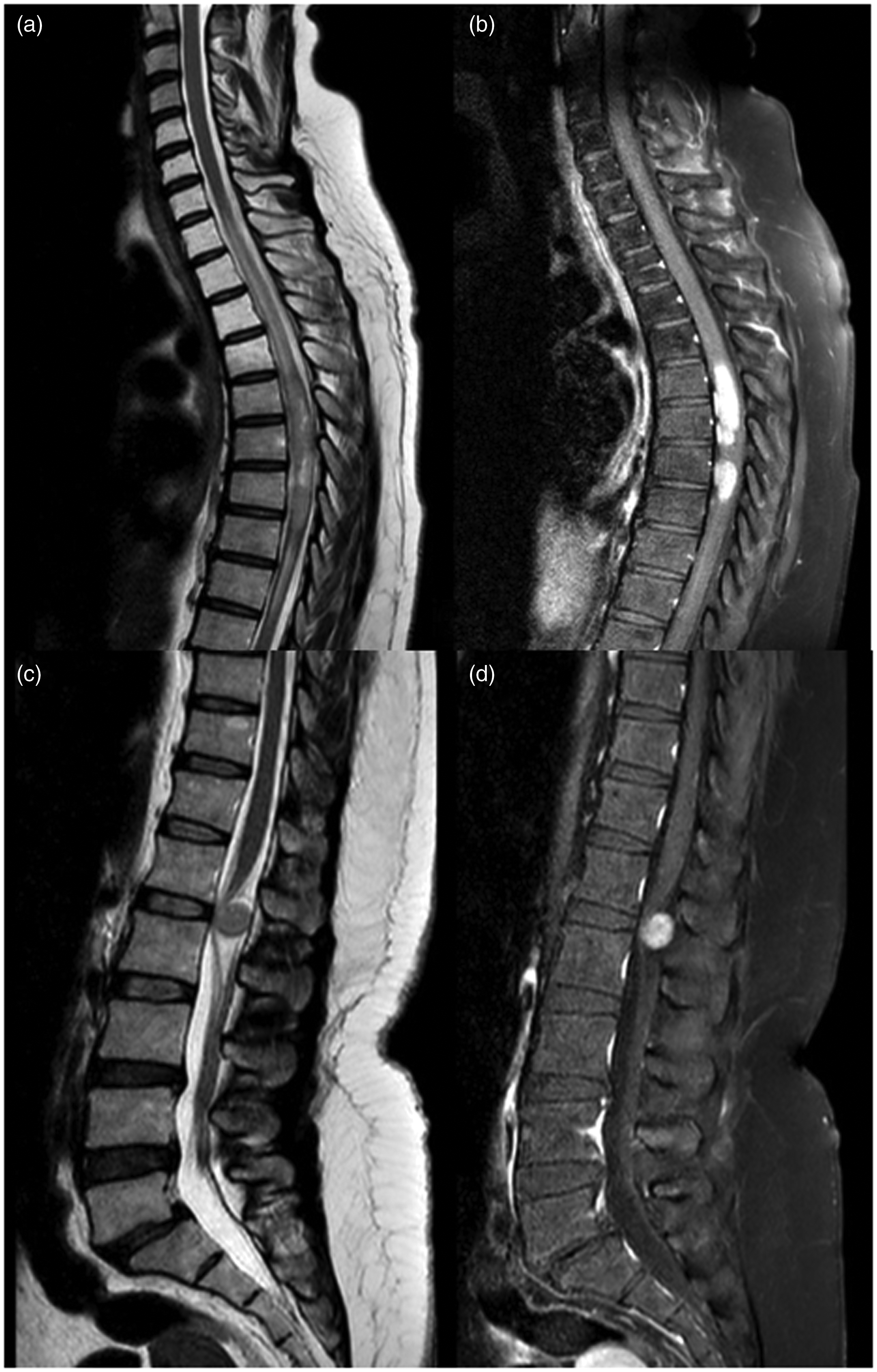
Sagittal T2-weighted MRI of the patient demonstrating an intramedullary lesion extending from T5 to T8 with hyperintense signals associated with increased spinal cord diameter (a) with scattered enhancement after gadolinium injection (b). Sagittal T2-weighted MRI of the lumbosacral spine showing a round intradural extramedullary isointense lesion compressing the conus medullaris (c) with homogenous enhancement after gadolinium injection.
The dura was opened, and midline myelotomy was performed in the thoracic region. The lesion was microscopically resected as much as possible (intralesional resection and multiple biopsies of the lesion). There was a soft, gray-to-white mass with poorly defined margins and poor consistency. Because the patient had appropriate spinal sagittal balance preoperatively, and we performed a limited laminectomy for tumor resection, no spinal fixation and fusion was performed. The patient’s neurological status did not improve after the surgery (ASIA impairment scale C), and she began rehabilitation and physical therapy postoperatively. She also received temozolomide (100 mg/m2/day orally) and irinotecan (40 mg/m2/day intravenously) in accordance with the standard protocol as adjuvant therapy for recurrent or metastatic Ewing sarcoma. Postoperative imaging of the thoracic spine revealed appropriate decompression (Figure 2a and b) and showed that the lumbosacral lesion had completely resolved (Figure 2c).

Postoperative sagittal T2-weighted cervicothoracic MR image of the patient showing an intramedullary lesion extending from T5 to T8 with hyperintense signals (a). Postoperative T-1 weighted MR image with gadolinium enhancement showing the lesion after internal resection and decompression (b). Post-chemotherapy T2-weighted MR image of the lumbosacral spine showing complete resolution of the extradural lesion (c).
Histopathological examination of the thoracic lesion revealed small round cells with moderate pleomorphism, nuclear atypia, and some true rosette formations in a pauci-vascular background (Figure 3a). Immunohistochemical staining of the tumor showed diffuse, strong immunoreactivity for cluster of differentiation (CD)99, and a high proliferative index (Ki-67: 85%–90%), with negative staining for leukocyte common antigen (LCA) (Figure 3b–d). The diagnosis of Ewing sarcoma was confirmed accordingly. At the 10-month follow-up, the patient’s neurological status had worsened, with bilateral lower extremity muscle strength scores of 0/5 (proximal/distal, respectively; ASIA impairment scale B), with spasticity and urofecal retention. She was alive with disease at the time of this report. The patient provided written informed consent for the publication of her case, and the reporting of this study conforms to the CARE guidelines. 38

(a) Histopathological sections showing sheets of small round cells with moderate pleomorphism, nuclear atypia, and some true rosette formations (arrows) in a pauci-vascular background (×400, H&E) and (b–d) Immunohistochemical staining of the tumor showing diffuse, strong immunoreactivity for CD99, and a high proliferative index (Ki-67) with negative staining for LCA (×100, ×100, ×40; b–d, respectively).
Discussion
Metastasis of Ewing sarcoma to the spinal cord is extremely rare, and the management and clinical significance is controversial, with no appropriate level of evidence. 2 Ewing sarcoma accounts for 10% to 15% of malignant bone tumors and 40% to 45% of all malignant tumors in children.1,2 Ewing sarcoma can arise in any bone or soft tissue; however, tumors in the spinal cord are rare. 2 We reviewed the current literature and identified 36 cases of intramedullary Ewing sarcoma that were either primary or metastatic (Table 1). Most of the cases were primary, and only five recurrent cases from other organs have been reported. We herein report a unique and rare case of metastatic Ewing sarcoma to both intramedullary and extramedullary sites in a single patient. The thoracic lesion was histologically revealed to be Ewing sarcoma in accordance with the immunohistochemical examination findings. The patient had been diagnosed with Ewing sarcoma 2 years prior to presentation and underwent surgery and chemoradiotherapy. She had no evidence of the disease 6 months prior to the current presentation. However, subsequently, she experienced a very progressive and malignant course of metastasis to the spinal cord. To the best of our knowledge, this is the only reported case of both intramedullary and extramedullary metastasis of Ewing sarcoma.
We performed a systematic review of the published literature in accordance with the preferred reporting items for systematic reviews and meta-analyses (PRISMA) guideline. 39 We searched the electronic databases, MEDLINE/PubMed, Embase, Scopus, Cochrane Library, and Web of Science (WOS) to 10 January 2022. We used the following Medical Subject Heading (MeSH) search terms, text words, and keywords: [‘Ewing sarcoma’ or ‘cord Ewing’ or ‘intramedullary Ewing’ or ‘extramedullary Ewing’] AND [‘recurrent’ or ‘metastatic’] in the English language, without date restrictions. We also manually searched the bibliographic lists of previously published review articles to identify related studies. All reference management was conducted using EndNote X8 for Windows (Clarivate Analytics, Philadelphia, PA, USA). We identified 36 articles reporting intramedullary or extramedullary Ewing sarcoma and these were included in the current systematic review.
The identified cases showed that intramedullary Ewing sarcoma was located mainly in the thoracic and lumbar regions, although cervical and sacral locations were also reported.4–36 The current case had a thoracic intramedullary lesion and a lumbar extramedullary lesion, both causing neural compression and deficits.
The clinical findings of spinal cord metastasis of Ewing sarcoma are similar to those of other lesions of the spinal cord, namely muscle weakness, sensory level, hyperreflexia, and involvement of sphincters in the form of spasticity. Chronic lesions cause spasticity and muscle wasting. 3 A review of the literature and a meta-analysis of the extramedullary cases of Ewing sarcoma by Saeedinia et al. 37 revealed that Ewing sarcoma should be considered among the differential diagnoses of intradural lesions despite their rarity, especially in patients with a previous history of the disease.
Gadolinium-enhanced spinal MRI remains the modality of choice for the diagnosis of Ewing sarcoma. 40 The lesions are mainly hyperintense in T2-weighted images and isointense in T1-weighted images. After contrast injection, segmental and scattered enhancement are observed.3,35 However, histology remains the gold standard for the diagnosis of Ewing sarcoma, and the examination should include immunohistochemical staining and molecular analysis comprising fluorescent in situ hybridization (FISH) and real-time polymerase chain reaction (RT-PCR) to identify and characterize EWSR1 translocation. 41
The treatment of Ewing sarcoma of the spinal cord lacks a standard and specific protocol, and the standard of care remains elusive. 12 Treatment comprises surgical resection (as much as possible while maintaining the neural anatomy) followed by chemoradiotherapy. 2 The prognosis and survival rate for patients with spinal cord Ewing sarcoma is not well known because of the rarity of the condition; however, based on previous cases (Table 1), the survival rate was 45% to 50% in primary cases and 30% to 35% in recurrent and metastatic cases.4–36 In surviving cases, rehabilitation and physical therapy should be considered part of the treatment program to improve the patient’s neurological status. In the current case, the patient improved markedly after 4 months of physical therapy and rehabilitation.
Conclusion
This is the first case of intramedullary and extramedullary spinal cord metastasis of Ewing sarcoma in a single patient. The patient was treated by surgical resection followed by adjuvant chemotherapy, physical therapy, and rehabilitation. Although rare, Ewing sarcoma should be considered among the differential diagnoses of spinal cord lesions in patients with neurological deterioration, especially in those with a history of Ewing sarcoma. Surgical resection and chemoradiotherapy are the main treatments; however, there is no standard guideline for the treatment of spinal cord Ewing sarcoma.
Supplemental Material
sj-pdf-1-imr-10.1177_03000605221108095 - Supplemental material for Metastatic thoracic and lumbar intramedullary and extramedullary Ewing’s sarcoma: a rare case report and literature review
Supplemental material, sj-pdf-1-imr-10.1177_03000605221108095 for Metastatic thoracic and lumbar intramedullary and extramedullary Ewing’s sarcoma: a rare case report and literature review by Seyed Reza Mousavi, Majid Reza Farrokhi, Keyvan Eghbal, Amirreza Dehghanian, Alireza Rezvani and Fariborz Ghaffarpasand in Journal of International Medical Research
Footnotes
Acknowledgement
We would like to acknowledge the editorial assistance of the Diba Negar Research Institute for improving the style and English of the manuscript.
Declaration of conflicting interest
The Authors declare that there is no conflict of interest.
Ethics statement
As a case report, no institutional review board (IRB) approval was required. The patient provided informed written consent to publish her case.
Funding
This research received no specific grant from any funding agency in the public, commercial, or not-for-profit sectors.
References
Supplementary Material
Please find the following supplemental material available below.
For Open Access articles published under a Creative Commons License, all supplemental material carries the same license as the article it is associated with.
For non-Open Access articles published, all supplemental material carries a non-exclusive license, and permission requests for re-use of supplemental material or any part of supplemental material shall be sent directly to the copyright owner as specified in the copyright notice associated with the article.