
Abstract
Objective
The effects of saliva on demineralized dentin and silver diamine fluoride (SDF) were investigated in vitro.
Methods
Dentin samples stored in deionized water (DIW), buffer solution (BS), basal medium mucin (BMM), and unstimulated whole saliva (UWS) were demineralized for 3 days and immersed in the same storage media. SDF as a 38 mass% solution was applied to the dentin samples for 3 minutes after they had been replaced in their respective medium. Surfaces were analyzed by scanning electron microscopy, energy-dispersive X-ray analysis (EDX), X-ray photoelectron spectroscopy (XPS), and X-ray diffraction (XRD).
Results
Scanning electron microscopy showed various surface deposits and coatings, including occlusion of dentinal tubules. DIW resulted in the thinnest coating, whereas BMM resulted in the thickest. EDX and XPS showed the formation of metallic silver and silver compounds in all four media, with the greatest formation in BS. XRD indicated that the main product was silver chloride except in DIW. Sulphur was found in BMM and UWS. EDX and XPS detected fluoride and XRD detected calcium fluoride and fluorohydroxyapatite in BS, BMM, and UWS.
Conclusion
The interaction between SDF and demineralized dentin was dependent upon the storage medium. BMM provided an outcome most similar to human saliva.
Introduction
Silver diamine fluoride (SDF) [Ag(NH3)2F], also spelled silver diammine fluoride, is an effective agent for preventing and arresting caries in both primary and permanent dentitions.1,2 The mechanism of action of SDF has been clarified in several studies.3,4 In 1974, Suzuki et al. 3 mixed enamel powder with SDF solution in synthetic saliva, which yielded calcium fluoride (CaF2) and trisilver phosphate (Ag3PO4). In 2011, Lou et al. 5 mixed SDF with hydroxyapatite (HAp) powder and gelatin (as a chemically representative protein) in deionized water (DIW), which yielded CaF2-like deposits and metallic silver (Ag). In 2017, Mei et al. 6 mixed SDF solution with apatite precipitation in Tris-buffered saline, which resulted in the formation of fluorohydroxyapatite and silver chloride (AgCl). Given these various interactions, storage medium should be chosen carefully when investigating the interaction of SDF with tooth components. Human saliva is clearly the most appropriate test medium; however, it is associated with difficulties in practice because of the large volumes required and the uncontrolled variations. Therefore, artificial growth media have been preferentially used. 7 Nevertheless, saliva substitutes should behave similarly to human saliva for successful simulation.
The use of saliva substitutes to assess the interaction of SDF and demineralized dentin should be explored from the viewpoints of both chemical and bacteriological effects. Buffer solution (BS) has been developed to establish a tooth tissue erosion–abrasion cycling model.8,9 This BS does not contain organic content, thus allowing isolated experimental factors to be tested.
In this study, we used BS, basal medium mucin (BMM),10,11 unstimulated whole saliva (UWS), and DIW (negative control) as four storage media to ascertain the effects of the reaction products of SDF on dentin in an in vitro model. The null hypothesis was that no difference would be observed among the saliva substitutes in the interaction between SDF and demineralized dentin.
Materials and methods
Sample preparation
The study protocol was approved by the institutional review board of the University of Hong Kong (UW 11-129), and the experimental design is illustrated in Figure 1. Caries-free human third molars were stored in 0.1% thymol solution at 4°C prior to preparation. Whole teeth were embedded in acrylic resin. Enamel was removed by sectioning with a Microslice 2 annular saw (Metals Research Ltd., Cambridge, UK) under water cooling. The exposed dentin block was drilled with a trephine (inner diameter of 3.8 mm). After examination, 20 qualified dentin discs (3.8-mm diameter, 1.0-mm thickness) were obtained. 12 These specimens were then stored in 0.1% thymol solution at 4°C. They were subsequently painted with two layers of acid-resistant nail varnish (Estée Lauder; New York, NY, USA) on one side, and the other surface was ground under running water on silicon carbide paper using grit sizes of 600, 800, and 1200 in sequence. They were then sonicated in a Biosonic UC100 sonicator (Coltene Whaledent, Inc., Cuyahoga Falls, OH, USA) for 3 minutes in DIW solution to remove debris. Finally, they were stored in DIW at 4°C prior to use from 1 to 3 days after cutting.

Flow chart of the experimental design.
Saliva preparation
UWS was collected by the vacuum filtration technique 13 from 10 healthy subjects aged 25 to 35 years after obtaining informed consent and institutional review board approval (UW 11-129). The saliva (pH, 6.2–7.5) was pooled and mixed. It was then vacuum filtered for 30 minutes using a bottle-top filtration set (Corning Inc., Corning, NY, USA) with a 0.22-µm-pore polyethersulfone membrane and stored at −18°C until use. Two storage media were also prepared: BS and BMM solution. DIW was used as a control. The BS (pH 7.0) contained 1.45 mmol/L Ca2+, 5.4 mmol/L PO43−, and 0.1 mol/L Tris buffer. 8 The BMM (pH 7.0) contained 2.5 g/L partially purified pig gastric mucin (type III; Sigma Chemical, St. Louis, MO, USA), 10.0 g/L proteose peptone (Oxoid, Unipath, Basingstoke, UK), 5.0 g/L trypticase peptone (BBL; Becton, Dickinson and Company, Franklin Lakes, NJ, USA), 5.0 g/L yeast extract (Difco Laboratories, Detroit, MI, USA), 2.5 g/L potassium chloride, 5.0 mg/L hemin, 1.0 mg/L menadione, 1.0 mmol/L urea, and 1.0 mmol/L arginine. 14
Twenty dentin discs were numbered in sequence, selected by a random number generator, and assigned to four groups. The dentin discs were stored individually in 5.0 mL each of UWS, BS, BMM, or DIW and agitated at 75 rpm in an incubator (Type 3157; Forma Scientific, Minneapolis, MN, USA) at 37°C for 24 hours for equilibration. Each disc was then individually immersed in a demineralization solution (50 mmol/L acetate BS to which 2.2 mmol/L each of KH2PO4 and CaCl2 had been added) at pH 4.5 and 37°C for 72 hours, 15 with agitation at 75 rpm. The specimens were then sonicated in a Biosonic UC100 sonicator in 20 mL of DIW for 20 minutes to reduce carryover of demineralization solution, followed by sonication in 8.0 mL of the respective medium for 5 minutes.
The test surface of each dentin disc was then gently air-dried with oil-free compressed air (Clean Ace Ne 530; Hakuba, Tokyo, Japan). Next, 10.0 µL of SDF solution (Saforide, 38 mass%, pH 12.5; J. Morita, Osaka, Japan) was applied to the demineralized dentin for 3 minutes. The SDF solution completely covered the dentin surface, and a 3-minute reaction time was allowed for the SDF to deeply infiltrate the dentin tubules and react with the storage medium saturated in the dentin tubules. The dentin discs were then briefly rinsed with 2 mL of the respective storage medium before storage in 8 mL of the same medium at 37°C for 5 days in a 15-mL plastic bottle (Corning Inc.). The medium was replaced daily, and light was excluded by wrapping each container in aluminum foil.
Scanning electron microscopy (SEM) and energy-dispersive X-ray (EDX) analysis
One dentin disc from each group was randomly selected (as mentioned above) on day 6 and cleaved diametrically using a dental chisel. The samples were prefixed twice in 2% glutaraldehyde in 0.1 M cacodylate BS titrated to pH 7.2 for 30 minutes, then rinsed several times with 70 wt-% ethanol and stored overnight. Next, the samples were dehydrated in ascending concentrations of ethanol (70%, 85%, 95%, and 100%) for 30 minutes in each solution and in 100% ethanol for 24 hours, then air-dried at room temperature. The samples were then sputter-coated with gold and analyzed using field-emission SEM (Hitachi S-4800 microscope; Hitachi, Tokyo, Japan) at 5 to 20 kV. Element analysis was performed by EDX spectrometry (Horiba EMAX EDX Detectors and EMAX Energy Software; Horiba, Japan).
X-ray photoelectron spectroscopy (XPS) analysis
One dentin disc from each group was selected at random on day 21 for XPS analysis (PHI 5400; PerkinElmer Inc., Waltham, MA, USA). XPS analysis was performed for detection of reaction products. Broad-range survey scans were performed to determine the atomic concentration (at%) at a pass energy of 89.45 eV and with an entrance slit width of 4 mm and resolution of approximately 1.3 eV. Binding energy calibration was performed with respect to C1s, from surface hydrocarbons, assuming the value of 284.8 eV for this. The binding energy and the chemical shifts of the Ag3d photo peaks and the kinetic energy of the so-called AgMNN Auger structures were then determined.16,17
Powder X-ray diffraction (XRD) analysis
One dentin disc from each group was selected at random on day 21 for step-scanned powder XRD (D8 Advance; Bruker Corporation, Billerica, MA, USA) using CuKα (λ = 1.5406 Å) at 1.6-kW radiation via a Göbel mirror. XRD analysis was performed for detection of the crystal characteristics. After data collection, the mounted sample was unloaded and the measurement was repeated with the same sample holder. The scan was performed over 20° to 60° (2θ) with a step size of 0.05° and scan speed of 40 s per step. Reproducible datasets of each sample were obtained. Patterns were compared with the International Centre for Diffraction Data database (PDF-2 Release 2004; International Centre for Diffraction Data, Newtown Square, PA, USA).
Results
SEM showed that the treated dentin surfaces had different morphological characteristics (Figure 2). The DIW group exhibited a thin, smooth, regular coating with some occlusion of dentin tubules (Figure 2(a)). The BS group showed uniform occlusion of polyhedral structures (30–80 µm) (Figure 2(b)). The BMM group showed a relatively thin coating with irregular deposits and some delineation of tubule orifices (Figure 2(c)). In the UWS group, the dentin surfaces were covered with a rougher coating that partially blocked some dentinal tubules, and some irregular deposits (50–200 µm) were present (Figure 2(d)). For comparison, Figure 2(e) and (f) shows chemically demineralized dentin (bank groups) samples after demineralization and before storage in saliva media. The SEM photographs showed that the dentin tubules were enlarged and completely open.

Scanning electron microscopy views of the dentin surface. After treatment: (a) DIW group. (b) BS group. (c) BMM group. (d) UWS group. Before treatment: (e) and (f).
Area-scan EDX analysis was performed for all four dentin samples. The results of the qualitative EDX analysis and subsequent quantitative XPS analysis are summarized in Table 1. All four groups were found to contain elemental silver and tooth tissue components such as C, O, N, Ca, P, and Na. In the BS group, the atomic concentration of Ag was 12.2% and that of Cl was 12.9%, and the ratio of Ag to Cl was 12.2/12.9 = 0.9, suggesting that AgCl had formed. In the BS group, the amounts of silver and chloride were relatively equal (ratio = 0.9). In the DIW and BMM groups, however, the amount of silver was much higher than the amount of chloride.
EDX and XPS analysis.

Note: For the qualitative EDX analysis, the background elemental peaks (i.e., Ca, P, O, N, C, and Na) were excluded from the EDX analysis report.
EDX, energy-dispersive X-ray analysis; XPS, X-ray photoelectron spectroscopy; DIW, deionized water; BS, buffer solution; BMM, basal medium mucin; UWS, unstimulated whole saliva.
A spot-scan EDX analysis was conducted in the UWS group to visually identify the major element peaks. Higher magnification (×5000) showed that crystal-shaped particles had blocked and were distributed along the dentin tubules (Figure 3(d2)). The crystal particles contained mainly silver and chlorine. Trace amounts of fluorine were also detected (Figure 3(d4)). At the same site, element mapping was also performed in the UWS group (Figure 4). Both Ag and AgCl had formed and covered the treated dentin surface (Figure 4(d2)). Ag was distributed along and extended downward into the tubules, and it blocked the exposed dentin tubules (Figure 4(d2)).

Element mapping analysis of the cross-sectional surface of a dentin sample in the unstimulated whole saliva group.
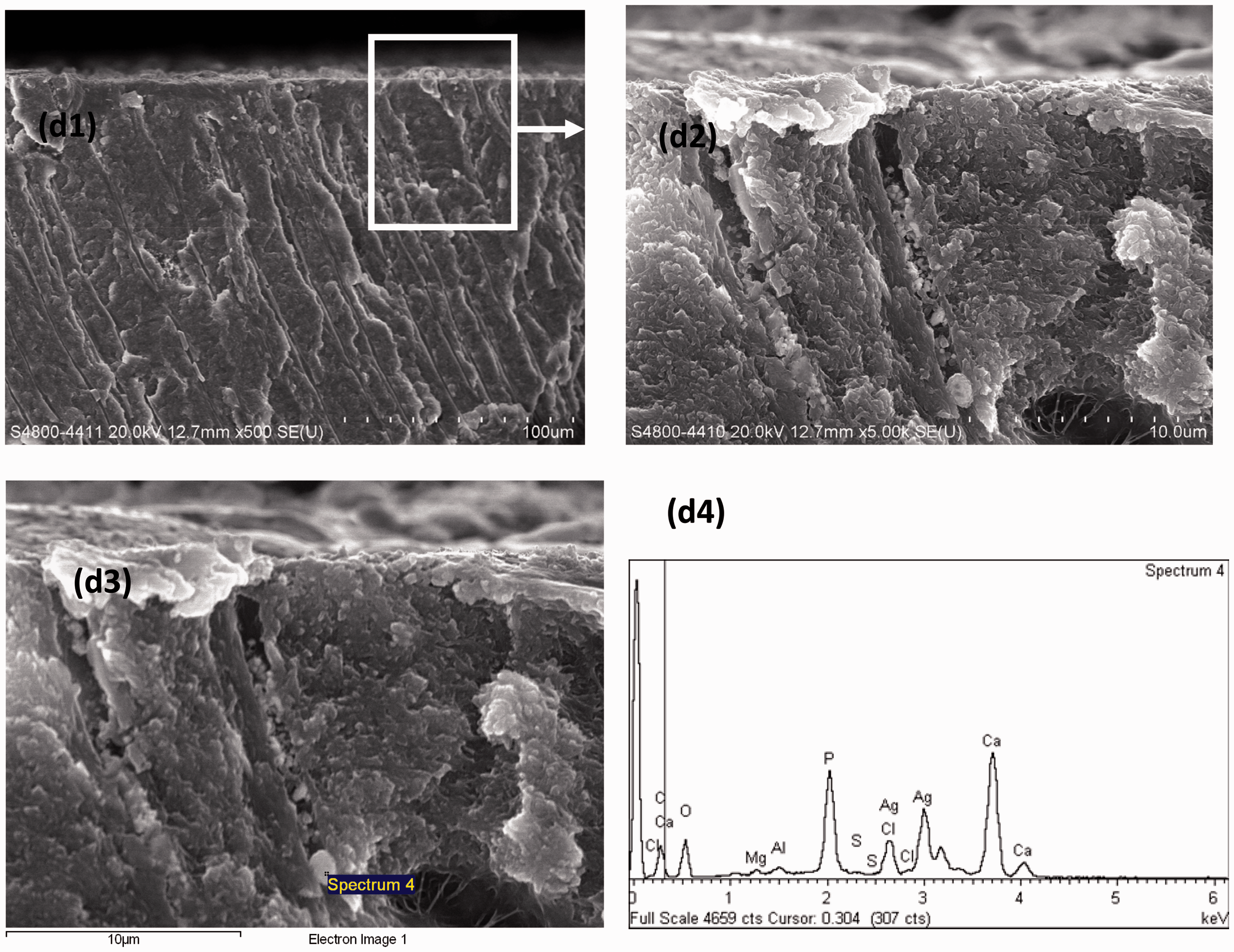
Cross-sectional scanning electron microscopy view of dentin samples in unstimulated whole saliva group. (d1) Low magnification (×500). (d2) High magnification (×5000) (d3) and (d4) Spot-scan energy-dispersive X-ray analysis of a crystal particle found in the high-magnification spectra.
XPS analysis has been expanded for chemical identification since establishment of the Auger data library. 17 Photoelectron and Auger line energy reports for Ag 3d5/2 and AgM4N4.5N4.5 lines are presented in Table 2. The reported values for binding energies in the DIW group were Ag3d5/2 = 368.0 eV, AgM4N4.5N4.5 = 357.9 eV, and Auger parameter = 725.9 eV, indicating good agreement with previously reported values for Ag (Ag3d5/2 = 368.3 eV, AgM4N4.5N4.5 = 357.7 eV, and Auger parameter = 726.0 eV). 18 In the BS group, the Auger transition exhibited a 2.5-eV shift from 723.4 eV to 725.9 eV compared with the DIW group, and the binding energy of Cl2p was 198.5 eV. These results are consistent with the parameters for AgCl in a previous report. 17 In the UWS group, the S2p peak was presented in the XPS analysis.
Photoelectron and Auger line positions of dentin samples in XPS.

XPS, X-ray photoelectron spectroscopy; DIW, deionized water; BS, buffer solution; BMM, basal medium mucin; UWS, unstimulated whole saliva.
Figure 5 shows the XRD patterns of all four groups. The X-axis represents 2θ (degrees), and the Y-axis represents intensity (number of counts). The XRD patterns of the four groups showed similar characteristics. However, the peaks in the BS group were sharper and had the strongest intensity. The 2θ peaks at 25.87° (002), 31.72° (211), 32.16° (112), 32.83° (300), and 46.64° (222) corresponded to the main reflection planes of HAp (JCPDS 46-0905). 19 The peaks at 25.86° (002), 31.94° (211), 33.13° (300), 32.27° (112), and 46.87° (212) corresponded to the main reflection planes of fluorapatite (FAp) (JCPDS 15-0876). The XRD data clearly confirmed that FAp and HAp crystals were present in all four groups. The reflections at 38.10° (111) and 44.37° (200) corresponded to Ag (JCPDS 01-1167). In this study, the peaks of Ag were present in the DIW, BMM, and UWS groups but not in the BS group.

X-ray diffraction patterns of samples in the (a) DIW group, (b) BS group, (c) BMM group, and (d) UWS group.
Discussion
SEM examination showed different surface morphologies in the four treated dentin groups (Figure 2). The treated dentin surfaces were covered with deposits and coatings that both partially and fully occluded the dentinal tubules. The DIW group showed the thinnest coating, whereas BS produced the thickest. EDX and XPS showed that silver and silver compounds remained as the major reaction products in all four groups after SDF application (Table 1). Based on the XRD findings, silver compounds existed in the form of Ag and AgCl but probably not as Ag3PO4, silver sulfide, or silver fluoride (Table 2).
In XRD analysis, sharper and more intense peaks are considered to represent more crystalline material. 20 The intensity in the DIW group was moderate, while the BMM and UWS groups showed complex and irregular peaks with the lowest intensity (Figure 5). Because of the similarity of their XRD patterns, it was difficult to distinguish FAp from HAp. Additionally, the HAp crystals in this study matched the pattern of calcium-deficient HAp in the XRD database, which most likely reflects the demineralization process of the dentin.
Silver fluoride, which has 2θ peaks at 31.37° (002), 34.47° (100), 38.02° (101), and 47.28° (102) (JCPDS 17-0325), was not observed. The absence of an obvious peak of Ag with BS might have been caused by the high degree of reactivity of silver ions to chloride ions to form AgCl. All four dentin groups showed peaks at 32.24° (200), 46.23° (220), 54.83° (311), and 57.48° (222), which correspond to AgCl (JCPDS 31-1238) as shown in Figure 5.
In the previous literature, Ag3PO4 and CaF2 were detected by XRD after applying SDF to powdered enamel in vitro.4,6 One of these studies revealed the subsequent changes after SDF application on powdered enamel. 4 Samples were stored in synthetic saliva for 1, 2, 4, 10, and 20 weeks. XRD analysis showed that CaF2 gradually disappeared. The authors explained that CaF2 was not stable in the presence of HPO4−, which is present in saliva, and slowly changed into FAp. In 2017, Mei et al. 6 mixed calcium phosphate with different SDF concentrations (0.38, 1.52, 2.66, and 3.80 mg/mL) to investigate apatite crystals and compared the findings with a calcium phosphate control. XRD analyses revealed that small localized fluoride anions substituted the hydroxyl anions in HAp crystals. The authors concluded that SDF reacted with calcium and phosphate ions and produced fluorohydroxyapatite. Although the above-mentioned studies omitted complex saliva environment, they all concluded the existence of fluorohydroxyapatite.
The current study revealed that although Ag3PO4 and CaF2 might exist, they could not be confirmed because the characteristic 2θ peaks of Ag3PO4 (38.99° (210) and 42.83° (211), JCPDS 01-1058) were similar and partially overlapped with the FAp peaks (33.13° and 46.87°). In addition, the CaF2 peaks (2θ peaks at 32.89° (111) and 55.22° (220), JCPDS 01-1274) could not be confirmed because they also partially overlapped with the FAp peak (33.13°). AgCl formed, which was consistent with the findings in the previous literature. However, we observed no formation of silver thiocyanate, which has 2θ peaks at 20.24° (012), 23.18° (101), 26.50° (111), 38.02° (101), 31.37° (002), 34.47° (100), and 47.28° (102) (JCPDS 36-0608). The reason for the lack of silver thiocyanate formation might be because thiocyanate ions were not present in any of the three recipes of substitutes (BS, BMM, or UWS).
Silver sulfide, with 2θ peaks at 26.19° (111), 29.06° (012), 34.74° (112), 36.81° (022), and 37.74° (200) (JCPDS 03-0844) was unlikely to have been formed. Although a sulfur element was confirmed by EDX and XPS in the UWS group, the treated dentin sample did not match the crystal pattern of silver sulfide in the XRD database. Silver and sulfur elements thus probably existed in the form of silver–protein complexes.
XPS analysis did not detect a chloride element with UWS. This might be explained by XPS detection being only performed to examine the most superficial dentin layer, and no chlorine was found in this area. EDX and XPS revealed a sulfur element in the BMM and UWS groups; however, XRD suggested that a free crystal form of sulfur was unlikely to have been present. Therefore, this sulfur probably existed in the form of a silver–protein complex.
EDX showed only trace amounts of fluorine in all four groups, and XRD suggested that fluorine exists in the form of CaF2 and FAp. XPS indicated trace amounts of fluorine in the DIW, BMM, and UWS groups but not in the BS group, as exhibited by the thickest layer of AgCl coating the dentin surface. Based on the above findings, fluoride does not appear to be a major reaction product after SDF application under the conditions tested. This may be supported by a previous report showing that CaF2 could be easily washed away after application of SDF in vitro. In addition, any FAp formed will not act as a reservoir for fluoride because of its low solubility. 5
This in vitro study was performed to assess the effect of saliva substitutes with SDF on dentin. Demineralized dentin instead of carious dentin was used. Dentin specimens were equilibrated with various storage media prior to demineralization. Pellicles and biofilms were not emphasized. Although a pellicle coating that has formed on enamel from protein-containing media has been shown to offer some protection of enamel surfaces against demineralization for short periods of time, it is not considered to be effective on dentin; 21 pellicles are reportedly ion-permeable rather than a barrier for dentin substitute. 22 To eliminate the effect of pellicles in future studies, the demineralized dentin surface should be examined under SEM after exposure to saliva media and before application of SDF.
The mineral content of BMM was not precisely controlled because of the complexity of its composition. 11 In the literature, defined medium with mucin was later introduced as a better protein-containing saliva than BMM because defined medium with mucin has a standardized composition. 10 Despite this limitation of the present study, the saliva media used in this trial produced differing outcomes of treatment with SDF. 23 The null hypothesis of this study was therefore rejected. That the BS group yielded relatively high amounts of Ag and AgCl shows that BS performs differently than human saliva. In addition, the presence of elemental sulfur in both the BMM and UWS groups indicates that BMM was closer to human saliva in this in vitro experimental model than the other test groups. This suggests that the selection of saliva substitutes should be considered in vitro investigations examining SDF and tooth interactions when using bacterial models. In most experiments to date, the researchers chose their saliva formulation compositions in a historical manner based on the model being used. No attempts have been made to clarify the effects of these components on the materials tested. Therefore, clear definitions of artificial saliva formulations and selective use of specific formulations for particular investigations may allow more authentic interactions between the test variables, thus affording more meaningful results.
Given the presence of the chlorine element in human saliva (average concentration of about 14 mmol/L), 24 the formation of AgCl would be expected to occur naturally after SDF treatment. The interaction between AgCl and oral tissues has been seldom reported. In one study, the authors compared the cytotoxic effects of several dental metals on human gingival fibroblasts in vitro. 25 The cytoxicity of SDF against human gingival fibroblasts was reportedly higher than the cytotoxicity of AgCl.
Conclusions
Under the limitations of the conditions tested, SDF treatment on demineralized dentin showed that Ag and silver compounds were the major reaction products. Different chemical reactions with SDF occurred depending on the composition of the medium. Different saliva substitutes produced different interactions between SDF and demineralized dentin and should therefore be considered in the design and outcomes of in vitro investigations.
Footnotes
Acknowledgement
We acknowledge Dr Markku Heinonen, Department of Physics and Astronomy, University of Turku, for the XPS analysis and Ching M. Che, HKU-CAS Joint Laboratory on New Materials, State Key Laboratory of Synthetic Chemistry and Department of Chemistry, the University of Hong Kong, Hong Kong Special Administrative Region, P.R. China.
Declaration of conflicting interest
The authors declare that there is no conflict of interest.
Funding
This study was supported by HKU Small Project Funding (#10212.10401066.26499.08007.323.01).