
Abstract
Objectives
Proanthocyanidins (PAs) have been widely used as effective agents for dentin collagen cross-linking to enhance the biomechanics and biostability of dentin in vitro. However, the effects and protective mechanisms of various tea root-derived PA components on dentin remain undefined. This study evaluated the effects of these tea root-derived PA components on dentin biomechanics and biostability.
Methods
In this study, ethyl acetate and n-butyl alcohol were used to extract PAs with different degrees of polymerization from tea roots; the effects of these PA extracts on dentin were evaluated.
Results
Dentin was treated with glutaraldehyde, ethyl acetate, n-butyl alcohol, or water. PAs with a high degree of polymerization, extracted using n-butyl alcohol, were able to more effectively improve dentin collagen cross-linking, increase resistance to bacterial collagenase digestion, and enhance dentin elasticity, relative to treatment with glutaraldehyde or PAs with a low degree of polymerization (extracted using ethyl acetate). Additionally, treatment with aqueous extract of tea roots was detrimental to dentin stability and function.
Conclusions
PAs with a high degree of polymerization were effective for dentin protection and restoration in vitro, suggesting clinical treatment potential for tea root-derived PAs.
Keywords
Introduction
Dentin is composed of a type I collagen-based organic phase and a hydroxyapatite crystal-based mineral phase.1,2 Dentin collagen cross-linking provides a scaffold for maintenance of dentin biomechanics and biostability. 3 For resin-dentin adhesive restoration, the cross-linking process can prevent dentin collagen-based organic degradation, which enables increased stability and durability of resin-dentin adhesion through protection of exposed dentin collagen in the hybrid layer.4–6 Therefore, enhancement of dentin collagen cross-linking is a valuable aspect of dentin repair and protection. 7 Demineralization leads to denuded collagen fibrils, which exhibit increased susceptibility to hydrolytic and enzymatic degradation.8,9 Because dentin collagen experiences demineralization-induced instability, collagen cross-linkers have been widely used to improve its stability and durability, thus achieving good performance in dental tissue repair.10,11
Proanthocyanidins (PAs) are secondary plant metabolites that reportedly have a positive effect on dentin collagen cross-linking, which enables improvement of dentin biomechanics and biostability.12–14 PAs constitute a group of oligomers and polymers based on the monomeric flavan-3-ol. 14 Therefore, the bioactivity of PAs is affected by their origin, extraction techniques, extraction reagents, and a variety of other factors. The proportions of monomers, oligomers, and polymers greatly determine the clinical effects of a given preparation of PAs. 15 In practice, the various components of PAs, with distinct molecular weights and degrees of polymerization, have been found to exert distinct effects on the cross-linking of dentin collagen.
The use of PAs in dentin bonding has been reported to cause multiple complications due to weaknesses in PAs, such as poor polymerization. It important to understand the effects of each component on the bioactivity of a given preparation of PAs. 16 Fractional solvent extraction is the most commonly used method for extraction of PAs with different degrees of polymerization; this method typically involves the use of acetone for preliminary extraction of PAs, followed by ethyl acetate for further extraction and n-butyl alcohol for isolation of PA components. 17 We previously found that tea roots contain abundant PAs. Furthermore, we used ethyl acetate and n-butyl alcohol for preliminary extraction of tea root-derived PAs, which enabled isolation of various PA components. 18 However, the biological effects of these PA components on dentin remain undefined. Therefore, this study was performed to evaluate the effects of PA components on the biomechanics and biostability of dentin. We hypothesized that PA components would exert distinct protective effects on the elasticity of dentin, as well as on its ability to resist degradation by bacterial collagenase.
Materials and methods
Tea root materials and extraction methods
Tea roots (37 g) used in this study were harvested from Mountain Dagong, Tongling County, Anhui Province, China. Sundried tea roots were ground and extracted four times in 80% aqueous acetone at room temperature, producing a water-soluble extract. This water-soluble extract was then mixed with petroleum ether (1:3, v/v) to provide a petroleum ether-soluble fraction (2.01 g) and water-soluble fraction A. Water-soluble fraction A was then mixed with ethyl acetate (1:3, v/v) to provide an ethyl acetate-soluble fraction (2.60 g) and water-soluble fraction B. Water-soluble fraction B was then mixed with n-butyl alcohol (1:3, v/v) to provide an n-butyl alcohol-soluble fraction (1.10 g) and water-soluble fraction C (0.61 g). To produce pretreatment solutions for dentin treatment assays, all three extracts (0.13 g each) were first dissolved in 20% dimethyl sulfoxide (DMSO) and then diluted in HEPES buffer (20 mM, pH 7.2) to achieve final concentrations of 0.65% (w/v) of the respective tea root extracts. Separately, glutaraldehyde was diluted in HEPES buffer to achieve a final concentration of 2% glutaraldehyde, with an equal volume of DMSO. HEPES buffer with an equal volume of DMSO served as a control solution.
Dentin sample preparation
Forty-five intact human molars were collected at the Affiliated Stomatological Hospital of Anhui Medical University and stored at −20°C before use. All patients who provided molars for this study were 20 to 30 years old. In the laboratory, a slow speed diamond saw was used to remove crowns with a water rinse. In total, 25 dentin discs of 5.0 mm width × 5.0 mm thickness × 5.0 mm length and 75 dentin beams of 1.0 mm width × 1.0 mm thickness × 8.0 mm length were produced. All dentin discs and beams were randomly divided into five groups (n = 5 dentin discs and n = 15 dentin beams per group), then treated with 10% phosphoric acid solution for 5 hours for demineralization. 19 Dentin discs and beams were soaked in either DMSO (control), ethyl acetate PA extract, n-butyl alcohol PA extract, aqueous PA extract, or glutaraldehyde for 1 hour. The study protocol was performed in accordance with the tenets of the Declaration of Helsinki and was approved by the Ethics Committees of Anhui Medical University. Written informed consent was obtained from each patient prior to provision of the molar for use in this study.
Reagents
Bacterial collagenase I was obtained from Sigma-Aldrich (St. Louis, MO, USA). A human hydroxyproline enzyme-linked immunosorbent assay kit was purchased from Nanjing Jiancheng Bioengineering Institute (Nanjing, China). Glutaraldehyde was purchased from Tianyu Chemical Reagent Inc. (Tianjing, China). Ethyl acetate and n-butyl alcohol were purchased from Tedia Co. Ltd. (Fairfield, OH, USA).
Fourier-transform infrared (FTIR) spectroscopy
An infrared spectrophotometer (Nicolet 6700, Thermo Scientific Inc., Waltham, MA, USA) with attenuated total reflectance was used to detect FTIR spectra of demineralized dentin discs in each treatment group. The resolution was 4 cm−1 and the transmission range of the diamond attenuated total reflectance crystal ranged from 400 to 4000 cm−1. Thirty-two scans were performed for each sample.
Bacterial collagenase-mediated collagen degradation
Dentin discs were incubated in bacterial collagenase I (100 U/mL) at 37°C for 7 days; the digest supernatant was then hydrolyzed with 6 M HCl at 98°C for 24 hours. Samples were lyophilized, then dissolved in 600 µL water; the human hydroxyproline enzyme-linked immunosorbent assay kit was used to examine the collagen enzymolysis-induced release of hydroxyproline.
Elasticity modulus test
The WDS-20 load tester (Fangyuan Test Instrument Co., Ltd., Jinan, China) was used to detect the elasticity of dentin beams at a crosshead speed of 0.5 mm/minute. Following treatment with DMSO, ethyl acetate PA extract, n-butyl alcohol PA extract, aqueous PA extract, or glutaraldehyde, dentin beams were evaluated by a three-point bending assay, in accordance with the method used in a previous report. 20 A constant load increase was applied to each treated dentin beam until failure was observed. The maximum load was recorded and used to generate a load-deflection curve, based on the following elastic modulus equation: E = (L3 m)/(4 b d3). In this equation, E indicates the modulus of elasticity in bending; L indicates the support span; b indicates the width of the tested beam; d indicates the depth of the tested beam; and m indicates the slope of the initial straight-line portion of the load deflection curve. 20
Statistical analysis
For all parameters, the mean ± standard deviation were determined. One-way analysis of variance was used for statistical analysis of dentin collagen degradation and the elasticity of dentin beams. The Tukey post hoc test was used for subsequent pairwise comparisons between groups. p values <0.05 were considered indicative of significant differences. Data are representative of at least three independent experiments.
Results
Effect of tea root-derived PA components on FTIR spectra of dentin collagen
We previously found that the tea root extract contains abundant PAs, including diverse oligomers and polymers with an average degree of polymerization of 5.41. Notably, catechin and epicatechin monomers are the predominant structural units of PAs. 18 Ethyl acetate is reportedly able to extract PAs with a low degree of polymerization, whereas n-butyl alcohol can extract PAs with a high degree of polymerization. The remaining PA polymers are primarily maintained in the aqueous phase.17,18,21 Glutaraldehyde is a high-efficiency dentin collagen cross-linker.22,23 Therefore, ethyl acetate PA extract, n-butyl alcohol PA extract, aqueous PA extract, and glutaraldehyde were used to treat demineralized dentin discs for examination of FTIR spectra. 24 Intact collagen exhibits a unique helical structure with a specific amide band in FTIR spectra. As shown in Figure 1, amides I, II, and III at 1648, 1547, and 1235 cm−1, respectively, were the prominent FTIR bands. The bands of amides I and II represent the main absorption bands of peptide groups in collagen. The amide I band is mainly attributed to the stretching vibration of the carbonyl group, which is very sensitive to the peptide bond-related hydrogen bond and coupling between transition dipoles. The amide II band is mainly attributed to the bending vibration of the N–H bond and stretching vibration of the C–N bond. The wider and higher bands of amides I and II indicate greater involvement of the hydrogen bond, thereby enhancing the cross-linking of dentin collagen. 24 Compared with the FTIR spectra of dentin collagen in the DMSO-treated control group, the FTIR spectra of dentin collagen in the groups treated with glutaraldehyde, ethyl acetate PA extract, n-butyl alcohol PA extract, and aqueous PA extract were all considerably altered. Specifically, the dentin collagen treated with n-butyl alcohol PA extract showed wider amide I and II bands, compared with those of other treatments, without any change in wave crest position or appearance of new wave crests. This finding suggested that treatment with PAs with a high degree of polymerization, extracted using n-butyl alcohol, had a more positive effect on dentin collagen cross-linking, relative to the effect of treatment with PAs with a low degree of polymerization, extracted using ethyl acetate, or treatment with glutaraldehyde. Moreover, the bands of amides I, II, and III were significantly lower in the spectra of the group treated with aqueous PA extract than in the spectra of the groups that received the other four treatments, which might be attributed to the degradation of dentin collagen induced by treatment with the aqueous PA extract. Overall, these findings indicate that tea root-derived PAs with a high degree of polymerization are more effective in cross-linking dentin collagen than glutaraldehyde or tea root-derived PAs with a low degree of polymerization.
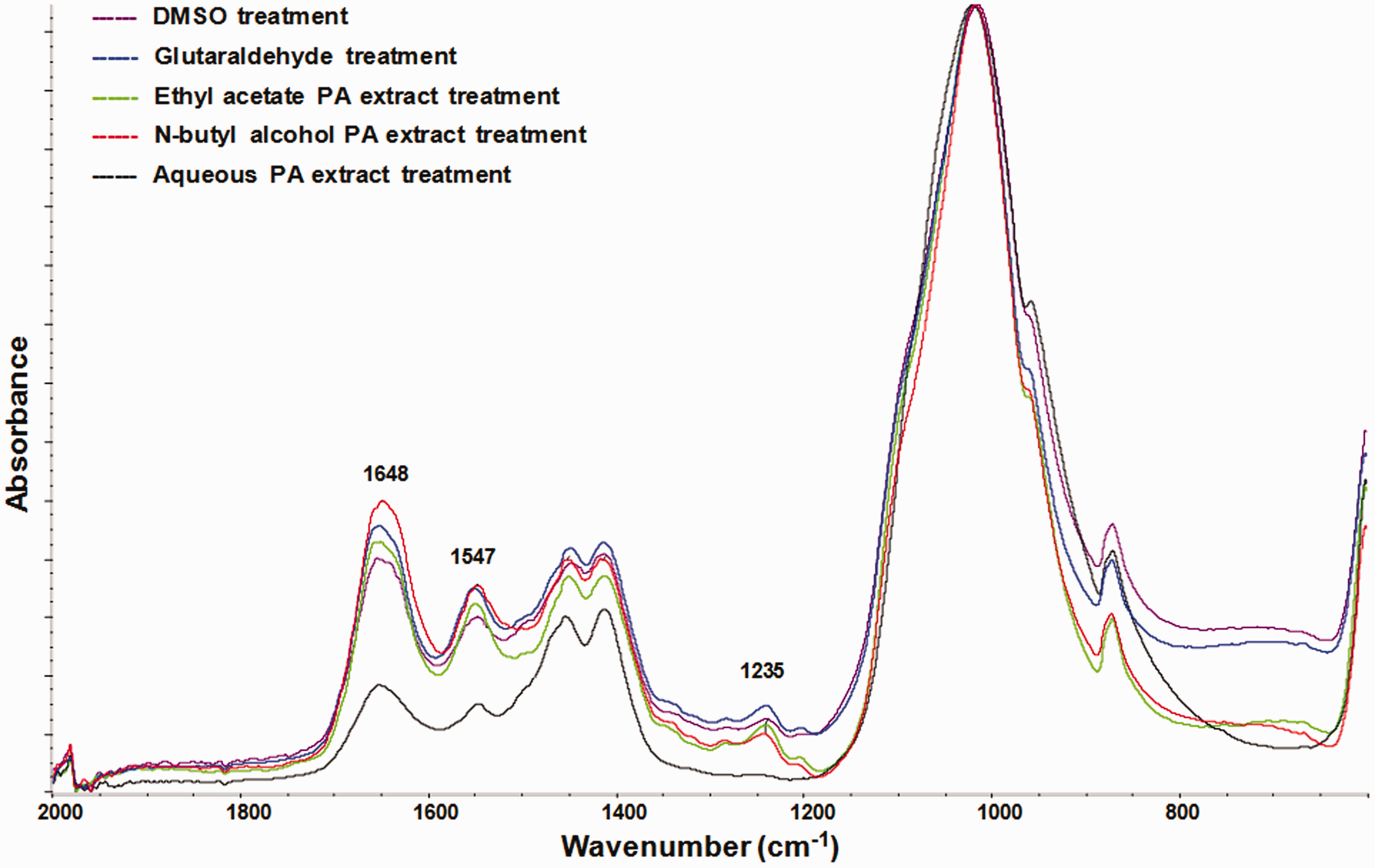
Fourier-transform infrared (FTIR) spectra of dentin films treated by dimethyl sulfoxide (DMSO), glutaraldehyde, ethyl acetate proanthocyanidin (PA) extract, n-butyl alcohol PA extract, or aqueous PA extract. Amides I, II, and III are indicated at 1648, 1547, and 1235 cm−1. DMSO treatment served as control.
Effect of tea root-derived PA components on resistance to bacterial collagenase digestion
Both bacterial collagenase and dentin proteases lead to the degradation of dentin collagen. Dentin proteases catalyze the enzymatic hydrolysis of dentin collagen in a single and specific site to produce two fragments; however, bacterial collagenase is able to hydrolyze dentin collagen at multiple sites to generate multiple small molecule peptides, which can be used to examine the degradation of dentin collagen and release of hydroxyproline.25,26 To examine the resistance of dentin to bacterial collagenase degradation, dentin discs treated with DMSO, ethyl acetate PA extract, n-butyl alcohol PA extract, aqueous PA extract, or glutaraldehyde were subjected to a bacterial collagenase-mediated test of collagen degradation. Figure 2 shows that the hydroxyproline level was significantly lower in dentin discs treated with n-butyl alcohol PA extract than in dentin discs treated with either glutaraldehyde (p < 0.05) or ethyl acetate PA extract (p < 0.0005), indicating that tea root-derived PAs with a high degree of polymerization exhibited better protection of dentin collagen from bacterial collagenase digestion, relative to the protection provided by glutaraldehyde or tea root-derived PAs with a low degree of polymerization. In addition, dentin discs treated with the aqueous PA extract showed the highest level of hydroxyproline among all five groups (p < 0.0005 compared with n-butyl alcohol PA extract), suggesting that components of the aqueous PA extract might exert a negative effect on the resistance of dentin collagen to bacterial collagenase degradation. Therefore, these findings indicate that tea root-derived PAs with a high degree of polymerization can inhibit bacterial collagenase degradation of dentin collagen more effectively than glutaraldehyde or tea root-derived PAs with a low degree of polymerization.

Enzyme-linked immunosorbent assay for bacterial collagenase digestion of dentin. Dentin discs treated with dimethyl sulfoxide (DMSO), glutaraldehyde, ethyl acetate proanthocyanidin (PA) extract, n-butyl alcohol PA extract, or aqueous PA extract were subjected to bacterial collagenase-mediated collagen degradation. Human hydroxyproline in the digest supernatant was detected by enzyme-linked immunosorbent assay. DMSO treatment served as control. Data are expressed as mean ± standard deviation from at least three independent experiments. n = 5, *p < 0.05, ***p < 0.0005.
Effect of tea root-derived PA components on elasticity of dentin
To examine whether treatment with tea root-derived PA components can cause changes in the elasticity of demineralized dentin, a previously published method was used in which demineralized dentin beams were treated with DMSO, ethyl acetate PA extract, n-butyl alcohol PA extract, aqueous PA extract, and glutaraldehyde. 20 As shown in Figure 3, demineralized dentin beams treated with n-butyl alcohol PA extract exhibited the highest elasticity among all five treatments. Compared with glutaraldehyde and tea root-derived PAs with a high degree of polymerization (i.e., ethyl acetate PA extract), tea root-derived PAs with a high degree of polymerization (i.e., n-butyl alcohol PA extract) significantly improved the elasticity of demineralized dentin (p < 0.05 compared with glutaraldehyde and p < 0.0005 compared with ethyl acetate PA extract), which was consistent with the results of FTIR spectra and bacterial collagenase digestion assays. In addition, treatment with the aqueous PA extract greatly weakened the elastic properties of demineralized dentin beams (p < 0.0005 compared with n-butyl alcohol PA extract), which might have been caused by the increased degradation of dentin collagen. Consequently, these findings indicate that tea root-derived PAs with a high degree of polymerization represent the most effective PA component for protection and promotion of the elasticity of dentin.
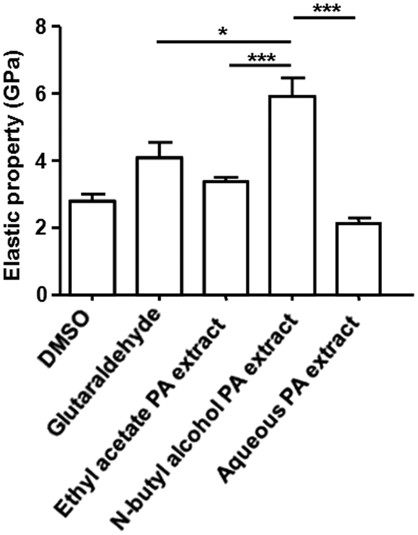
Tea root-derived proanthocyanidin (PA) components affect dentin elasticity. Demineralized dentin beams treated with dimethyl sulfoxide (DMSO), glutaraldehyde, ethyl acetate PA extract, n-butyl alcohol PA extract, or aqueous PA extract were subjected to the elasticity modulus test. DMSO treatment served as control. Data are expressed as mean ± standard deviation from at least three independent experiments. n = 15, *p < 0.05, ***p < 0.0005.
Discussion
As a natural product extracted from a variety of plants, including grape seeds, PAs have been identified as effective reagents to protect, strengthen, and repair dentin. 27 PAs are a class of complex components that consist of multiple secondary metabolites with various degrees of polymerization. Previous studies have shown that PAs from different sources exerted distinct protective effects on dentin.28–30 The proportion of PA oligomers is often regarded as a key indicator of their effects, based on their abilities to scavenge radicals and resist oxidation. However, oligomers with a low degree of polymerization exhibit insufficient convergence capacity and weak binding to dentin collagen.15,31 In the present study, tea root-derived PAs with a high degree of polymerization (extracted using n-butyl alcohol) were shown to achieve better dentin collagen cross-linking, prevent bacterial collagenase degradation, and improve dentin elasticity, compared with tea root-derived PAs with a low degree of polymerization (extracted using ethyl acetate). The molecular weight and degree of polymerization of PAs have been proven to substantially affect dentin collagen cross-linking.28,30 Low-molecular-weight PAs mainly promote intramolecular cross-linking, whereas high-molecular-weight PAs primarily mediate intermolecular cross-linking of collagen. 32 Moreover, PAs with a high molecular weight and a high degree of polymerization may shield some sites of dentin collagen fibers that are susceptible to bacterial collagenase-mediated collagen degradation, thereby reducing sensitivity to bacterial collagenase and enhancing dentin collagen stability. 33 It has been reported that (−)-epigallocatechin gallate, a type of PA monomer, exhibits a substantial protective effect on dentin collagen, due to the increased intramolecular cross-linking of dentin collagen mediated by the six phenolic hydroxyls of (−)-epigallocatechin gallate.15,30,34 Overall, we presume that the number of phenolic hydroxyls and the degree of polymerization are the two critical factors that influence the effects of PAs on dentin collagen cross-linking and stability.
Glutaraldehyde is an effective dentin adhesive for enhancement of the bond strength of dentin and resin.22,23 However, the clinical applications of glutaraldehyde are greatly limited by its toxicity; thus, there is an urgent need for less toxic alternatives. Some natural products with low toxicity have been shown to exhibit biological functions that could enhance dentin strength and dentin collagen cross-linking, thereby attracting attention from researchers. In addition to grape seeds, new dentin biomodification agents can be extracted from Anacardium occidentale, Myracrodruon urundeuva, and other plants.7,35,36 Currently, grape seeds are the major source of PAs.37,38 In practice, ethyl acetate has been mainly used to extract highly purified PAs with a low degree of polymerization from grape seeds. 17 The results of the present study showed that tea root-derived PAs with a high degree of polymerization, extracted using n-butyl alcohol, exhibit better protection of dentin collagen, compared with the protection mediated by ethyl acetate PA extract. Consequently, appropriate PA extraction solvents should be selected to achieve better clinical effects.
The results of this study suggest that use of the aqueous PA extract was detrimental to the cross-linking of dentin, impaired the resistance of dentin to bacterial collagenase degradation, and did not maintain the elasticity of dentin. The aqueous PA extract is composed of polysaccharides, phenolic acids, amino acids, and other water-soluble components. The negative effect of the aqueous PA extract on dentin was potentially due to the residual phenolic acids in the extract, which were able to trigger the degradation of dentin collagen. Therefore, to maximize the positive effect of PAs, tea root and other PA-related plants should be refined to remove the water-soluble phenolic acids. The findings in this study demonstrate that tea root-derived PA fractions, extracted using organic solvents, show distinct biological effects on dentin, thus providing a new natural source of PAs; however, clinical applications of tea root-derived PAs require further clinical research.
In conclusion, our extraction of tea root-derived PAs yielded different PA components with distinct degrees of polymerization, using an organic solvent extraction method. The results of FTIR spectra analysis, bacterial collagenase-mediated collagen degradation assays, and dentin collagen elasticity assays showed that tea root-derived PAs with a high degree of polymerization (extracted using n-butyl alcohol) were able to more effectively improve dentin cross-linking, through protection and strengthening of dentin collagen, relative to treatment with glutaraldehyde or tea root-derived PAs with a low degree of polymerization (extracted using ethyl acetate). Finally, the findings of this study indicate that tea root can serve as a natural source of PAs for the protection and repair of dentin, which greatly increases the utility of tea root in clinical applications.
Footnotes
Declaration of conflicting interest
The authors declare that there is no conflict of interest.
Funding
This work was supported by funds from National Natural Science Foundation of China to Guanhu Bao [Grant number 31972462], Anhui Provincial Key Research and Development Plan [Grant number 201904a06020011], and a Key Grant from the Department of Sciences and Technology of Anhui Province [Grant number 11080001].