
Abstract
Objective
Iterative decomposition of water and fat with echo asymmetry and least-squares estimation-iron quantification (IDEAL-IQ) is a noninvasive and objective method used to quantitatively measure fat content. Although this technique has been used in the entire abdomen, IDEAL-IQ findings in the sacroiliac joint (SIJ) have rarely been reported. This preclinical study was performed to quantify the amount of fat in the SIJ in healthy volunteers by IDEAL-IQ.
Methods
From April to November 2017, 60 healthy volunteers with low back pain were included in this retrospective study. The participants were allocated into groups by age (15–30, 31–50, and ≥51 years), sex (male and female), and body mass index (BMI) (<18.5, 18.5–23.9, and ≥24.0 kg/m2). The iliac-side (Fi) and sacral-side (Fs) fat fractions were obtained in all groups. Two- and three-factor multivariate analyses were performed to analyze the effects of sex, age, and BMI on the Fi and Fs.
Results
The interaction among sex, age, and BMI had no statistically significant effect on the dependent variable. Both Fi and Fs were significantly influenced by age. Fs was significantly influenced by sex.
Conclusion
The IDEAL-IQ sequence can be used to quantitatively assess the SIJ fat content in healthy volunteers.
Keywords
Introduction
Patients with ankylosing spondylitis (AS) generally have osteoporosis or osteopenia. 1 As the disease progresses, new bone forms at sites of inflammation, and this new bone formation is accompanied by an increased risk of osteoporosis and vertebral fractures.2–4 Osteoporosis is present in as many as 25% of patients with AS. 5 A Chinese study of 1051 patients with AS showed that the incidence of osteoporosis was 34% overall and 39.32% among men with AS. 6 With their increased rates of disability and mortality, patients with AS have large social and family burdens related to medication and healthcare expenses. Therefore, it is important to detect osteoporosis to treat and prevent vertebral fractures.
Magnetic resonance imaging (MRI) has a unique advantage in quantitatively detecting changes in bone marrow fat. Magnetic resonance spectroscopy (MRS) is a widely accepted nontraumatic imaging method 7 for studying the metabolism, biochemical compositions, and compound quantification in living tissues.8–10 However, long scan times, respiratory movement, and uneven magnetic fields can affect the scanning success rate of MRS, and the high uniformity of the main magnetic field and the complexity of postprocessing are limiting factors.9,11
Iterative decomposition of water and fat with echo asymmetry and least-squares estimation-iron quantification (IDEAL-IQ) can be used for water and fat imaging. This method is more reliable, provides better image quality, and has a shorter scan time than MRS. It is a noninvasive and objective method used to quantitatively measure the fat content and provide information on the fat spatial distribution.12–14 Previous studies of fat/water signal ratio images in the entire abdomen have been reported.12,15 However, IDEAL-IQ findings for the sacroiliac joint (SIJ) have rarely been reported. Therefore, the present study was performed to quantitatively analyze the SIJ fat content in healthy volunteers by IDEAL-IQ.
Materials and methods
Ethical approval
This preclinical study was approved by the institutional review board, and written informed consent was obtained from each participant.
Patients
This study included patients with SIJ pain who were referred for an MRI examination from April to November 2017. The inclusion criteria were symmetrical development of the SIJ with no lesions involving the blood system, no history of lumbosacral pain or trauma of the SIJ, and no metal or pacemaker in the body. The exclusion criteria were SIJs with a primary or metastatic tumor, tuberculosis, or other pathological changes; bilaterally abnormal SIJ morphology, such as rotation or tilt; significant endocrine or metabolic disease; the use of drugs that affect bone metabolism; and similar diseases (such as rheumatoid arthritis, gouty arthritis, or psoriatic arthritis) that may lead to SIJ arthritis.
According to the body mass index (BMI) threshold of overweightness and obesity in Chinese adults proposed by the Working Group on Obesity in China in 2003, 15 the patients were divided into the following three groups: obese group (BMI of ≥24.0 kg/m2), normal weight group (BMI of 18.5–23.9 kg/m2), and underweight group (BMI of <18.5 kg/m2). According to their age, the patients were divided into the following three groups: age of 15 to 30, 31 to 50, and ≥51 years.
MRI techniques
All participants underwent regular MRI and IDEAL-IQ examinations of their SIJs with a 3.0 T system (Discovery 750; GE Healthcare, Chicago, IL, USA) and eight-channel body phase control matrix coils. All sequences recorded the SIJs bilaterally (Table 1). Standing MRI sequences were obtained: oblique axial (perpendicular to the long axis of the sacrum) T1-weighted fast spin-echo, and oblique axial T2-weighted fat-suppression fast spin-echo. The scanning parameters of IDEAL-IQ were as follows: axial: repetition time/echo time, 2.6/6.0 ms; echo train length, 3; number of excitations, 0.5; field of view, 38 × 38 cm; matrix, 192 × 192; thickness/gap, 4.0/0.4 mm; band width, 111.11 kHz; time, 42 s; and flip angle, 3°.
Comparison of Fi and Fs in the same age group and across different age groups (±s).

Fi, fat fraction on the iliac side; Fs, fat fraction on the sacral side; 95% CI, 95% confidence interval.
**P < 0.01, *P < 0.05.
Superscripts a, b, and c indicate different P values in different age groups.
Image postprocessing and region of interest
The IDEAL-IQ images were transferred to a GE Advantage Windows 4.6 workstation (GE Healthcare). Two radiologists with 5 and 8 years of experience in musculoskeletal system imaging read the films without knowledge of the clinical situations or grouping of the patients. The region of interest (ROI) was manually selected by the same radiologist. The standard MRI sequences were interpreted before IDEAL-IQ. The fat fraction (FF) was automatically generated by the viewer function on the workstation. The ROI on the FF sequence was selected according to conventional MRI sequences, and the ROIs were manually identified on the iliac side and sacral side with the same area (three regions) and same size on both SIJs. The FF of the iliac side was recorded as Fi, while that of the sacral side was recorded as Fs. These ROIs were adjacent to cortical bone and had a mean area of 27 mm2 (range, 15–30 mm2). The data on three consecutive sections were obtained and then averaged as the final result for subsequent analyses.
Statistical analysis
All statistical analyses were performed using SPSS Statistics version 22.0 for Windows (IBM Corp., Armonk, NY, USA). The interclass correlation coefficient (ICC) was used to analyze the consistency of the parameters of two repeated measurements, and an ICC of >0.75 was considered to indicate good consistency. The differences in the Fi and Fs between the two sexes were analyzed by the independent-samples t test, and the differences in the Fi and Fs within age groups were analyzed by the paired-samples t test. The correlation between the FF of different groups (sex, age, or BMI) was analyzed by Kendall’s tau-b correlation. Three-factor analysis of variance (ANOVA) was used to analyze the interaction effects of age, sex, and BMI on the Fi and Fs. The effects of age and sex on the Fi and Fs were analyzed by two-factor ANOVA, and P values of <0.05 were considered statistically significant.
Results
Patients
Sixty participants were involved (32 men and 28 women). Their ages ranged from 15 to 82 years (mean, 38.83 ± 16.40 years). The obese, normal weight, and underweight group comprised 22, 33, and 5 participants, respectively. The 15- to 30-year age group (mean age, 23.77 ± 3.87 years) comprised 26 patients (15 men and 11 women) with an average BMI of 23.27 kg/m2 (range, 18.26–38.82 kg/m2). The 31- to 50-year age group (mean age, 38.6 ± 6.40 years) comprised 15 patients (9 men and 6 women) with an average BMI of 22.55 kg/m2 (range, 17.72–26.56 kg/m2). The ≥51-year age group (mean age, 59.63 ± 6.69 years) comprised 19 patients (8 men and 11 women) with an average BMI of 21.98 kg/m2 (range, 16.90–30.04 kg/m2).
Consistency analysis of Fi and Fs measurements
The intragroup ICC of the Fi calculated by the two radiologists was 0.843 [95% confidence interval (CI), 0.751–0.903], and the intragroup ICC of the Fs was 0.867 (95% CI, 0.787–0.918).
Comparison of Fi and Fs among age groups
According to Levene’s test, the variances of the Fi and Fs were homogeneous and the differences in the Fi (F = 5.289, P = 0.008) and Fs (F = 12.680, P < 0.001) among the age groups were statistically significant (Table 1).
Comparison of FF in men and women among different age groups
The paired-samples t test showed a significant difference in the Fi and Fs between men and women aged ≥51 years. There was no significant difference in the Fi and Fs between men and women in the 15- to 30-year age group or 31- to 50-year age group (Table 2).
Comparison of Fi and Fs between men and women in different age groups.

Data are presented as mean ± standard deviation.
Fi, fat fraction on the iliac side; Fs, fat fraction on the sacral side.
**P < 0.01
Comparison of BMI among different age groups and between different sexes
The mean BMI in the 15- to 30-year age group, 31- to 50-year age group, and ≥51-year age group was 23.28 ± 4.15, 22.55 ± 2.69, and 21.98 ± 3.53 kg/m2, respectively. The mean difference in the BMI among the three groups was not statistically significant (F = 0.717) according to single-factor ANOVA (Table 3).
Comparison of BMI between men and women in different age groups.

Data are presented as mean ± standard deviation.
BMI, body mass index; Fi, fat fraction on the iliac side; Fs, fat fraction on the sacral side.
Correlation of FF with sex, age, and BMI
The correlation between Fi and sex was statistically significant (r = 0.491, P = 0.013) in the ≥51-year age group. The correlations of Fs with sex and age were also statistically significant (r = 0.589, 0.453 and P = 0.003, 0.008, respectively) (Table 4).
Correlation of Fi and Fs with sex, age, and BMI.
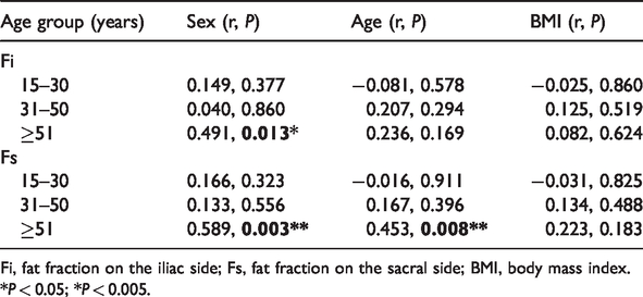
Fi, fat fraction on the iliac side; Fs, fat fraction on the sacral side; BMI, body mass index.
*P < 0.05; *P < 0.005.
Analysis of interaction effects of sex and age on Fi and Fs
The three-factor ANOVA showed no significant three-factor interaction effects on the Fi [F (2,45) = 1.538] across the sex, age, and BMI groups. The simple two-factor interaction analysis results showed no significant two-factor interaction effects across the sex and age groups [F (2,45) = 0.110]. There was no significant interaction effect between the sex and BMI groups [F (2,45) = 2.879] or between the age and BMI groups [F (3,45) =2.363]. The three-factor ANOVA showed no significant three-factor interaction effect on the Fs [F (2,45) = 0.371] across the sex, age, and BMI groups. The simple two-factor interaction analysis results showed no significant two-factor interaction effect between the sex and age groups [F (2,45) = 2.106]. There was no significant interaction effect between the sex and BMI groups [F (2,45) = 2.391] or between the age and BMI groups [F (3,45) = 1.387].
Multivariate ANOVA showed that the age group influenced both the Fi and Fs (F = 4.252, P = 0.019, partial η2 = 0.136 and F = 12.694, P < 0.001, partial η2 =0.320, respectively). The differences in Fi between the 15- to 30-year and 31- to 50-year age groups and between the 31- to 50-year and ≥51-year age groups were −5.54 (95% CI, −16.02 to 4.95) and −8.08 (95% CI, −19.25 to 3.09), respectively, with no statistical significance. The difference in Fi between the 15- to 30-year and ≥51-year age groups was −13.62 (95% CI, −23.38 to −3.86, P = 0.004), with statistical significance (Table 2).
Multivariate ANOVA showed that sex had a statistically significant influence on Fs (F = 9.007, P = 0.004, partial η 2 =0.143). The difference in Fs between the women 15- to 30-year and ≥51-year age group was −28.493 (95% CI, −42.129 to 14.856, P < 0.001), and the difference was statistically significant. The comparison of Fi and Fs between the other groups showed no statistically significant differences.
Discussion
IDEAL-IQ is an important approach for evaluation of the bone marrow fat content. In the present study, we used IDEAL-IQ to evaluate the main and interaction effects of age, sex, and BMI on the Fi and Fs in the SIJ in normal adults by two- and three-factor multivariate analyses. We found that the age group had an influence on both the Fi and Fs and that a three-factor interaction effect among sex, age, and BMI was not present. When the difference in BMI was not considered, the Fi and Fs gradually increased with age, and the effect of age on the Fi and Fs was statistically significant. We speculate that increases in fat deposition in the SIJ may be important for the early diagnosis of AS. In our future work, we plan to compare the fat content in the SIJ between patients with AS and healthy volunteers and determine whether the FF in the SIJ can reflect the activity of SIJ arthritis to some extent.
Feasibility of IDEAL-IQ sequence in quantitative measurement of FF in SIJ
Several studies have evaluated the fat content in the head and neck,16,17 breast, 17 heart,17,18 abdomen,16,19 pelvis, 16 and extremities,16,21,22 and the accuracy of quantitative analysis of the fat content in the liver and bone marrow has been widely recognized. 1 IDEAL-IQ technology showed a negative correlation between the vertebral bone density and vertebral body fat in women. 23 Other scholars24,25 compared the quantitative results of IDEAL-IQ with the pathological examination results and found that the results of the separation of fat and quantitative hydrops were highly correlated with the pathological results, and the quantitative results obtained using this technology were found to be stable and reproducible regardless of the machine field intensity. 26
FF value of SIJ in normal people of different ages
The present study showed that the SIJ fat content was lowest in the 15- to 30-year age group, which is consistent with the results of previous studies. 27 This is likely explained by the fact that the transformation process of red marrow to yellow marrow is slower in women than men in this age group. 28
One study showed that the adipose tissue content increased from 15% to approximately 60% in people aged 20 to 65 years with pathological changes in the anatomy of the ilium. 29 The transformation process of human bone marrow is basically complete at the age of approximately 25 years, and red bone marrow and trabecular bone marrow are abundant in women and men of this age; however, no significant difference in fat signals between male and female bone marrow have been found to date.
Consistent with the findings reported by Aoki et al., 30 we found that the FF was higher in menopausal and perimenopausal women because of their decreased levels of estrogen, which lead to an increased bone turnover rate, significant loss of calcium, 31 corresponding adjustments in osteoblast and osteoclast activity, gradual trabecular bone thinning, and an associated increase in bone marrow fat content. 32 Studies have shown that women in this age group have accelerated yellow myelosis and significantly higher fat content than men. 9 In the present study, the FF value in the ≥51-year age group was close to the proton density FF value in 60-year-old patients with osteoporosis in a previous study. 33
The fat content in the ilium and sacrum in the women in all age groups was slightly higher than that in the men. This can be explained by the fact that women have more hematopoietic tissues than men of the same age before menopause and that periodic physiological blood loss stimulates the proliferation and activity of hematopoietic tissues. 34
Clinical significance and prospect of this study
Previous studies have suggested that the fat metaplasia of the bone marrow and bone erosion sites in the upper subcortical region, evident in T1-weighted images, is a key part of the pathophysiological process of new bone formation related to spondyloarthritis.34,35 In the present study, fat deposition lesions could be found regardless of whether the condition was in an active or inactive stage, regardless of whether the patient was male or female, and regardless of whether both fat deposition and bone erosion occurred simultaneously in patients with AS. Therefore, we conclude that the FF in the SIJ in healthy volunteers is lower than that in patients with AS but that the activity of SIJ arthritis and sex have no significant effect on fat deposition.
IDEAL-IQ was recently used to evaluate the effect of laterality, sex, age, and BMI on FF measurements of both the parotid glands and submandibular glands. 36 The results showed that the FFs of both types of glands were correlated with age and BMI but not with sex. Another study concluded that BMI was correlated with extracellular lipids by MRS. 37 In the present study, we found no certain correlation between the BMI and FF, but we also found the same increasing tendency with age, which is consistent with the results of a previous study. 38 The clinical relevance of these findings needs to be validated with a larger sample size.
Limitations
The present study has several limitations. First, this was a preclinical study with a small sample size. Second, we had no pathological results for use as a reference. Third, the clinical relevance is unclear because the rationale for applying this technique for assessment of the SIJ is unclear; this issue needs to be addressed through additional research to obtain more conclusive findings. Finally, we did not consider the effect of drug treatment on the FF value.
IDEAL-IQ is a short-term and highly effective MRI scanning sequence that may be used to quantitatively analyze the FF value in the SIJ.