
Abstract
Minipigs are valued alternatives to dogs and non-human primates in non-clinical safety and toxicity studies, and Göttingen minipigs are bred specifically for experimental purposes. They are bred under barrier conditions and monitored regularly for many pathogens and opportunistic agents, and spontaneous disease is rare when compared to what is seen in production pigs. Knowledge of spontaneous background lesions is important when toxicological pathologists evaluate microscopic findings in pre-clinical toxicity studies to avoid interference with study data interpretation. In this brief communication, intra-abdominal granulomas/abscesses were seen in Göttingen minipigs. The minipigs did not show any clinical signs, but nodules were present in the abdominal peritoneum at necropsy. Microscopic evaluation revealed chronic inflammation, with abscess or granuloma formation. Areas of inflammation, occasionally associated with the presence of the Splendore-Hoeppli material, were surrounded by a fibrotic capsule. Special stains were applied to investigate for the presence of microorganisms.
The Göttingen minipig is widely accepted as a non-rodent alternative to dogs and non-human primates in pre-clinical safety studies, and awareness of spontaneous pathology is essential when interpreting microscopic findings. In this brief communication, we report two cases of intra-abdominal abscesses/granulomas. Intra-abdominal abscesses occur following extension of infection/inflammation present in abdominal organs, or due to perforation, causing a localized area of inflammation of the serosal membrane (peritoneum) that lines the abdominal cavity and the organs (ie, peritonitis). Peritonitis is thus most often caused by the introduction of an infection, but it may also result from other irritants (eg, foreign bodies, gastric acid from a perforated ulcer).
Abscesses are well known to occur in pigs and are mostly subcutaneous caused by fighting, castration, tail bites, and other damages to the skin, such as after administration of substances via injection. Abscesses in the skin are most often associated with bacteria such as Arcanobacterium pyogenes, streptococcal spp., etc. 2
Intra-abdominal abscesses result from local peritonitis and may be seen after extension of infection or inflammation from disease processes in abdominal organs and due to penetration of hollow organs. In production pigs, umbilical infection and castration of young pigs may also be followed by the spread of bacteria within the abdominal cavity, resulting in abscesses in various organs.
Göttingen minipigs are today widely recognized as a useful alternative non-rodent species in non-clinical toxicity studies, and gross and microscopic findings occurring as background findings have been reported by various authors.5,4,7 In Göttingen minipigs used for experimental purposes, the presence of intra-abdominal abscesses has not been published previously, to the best knowledge of the authors.
Here, we report incidental intra-abdominal abscesses in two Göttingen minipigs. These were the only cases seen in the studies, and they are therefore suggested to be of spontaneous origin. The housing of the animals was in accordance with EU Directive 2010/63/EU of September 22, 2010, on the protection of animals used for scientific purposes, and experimental procedures were carried out under a license approved by the National Animal Experiments Inspectorate under the Ministry of Food, Agriculture and Fisheries of Denmark. Procedures were performed in accordance with laboratory standard operating procedures and/or established laboratory best practices.
Case 1 was a 7.5-month-old female Göttingen minipig in the high-dose group of a 3-month study, with oral administration of the test article. Case 2 was an almost 8-month-old female Göttingen minipig from a two-week local tolerance study with subcutaneous administration of test article.
Case 1 had a small wound on the ear before the study started, but no other clinical signs were seen in any of the two pigs at the breeder or during the in-life phase of the studies. At necropsy, nodules were seen in the abdominal cavity. In case 1, a single nodule was seen in the peritoneum covering the apex of the urinary bladder, and a few nodules were recorded as being located on a tissue string/adhesion running from the apex of the urinary bladder to the liver. Nodules were between 2 and 5 mm in diameter. In case 2, a single nodule, more than 10 mm in diameter, was observed to be attached to the peritoneum lining the abdominal cavity.
In both studies, abnormalities were sampled, fixed in 10% neutral-buffered formalin, embedded in paraffin, and sectioned at approximately 5 µm. Tissue sections were placed on glass microscope slides and stained with a hematoxylin and eosin (H&E) stain, a periodic acid-Schiff (PAS) stain, a Masson trichrome (MT) stain, a silver (modified Grocott Methanamine Silver) stain, and a Gram stain.
Microscopically, the nodules corresponded to the presence of chronic-active inflammation (abscess/granuloma formation) surrounded by a thick fibrotic capsule (Figure 1A). The abscesses/granulomas were characterized by a central area of necrosis and the presence of mixed inflammatory cells, including degenerate neutrophils and more peripherally also including macrophages and lymphocytes. The outer rim of the capsule consisted of mature fibrosis, whereas the inner rim showed organization with ingrowth of capillaries and fibroblasts. In case 1, the presence of Splendore-Hoeppli material was occasionally seen in the reported lesions (Figure 1B), whereas the Splendore-Hoeppli phenomenon was not observed in case 2. The Splendore-Hoeppli phenomenon, also called asteroid bodies, is often seen in relation to bacterial infections1,6 and microscopically is seen as strongly eosinophilic, amorphous material with star-like or club-shaped figures at the periphery of the amorphous material. 3 The fibrotic capsules of the intra-abdominal granulomas/abscesses were of considerable thickness, and the thickness of the capsule indicates a process of longer duration.
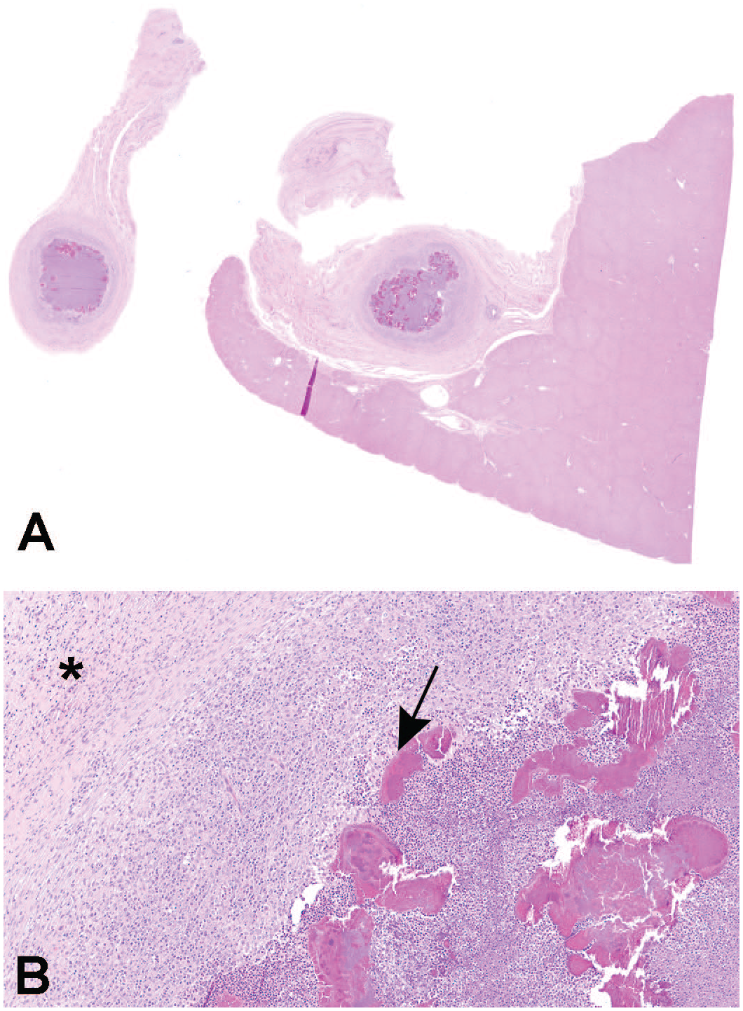
Microscopic features of the abdominal abscesses/granulomas, stained with hematoxylin and eosin. (A) The microscopic lesion is well-demarcated, with a focus of chronic-active inflammation surrounded by a fibrotic capsule, original objective x1.25. (B) Chronic active inflammation in the center of the lesion, with Splendore-Hoeppli phenomenon (arrow), surrounded by a fibrous capsule (asterisk), original objective ×10.
Special stains were applied to the lesions to highlight the microscopic features of the lesions: The MT stain for collagen was used to highlight the fibrous capsule surrounding the lesion (Figure 2A), and Gram, PAS, and silver (modified GMS) stains were used to investigate for bacteria or other infectious agents, such as fungi. Organisms consistent with Gram-positive bacteria were identified (Figure 2B).
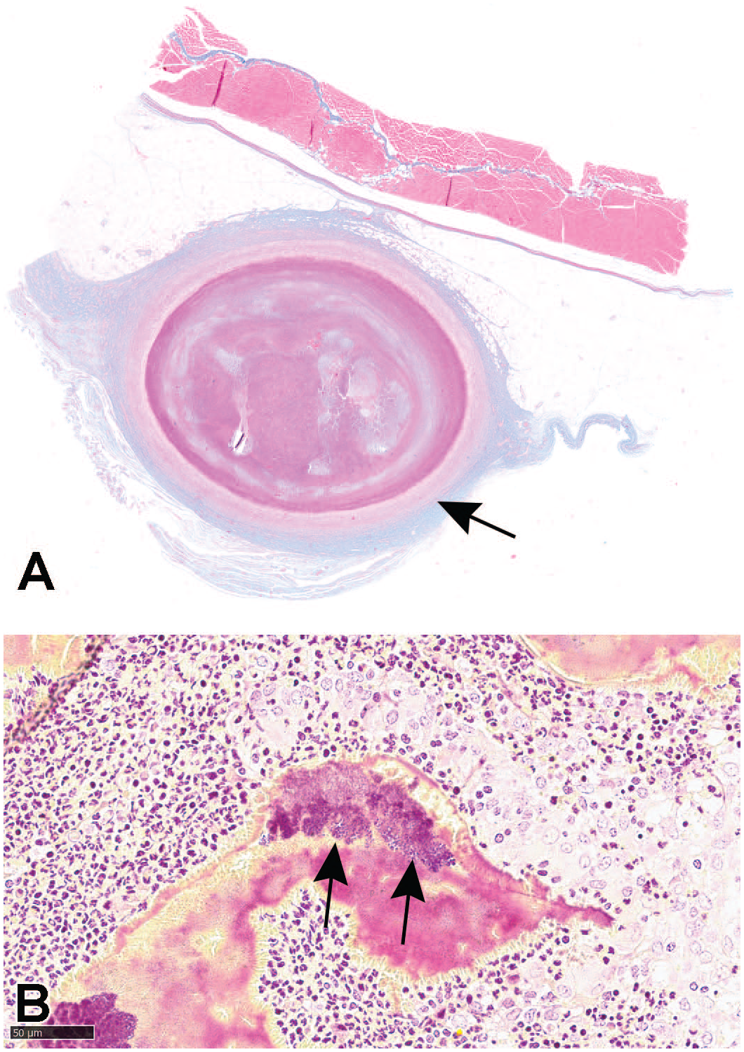
Masson trichrome and Gram histochemical stains. (A) Microscopic lesion stained with the Masson trichrome stain for collagen, to visualize the fibrous capsule (arrow), original objective ×1.25. (B) Microscopic lesion stained with a Gram stain to identify bacteria (arrows identifying Gram-positive bacteria), original objective ×40.
In summary, the Göttingen minipig is a valuable non-rodent model in pre-clinical safety investigations of potential new pharmaceutical drug candidates. Knowledge of spontaneous microscopic background findings is important when performing the histopathological evaluation of these pre-clinical studies. Spontaneous findings of abscesses/granulomas with or without the presence of Splendore-Hoeppli material are well known to occur with certain microbial infections in various species but are not considered to be within the range of the normal microscopic findings observed in the Göttingen minipig. The Göttingen minipig is bred for experimental use only and kept under strict barrier conditions at the supplier, and this limits the presence of spontaneous background findings, including findings associated with microbial infections, in this laboratory minipig species. Here, we report two cases of spontaneously occurring abdominal abscesses/granulomas. In one case, the abscesses/granulomas were associated with Splendore-Hoeppli material. In both tested studies, the tested drugs did not have any immunosuppressive effects in immune system organs, further supporting the spontaneous origin.
Footnotes
Acknowledgements
The authors would like to acknowledge Prof. Yuval Ramot for his valuable input and review during the preparation of this brief communication, the Scantox A/S pathology and histology team, and Dr. Kathleen Funk and the histology team of EPL® pathology for their precious collaboration and support.
Declaration of Conflicting Interests
The author(s) declared no potential conflicts of interest with respect to the research, authorship, and/or publication of this article.
Funding
The author(s) received no financial support for the research, authorship, and/or publication of this article.