
Abstract
Minipigs are now used in greater numbers in contract research organizations (CROs) as well as in the pharmaceutical industry. Most CROs or pharmaceutical companies use the Göttingen minipig, which displays a number of important background lesions. This review will discuss some of the more infrequent minipig background changes. Porcine stress syndrome is an autosomal recessive pharmacogenetic disorder in minipigs causing malignant hyperthermia and muscle necrosis. Possible triggers, clinical pathology as well as heart, muscle, liver, lung, and kidney histopathology are discussed. Additional spontaneous changes, background findings, and peculiar anatomical and histological features include thrombocytopenic purpura syndrome, spontaneous glomerulonephritis, osteochondritis, ellipsoids, or Schweigger–Seidel sheaths in the spleen, as well as the presence of a perimesenteric plexus adjacent to mesenteric lymph nodes, squamous epithelial metaplasia of the salivary gland, and cupping of the optic disk in the minipig eye. In order to maximize the data gained from minipig studies, the interpretation of pathology findings requires the input of experienced pathologists who understand the significance of artifacts and spontaneous, background lesions in minipigs and can distinguish these from induced lesions.
Introduction
Minipigs are now used in greater numbers in contract research organizations (CROs) as well as in the pharmaceutical industry. They provide significant advantages over dogs due to their greater tolerance of nonsteroidal anti-inflammatory drugs, antihypertensive agents, and sympathomimetic drugs (Dincer 2007). In addition, minipigs are good models for human drugs since their digestion is similar to that of humans (Dincer and Svendsen 2006), and their smaller size makes them more manageable laboratory animals than domestic pigs. The minipig offers advantages as a model in dermal, cardiovascular, and reproductive toxicity studies because of the similarity between the porcine and corresponding human systems (Mortensen, Brinck, and Lichtenberg 1998; Lavker, Dong, and Zheng 1991). Finally, as a food animal, the use of minipigs for compound testing may be more acceptable to the public than animals such as dogs or monkeys (Forster et al. 2010). Most CROs or pharmaceutical companies use the Göttingen minipig (GMP). GMPs derive from a triple cross of Minnesota minipig (60%), Vietnamese pot-bellied pig (33%), and German Landrace (7%) developed at the University of Göttingen, Germany (Bollen and Ellegaard 1997). This review will discuss findings associated with spontaneous conditions, peculiar anatomical microscopic features, and potential artifacts that may confound the interpretation of toxicity studies in the GMP. These include thrombocytopenic purpura (TP) syndrome, spontaneous glomerulonephritis, porcine stress syndrome (PSS), ellipsoids, or Schweigger–Seidel sheaths in the spleen, perimesenteric plexus adjacent to mesenteric lymph nodes, squamous epithelial metaplasia of the salivary gland, ectopic cartilage, and cupping of the optic disk in the eye.
TP syndrome
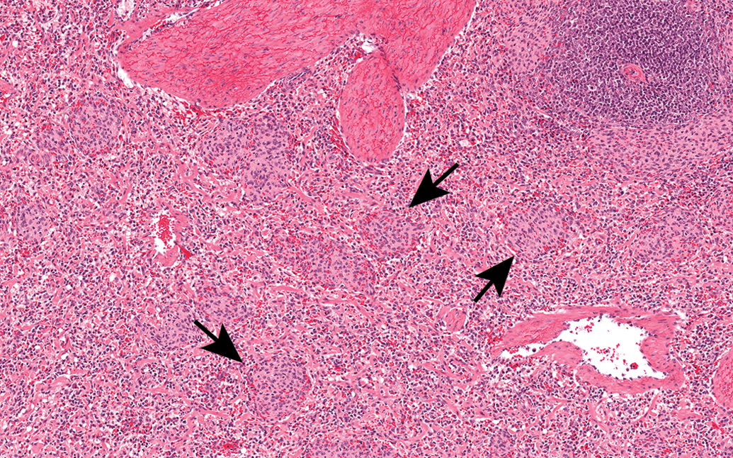
TP syndrome, which is thought to be immune mediated, has been described in GMPs (Carrasco et al. 2002; Maratea, Snyder, and Stevenson 2006). Carrasco and coworkers (2002) suggest that the cause of this condition is a type III hypersensitivity. Andersen and Nielsen (1973) state that TP may be caused by the colostral transfer of antiplatelet isoantibodies. The disease is characterized by the presence of severe subcutaneous hemorrhages, thrombocytopenia, and anemia (Maratea, Snyder, and Stevenson 2006). In addition, the syndrome may include membranoproliferative glomerulonephritis and vascular lesions. Widespread hemorrhage is a feature of the syndrome, which generally includes cutaneous purpura (petechiae, ecchymoses, and hematomas in the skin; Figure 1), and hemorrhage into the heart (Figure 2), urinary bladder (Figure 3), ureter, intestine, kidney, and lung, often with the accompanying presence of hemosiderin. In the bone marrow, there are increased numbers of immature and apoptotic megakaryocytes and increased hemopoiesis (i.e., an increase in foci of erythroid precursors; Figure 4). Occasional mixed inflammatory infiltrates are noted in conjunction with hemorrhage in the skeletal muscle (Carrasco et al. 2002). Extramedullary erythropoiesis in the liver and spleen is also common (Maratea, Snyder, and Stevenson 2006). In addition, vascular lesions in the arterioles of the kidney and heart have been reported (Maratea, Snyder, and Stevenson 2006) including neointimal proliferation and amphophilic myxoid material in the tunica media. Cardiac lesions include hemorrhage and myocardial necrosis. The carcase of the affected animal is pale white except for the areas of hemorrhage. Generally, porcine thrombocytopenia occurs in neonatal piglets; however in minipigs, the thrombocytopenic syndrome often occurs in sexually mature male and female minipigs (Carrasco et al. 2002). An incidence of 1 of 332 minipigs less than 6 months old with TP has been reported (Helke et al. in press ).
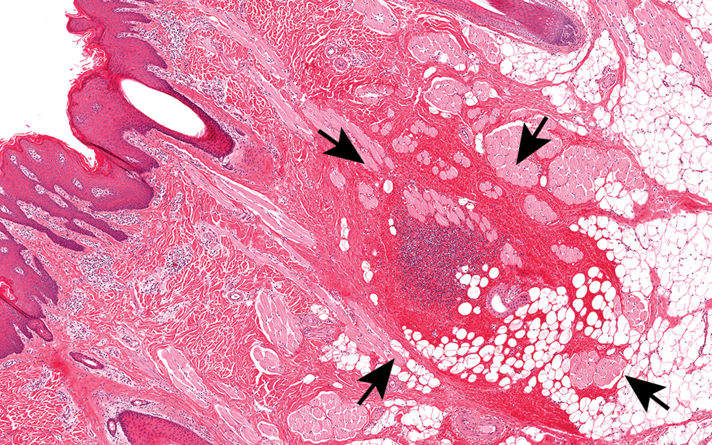
Hemorrhage (arrows) into the subcutaneous tissues of the skin observed in thrombocytopenic purpura in the minipig.
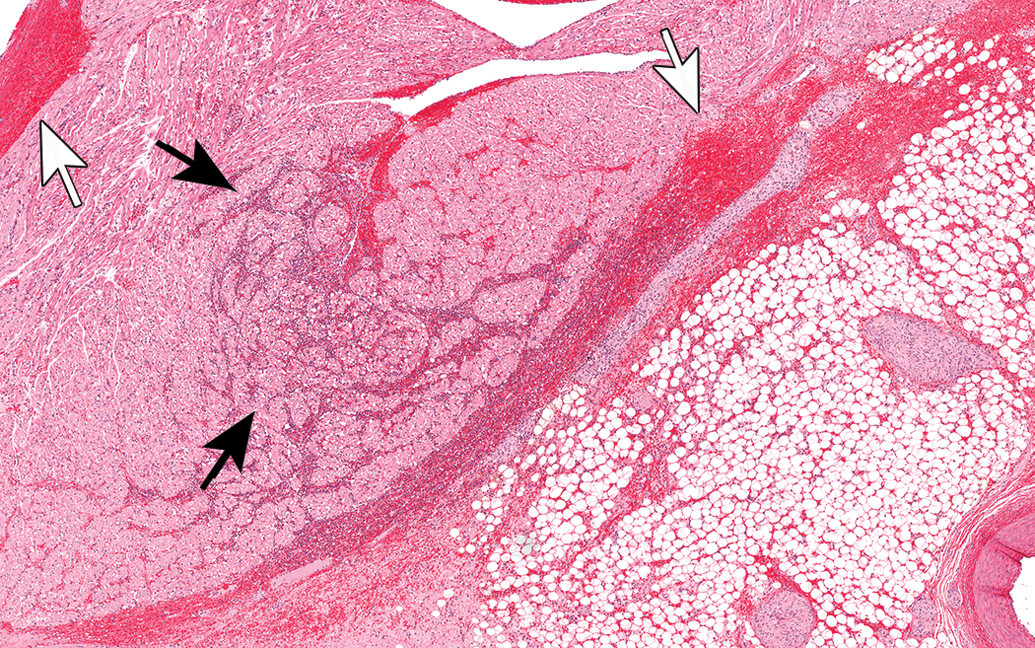
Thrombocytopenic purpura in the minipig heart with hemorrhage (white arrows) and inflammation (black arrows).
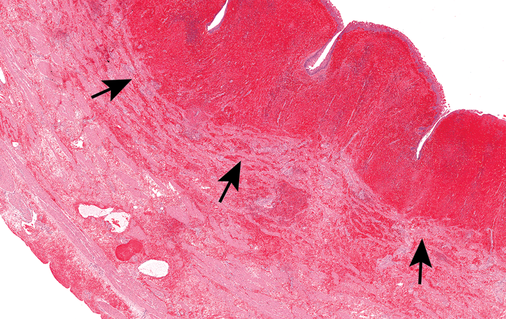
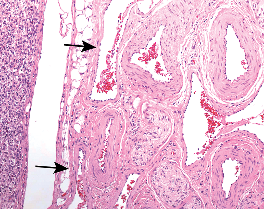
Hemorrhage (arrows) below the mucosa of the urinary bladder in thrombocytopenic purpura in the minipig.
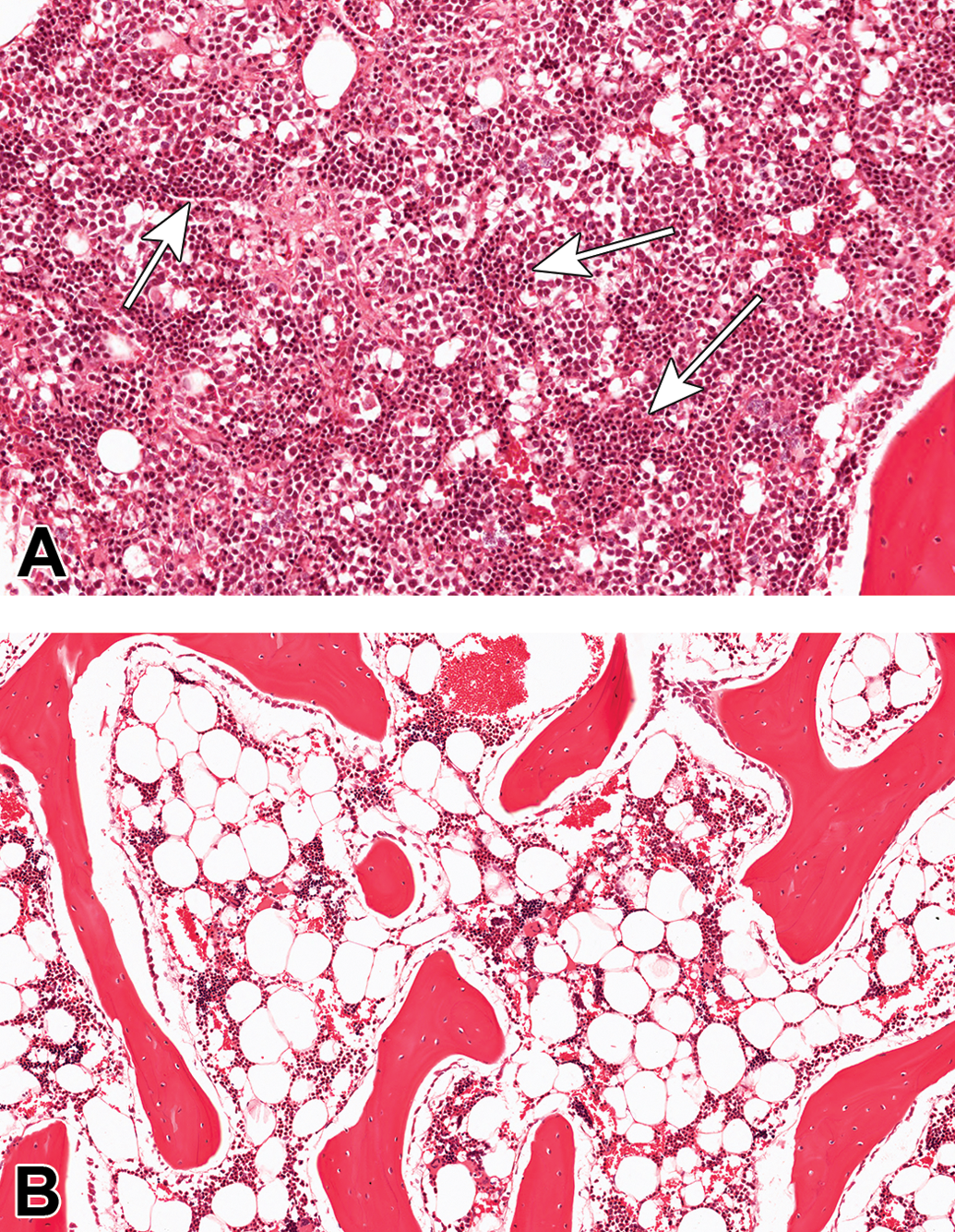
(A) Increased erythropoiesis (arrows) observed in the bone marrow taken from a minipig with thrombocytopenic purpura. (B) Normal bone marrow in minipig for comparison.
Spontaneous Glomerulonephritis
Glomerulonephritis in the minipig has been reported previously (Madsen, Jensen, and Larsen 1998; Dincer and Svendsen 2006; Vezzali et al. 2011; Jeppesen and Skydsgaard 2015). The glomerular lesion is characterized by hypercellular glomerular tufts and capillary basement membrane thickening due to periodic acid–Schiff-positive deposits as well as mesangioproliferative glomerulonephritis (Vezzali et al. 2011; Figure 5). These authors state that the glomerulonephritis is unlikely to be a colony-related problem and the supplier rarely observes clinical signs (Svendsen 2006). Vezzali and coworkers (2011) report an incidence of 2.5% of glomerulonephritis, that is, 4 of 154 minipigs examined were affected. Dincer and Svendsen (2006) report an incidence of 1 case of glomerulonephritis of 150 animals. Interestingly, Maratea and coauthors (2006) report that in a group of 9 minipigs displaying TP syndrome, 3 animals also showed glomerulonephritis, so the 2 syndromes may be linked.
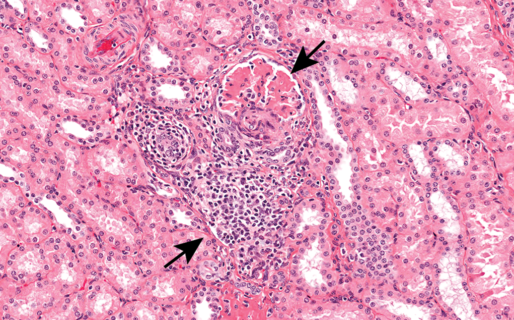
Glomerulonephritis in the kidney of a minipig consisting of glomerular and interstitial inflammation (arrows).
Vezzali and coworkers (2011) report occasional tubular/interstitial involvement as well as systemic involvement including hyperemia and hemorrhage in multiple organs, myocarditis, and peritonitis. The cause of the glomerulonephritis is not known. Chemically induced glomerulonephritis is common (Newman, Confer, and Panciera 2007), however in these recent reports the described lesions were present in untreated control animals. In farmed pigs, the cause of glomerulonephritis is thought to be immune mediated (Bourgault and Dorlet 1995) or genetic (Hegasy et al. 2002) or infectious (hog cholera, classical and African swine fever, cytomegalovirus, or circovirus; Choi and Chae 2003; Maxie and Newman 2007; Segalés, Allan, and Domingo 2005). Ascending urinary infections and septicemia are also theoretical causes of glomerulonephritis in minipigs, but these syndromes would generally display accompanying clinical signs.
PSS
PSS is an autosomal recessive pharmacogenetic disorder caused by a defect in the halothane (Hal) gene that produces pale, soft, and exudative (PSE) meat of inferior quality that results in significant losses to the meat industry. The biological cause of PSS, which leads to PSE meat, is the excessive release of calcium ions, which is promoted by a genetic mutation in the ryanodine receptors (RyR) located in the sarcoplasmic reticulum of the skeletal muscle cells (Fujii et al. 1991; Ziober et al. 2010). Both malignant hyperthermia in humans and PSS are caused by mutations in these receptors (Missiaen et al. 2000). Molecular tests such as the RYR-1 or HAL-1843® (University of Toronto Innovations Foundation, Ontario, Canada) stress gene test have been developed to test for the mutation associated with PSS (Fujii et al. 1991; O’Brien et al. 1993). In addition, a Hal susceptibility test has been developed to test for this syndrome in live animals; however, the assay for the RYR-1 mutation more accurately predicts both the homozygous and heterozygous forms of the PSS gene than does the Hal challenge test (Rempel et al. 1993). PSS causes lowered protein solubility and increased rate of postmortem degradation of structural proteins in the skeletal muscle (Boles et al. 1992). Possible triggers of PSS include exposure to Hal, increased physical activity, stress (Hall and Lucke 1985), handling and transport (Johansson and Jönsson 1977), fighting (Mitchell and Heffron 1982), high environmental temperature (Johansson and Jönsson 1977), high energy levels in diet, vitamin D deficiency, and caffeine (Watson et al. 1980).
Recently, PSS was described in a GMP at a CRO based in the United Kingdom (McKeag 2014). Due to the genetic makeup of the GMP, it is not surprising that this disease (probably derived from the Landrace strain) is evident in the minipig population. One male minipig aged 3 months presented with sudden death shortly after arrival (McKeag 2014). This animal presented with dyspnea, rigidity, distress, and variably congested skin (McKeag 2014). Further clinical signs reported in the literature may include cyanosis, hyperthermia (greater than 41°C; Johansson et al. 1974) and erythema, and blanching of the skin (Johansson and Jönsson 1977). Additional features of the disease reported in the literature include sudden onset, arrhythmias, rapid death (less than 30 min), high mortality, rapid rigor mortis (within 5 min), and a definite relationship to stress. The clinical pathological findings of PSS include an increase in serum creatinine phosphokinase activity (Bradley, Wells, and Gray 1979) as well as metabolic and respiratory acidosis (Gueorguieva et al. 1999), hemoglobinuria and myoglobinuria, altered cardiac myosin isoenzymes (Liou, Jiang, and Wu 2000), renal failure, and impaired blood coagulation. Clinical pathology evaluation was not completed in the single case of PSS (McKeag 2014).
At necropsy, macroscopic and microscopic changes in the single case of PSS reported from the CRO included white swollen wet muscles, epicardial and subendocardial hemorrhages of the heart (Figure 6), as well as acute renal tubular necrosis (Figure 7), pulmonary hemorrhage, edema (Figure 8), congestion and phagocytosed erythrocytes, and congestion of the liver and swollen skeletal muscle fibers (Figure 9; McKeag 2014). The literature reports pale white, wet, swollen muscles (Lee and Choi 1999); visceral congestion, particularly hepatic congestion, pulmonary edema and congestion; hydrothorax, hydropericardium, and edematous biceps femoris muscles (Johansson and Jönsson 1977); intramural as well as focal, pale, white areas of necrosis in the cardiac papillary muscles (Johansson et al. 1974) in PSS.
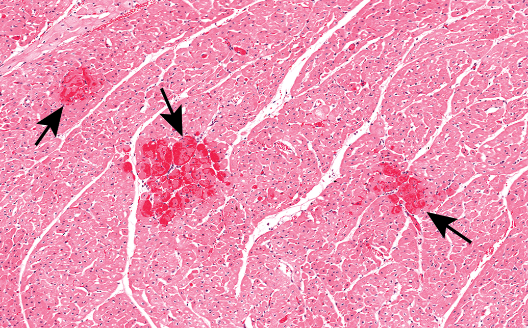
Myocardial degeneration, necrosis, and hemorrhage (arrows) noted in the heart taken from a minipig with porcine stress syndrome.
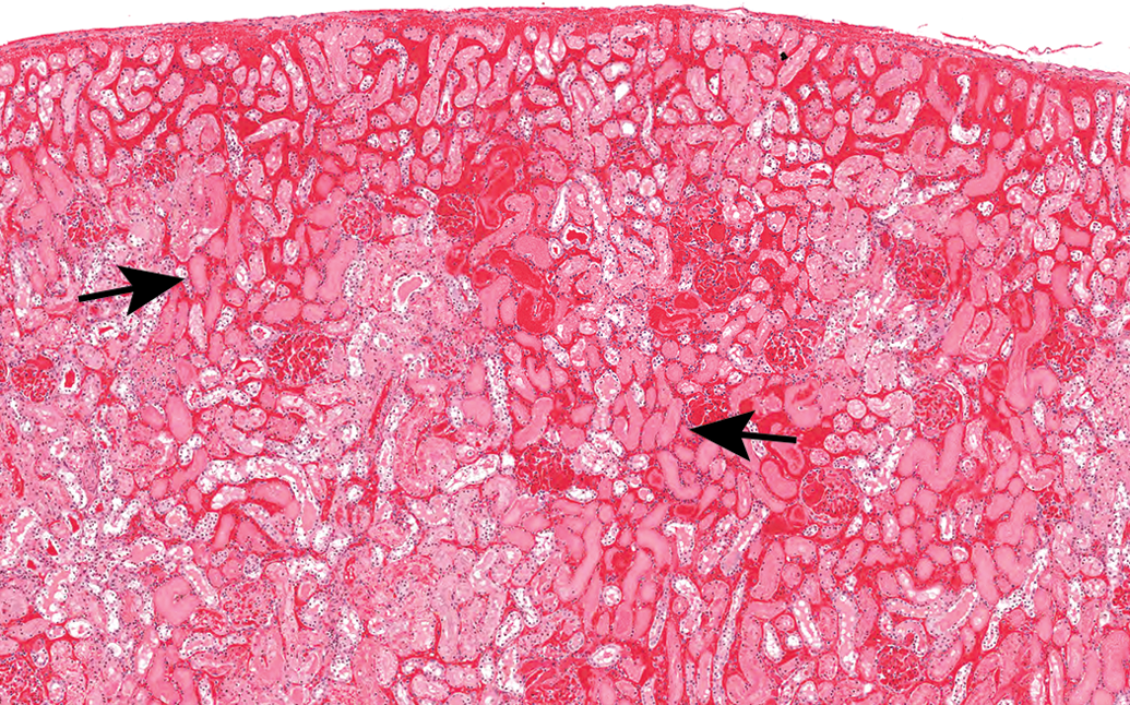
Acute tubular necrosis (arrows) of the kidney seen in porcine stress syndrome in the minipig.
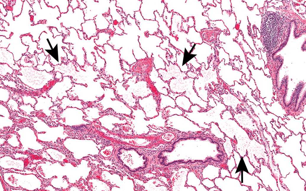
Edema (arrows) and alveolar hemorrhage in the lung taken from a minipig with porcine stress syndrome.
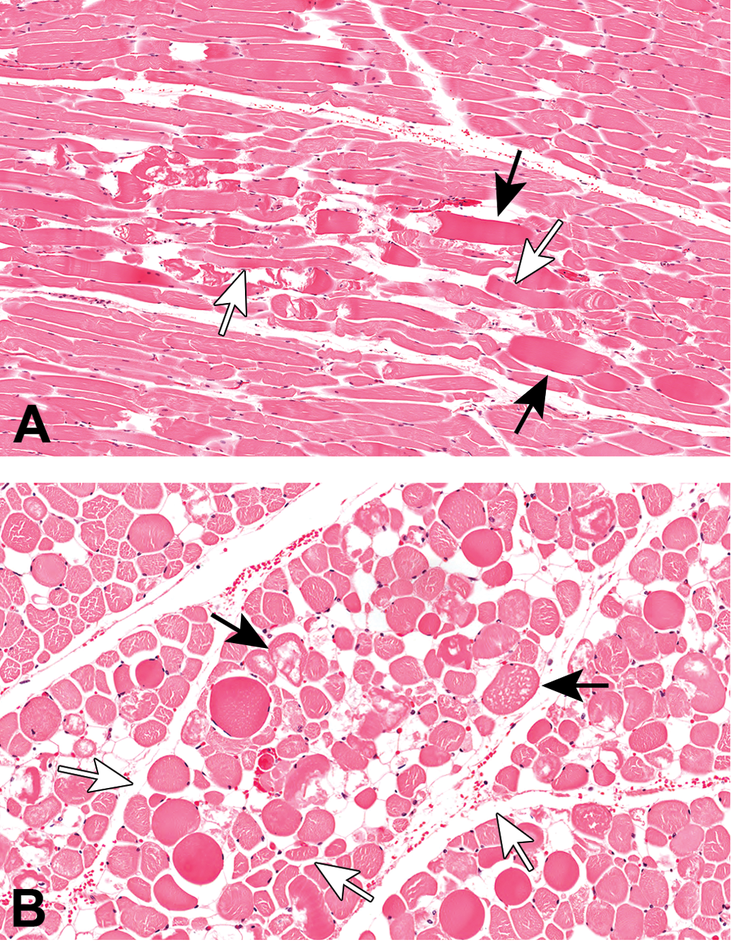
(A) Swollen fibers (black arrows) and floccular degeneration (white arrows) and (B) floccular degeneration (black arrows) and edema (white arrows) in longissimus dorsi muscle in minipig with PSS.
The literature describes histopathological lesions in the heart including myocardial degeneration and necrosis (Johansson et al. 1974), however, this is often restricted to only 1 or 2 cardiomyocytes (Johansson et al. 1974). Loss of striation, segmentation, and fine and coarse granular disintegration of the cytoplasm are also observed (Johansson et al. 1974). Scattered minimal neutrophil, fibroblast, and histiocyte infiltrates into the cardiac muscle are also a feature and the predilection site for myocardial lesions is the left, inner ventricular wall and papillary muscles (Johansson et al. 1974). The atrial muscles are seldom affected (Johansson and Jönsson 1977).
Electron microscopy was not performed in the single case (McKeag 2014), however, the literature reports that electron microscopy reveals an early reduction in glycogen granules within the cardiomyocytes and an increase in lipid droplets between the myofibrils and within the perinuclear sarcoplasm (Johansson and Jönsson 1977). Swelling of the sarcoplasmic reticulum and T-system is observed with faint or absent striations, electron dense deposits, and occasional complete lysis of myofilaments. Mitochondria are either swollen or display reduced cristae numbers (Johansson and Jönsson 1977).
Diagnosis of the disease requires identification of susceptible pigs, exposure to Hal (homozygous only), the polymerase chain reaction test for HAL-1843 (Ibáñez-Escriche et al. 2014), or histopathology and clinical history, which were used to confirm diagnosis in the described case.
Spontaneous Cutaneous Purpura and Bullous Pemphigoid
Familiarity with background skin lesions in the GMP is important in order to be able to differentiate between potential drug-induced skin lesions and spontaneous lesions. A generalized condition called spontaneous cutaneous purpura has been reported in GMPs (Maratea, Snyder, and Stevenson 2006; Figure 10), and this forms part of the TP discussed above. Perivascular hemorrhages and small aggregates of macrophages containing hemosiderin are observed in the skin. Vascular lesions are associated with the TP syndrome and are observed in young minipigs from 7 weeks to 1 year of age. The small to medium arteries in the skin and in some organs display degenerative lesions and arteriosclerosis. The arteriosclerosis is characterized by luminal narrowing, intimal proliferation, medial thickening, thrombosis, necrosis of the tunica media, myxoid deposits in the media, and fragmentation of the elastic lamina. Arterioles display similar lesions and perivascular neutrophils, eosinophils, lymphocytes, and macrophages are visible around the affected blood vessels (Maratea, Snyder, and Stevenson 2006).
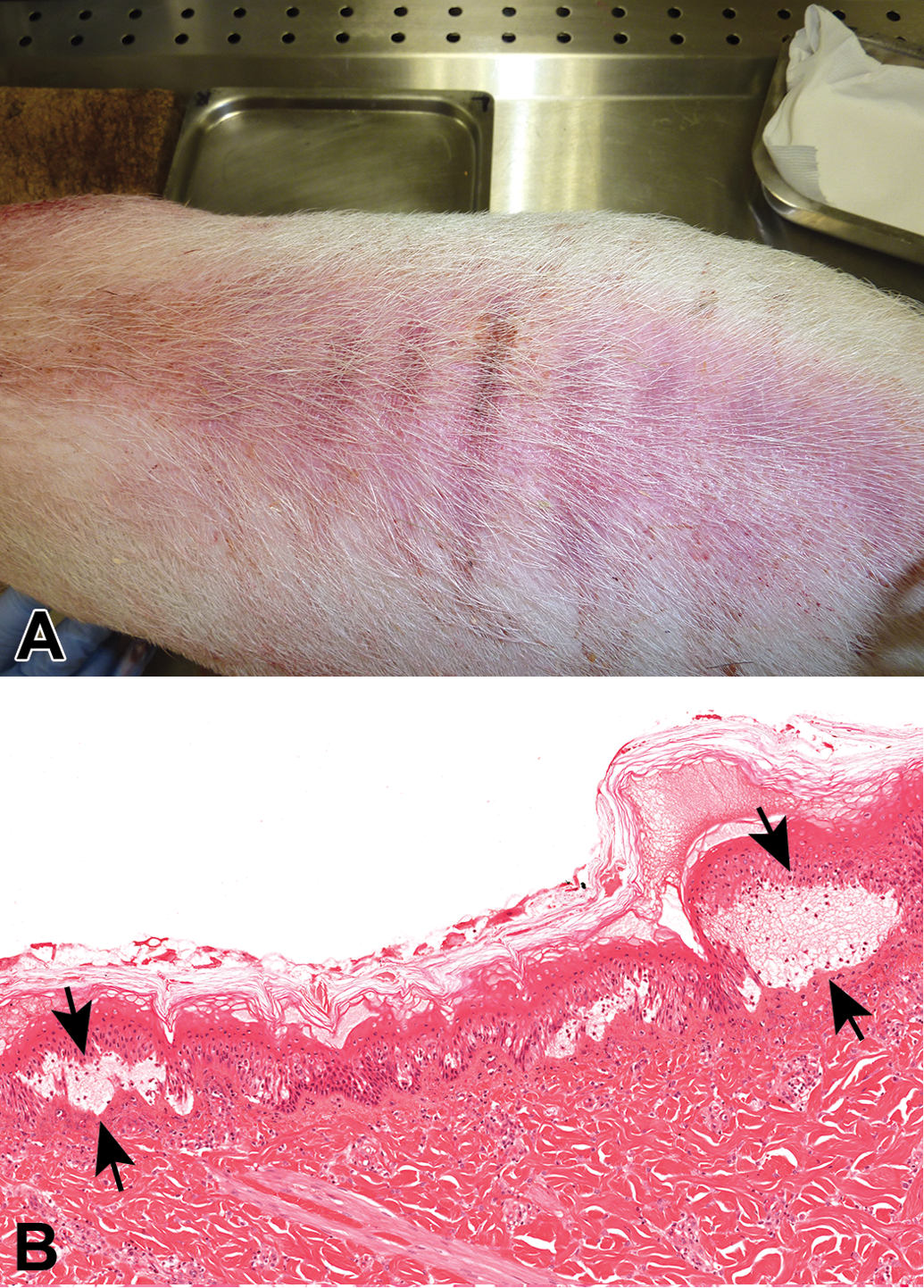
Bullous pemphigoid in minipig skin displaying the congestion noted in the skin (A) and separation of the epidermis and dermis (B, arrows).
Mirsky and coauthors described a spontaneous bullous pemphigoid in Yucatan minipigs in 2000, although this disease may be slightly different to that described in the minipig. This disease was also described in 2009 (Veronin et al. 2009), and the lesion consisted of epidermis separated from the dermis by a predominantly clear space that contained a small amount of cellular debris and necrotic mixed inflammatory cells (Figure 10). The superficial dermis contained a mixed, predominantly mononuclear, inflammatory cell population in and around foci of small arterioles, venules, and capillary vessels (Veronin et al. 2009). The disease occurs due to the deposition of immunoglobulin (IgG) autoantibodies directed against major hemidesmosomal antigens (Veronin et al. 2009). In the Yucatan minipig, IgG autoantibodies similar to those in humans directed against hemidesmosomes (bullous pemphigoid antigen) have been identified both in the skin and in circulation (Olivry et al. 2000). These antibodies are thought to fix complement, and this serves as a local chemotactic and proinflammatory stimulus resulting in mast cell activation, the attraction and degranulation of granulocytes, particularly eosinophils, and breakdown of the proteins responsible for maintaining the attachment of the epidermis and dermis (Mirsky, Singleton, and Olivry 2000).
Osteochondrosis (OC)
OC is a noninfectious, degenerative condition of the articular cartilage and growth plate with secondary changes in the bone (Ytrehus et al. 2004), which results in lameness in pigs. OC is seen in growing animals and macroscopically visible lesions include local thickening of articular cartilage, irregular cartilage surfaces, fissures between cartilage and subchondral bone, OC dissecans, and necrosis of subchondral bone (Reiland 1978; Busch and Wachmann 2011). Many factors are thought to contribute to OC, and these include genetic factors, growth rate, lean meat percentage, and mechanical stress (Kadarmideen et al. 2004). The fundamental cause is said to be a focal disturbance of endochondral ossification (Ytrehus, Carlson, and Ekman 2007). Histopathological lesions include focal epiphyseal cartilage necrosis, cleft formation extending from the articular surface to the subchondral bone, synovitis, joint effusion, and subchondral bone cyst formation (Ekman and Carlson 1998). Recently, McKeag (2014) described a case of osteochondritis in the tarsal joint of a male, 3-month-old minipig exhibiting lameness characterized by histopathological lesions of cartilage necrosis and cartilage flap formation (Figure 11).
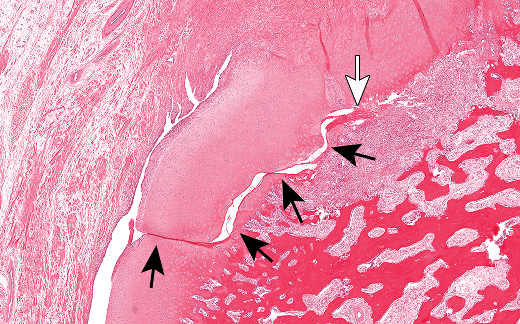
Cartilage necrosis (white arrow) and separation (black arrows) noted in osteochondrosis in a minipig.
Other Incidental Findings
Ellipsoids are a normal histological feature of the minipig spleen. Ellipsoids are pale, eosinophilic structures arranged concentrically around capillaries or small arterioles in the spleen which may prove confusing to the novice pathologist (Figure 12). The ellipsoids are prominent, consist of phagocytic cells and reticular fibers, and have been referred to in the literature as Schweigger–Seidel sheaths (Charles 1996; McInnes 2011). The perimesenteric plexus is a normal anatomical feature of the minipig mesenteric lymph nodes. In the mesenteric lymph node, the presence of a perimesenteric plexus can sometimes be confusing to pathologists (Figure 13). The vascular supply to the lymph nodes differs from that of other mammals. Major nodal arteries subdivide into branches that envelop the node as a capsular network from which further branches penetrate the parenchyma (McInnes 2011; Charles 1996). Foci of squamous epithelial metaplasia may be noted in the cuboidal epithelium of the salivary gland ducts (Figure 14; McInnes 2011). In the tongue, focal chondrocytes are occasionally observed, unassociated with inflammation (Figure 15). The origin of the cartilage cells is thought to be ectopic (McInnes 2011). Cupping of the optic disk is often noted as a background finding in the minipig eye (Figure 16; McInnes 2011). This lesion may be an artifact caused by excessive pressure on the eye and optic nerve at necropsy.
Schweigger–Seidel sheaths (arrows) adjacent to the periarteriolar lymphoid sheaths in the spleen of a minipig.
Perimesenteric plexus (arrows) adjacent to a mesenteric lymph node taken from a minipig.
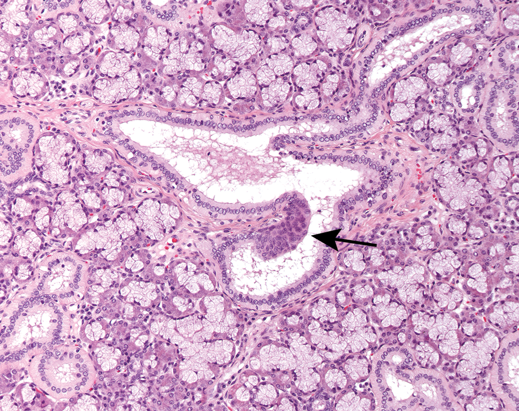
Squamous epithelial metaplasia (arrow) in cuboidal epithelium of salivary gland taken from a minipig.
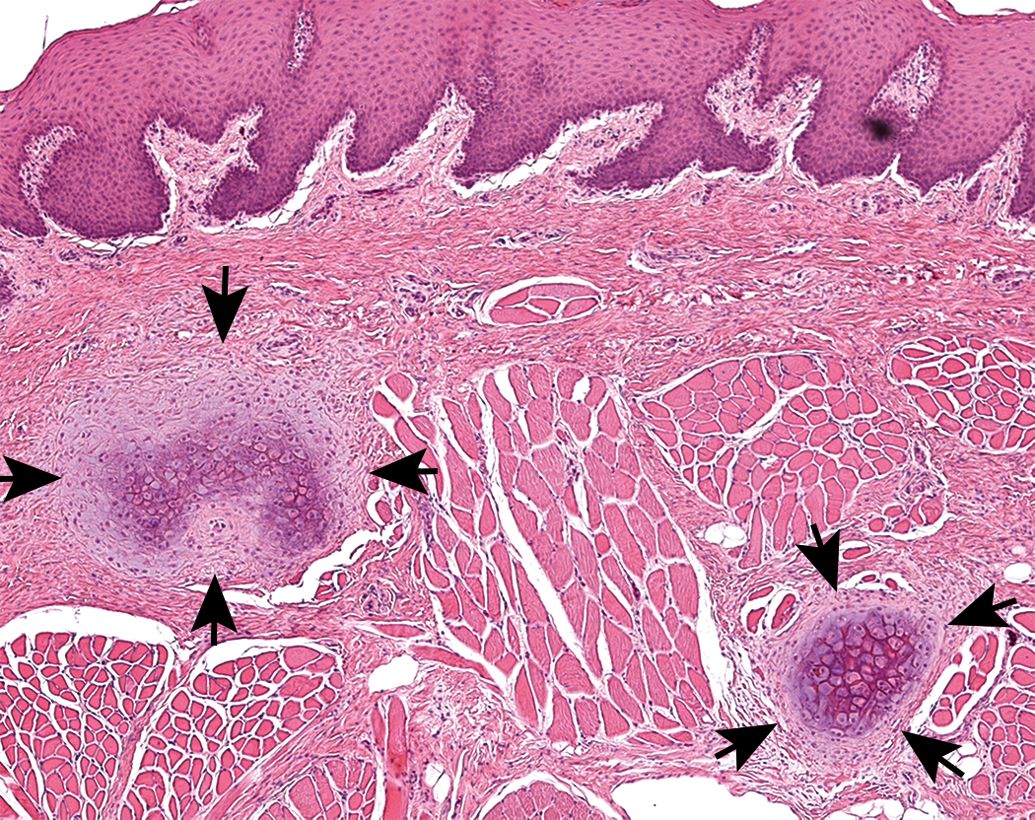
Focal chondrocytes (arrows) in tongue of a minipig.
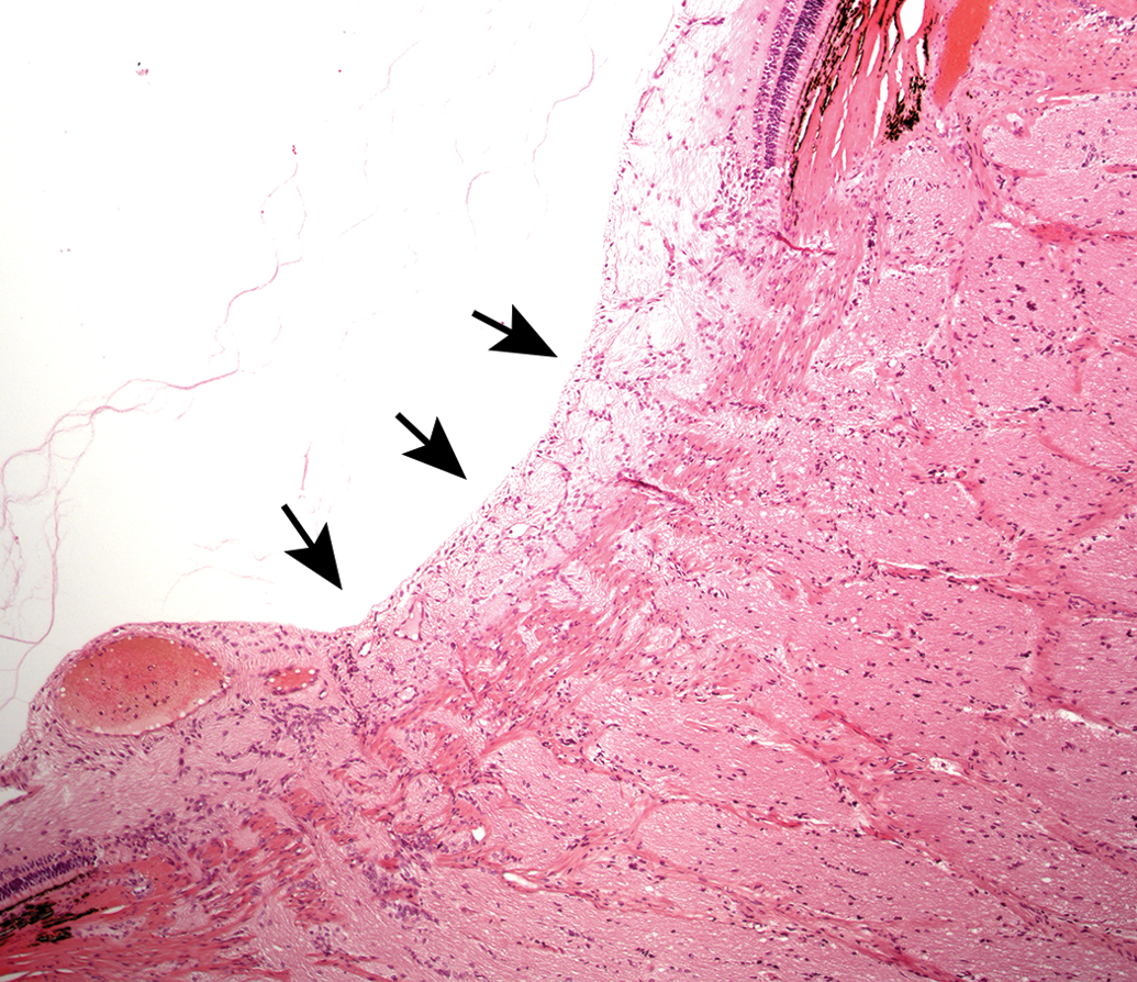
Optic cupping (arrows) in the eye of a minipig.
Conclusion
We have discussed a number of infrequent background lesions found in minipigs including PSS and OC. In order to maximize the data gained from minipig studies, the interpretation of pathology findings requires the input of experienced pathologists who understand the significance of artifacts and spontaneous background lesions in minipigs and can distinguish these from induced lesions.
Footnotes
Authors’ Contribution
All authors (EM and SM) contributed to conception or design; data acquisition, analysis, or interpretation; drafting the manuscript; and critically revising the manuscript. All authors gave final approval and agreed to be accountable for all aspects of work in ensuring that questions relating to the accuracy or integrity of any part of the work are appropriately investigated and resolved.
Declaration of Conflicting Interests
The author(s) declared no potential conflicts of interest with respect to the research, authorship, and/or publication of this article.
Funding
The author(s) received no financial support for the research, authorship, and/or publication of this article.