
Abstract
A sporadic, diffuse, interstitial mixed cell epididymitis of unknown etiology was noted in the epididymal cauda and distal corpus of young control Sprague-Dawley (SD) rats. Rats from 2 different suppliers were examined as part of routine toxicology studies. The incidence of this finding was 5/5 (study 1), 2/7 (study 2), and 2/7 (study 3). Although 2 of these studies partially overlapped temporally, none of the affected animals from any study was maintained concurrently with affected animals from any of the other 2 studies, and infectious causes, control article toxicity, or autoimmune processes were considered unlikely etiologies. Inflammation similar to that noted in the epididymides of these young rats was not present in other tissues and was not noted in study cohorts sacrificed at ages older than approximately 11 weeks or in rats of similar age from other concurrent studies. Similar findings were noted sporadically in historical control data, and consequently an age-related finding of unknown etiology and occurring in sporadic clusters is reported in SD rats ≤11 weeks old.
Introduction
Differentiation of the epididymis takes place between 28 and 96 days of age, with spermatozoa entering and filling the epididymal ducts at 72 days. Fluid begins flowing from the testis into the epididymis at 32 days. During differentiation, duct-lining cells change from an undifferentiated single cell layer to the adult cell types, including ciliated and halo cells, and the volume of ducts in the caput and cauda particularly increases. The interstitial connective tissue also increases variably between different parts of the epididymis (Reid 1958).
Lesions seen commonly in epididymides of young rats include sloughed germ cells and debris originating in the testes, focal lymphoid infiltrates, sperm granulomas, and cribriform change (Creasy 2012). Focal infiltrates of lymphocytes and other inflammatory cells are commonly noted in the interstitium of the caput of the epididymis of control Sprague-Dawley (SD) rats, especially surrounding vessels, and are generally of unknown etiology (Creasy et al. 2012). Sperm granulomas are occasionally noted in epididymides when sperm, which are immunogenic, are exposed to the immune system outside the protective space of the epididymal ducts; this may be a consequence of vas deferens obstruction, damage to the epididymal epithelium, rupture of the duct, or a chronic sequel to sperm stasis (Creasy et al. 2012). Cribriform change is caused by the infolding and bridging of the ductular epithelium in segments that have undergone contraction, forming pseudoglandular structures, and is typically noted in the distal corpus/proximal caput accompanied by decreased/absent luminal sperm and ductular atrophy (Creasy et al. 2012).
Edema of the epididymal interstitium with concomitant neutrophil infiltration has been observed in pubertal rats following intraperitoneal administration of
We have observed a lesion in the epididymal interstitium of young control rats that was histomorphologically similar to that reported with intraperitoneal
Method
The 3 studies described in this report were performed in compliance with the Guide for Care and Use of Laboratory Animals published by the U.S. National Institutes of Health (National Institute of Health Publication No. 85-23, revised 1996), and in accordance with the guidelines set by the Covance Laboratories Animal Care and Use Committee. The studies were conducted at Association for Assessment and Accreditation of Laboratory Animal Care International accredited sites.
Crl:CD(SD) rats obtained from Charles River Laboratories (Portage, MI, or Hollister, CA) were given ad libitum access to standard rodent diet and water. Rats were between 6 and 9 weeks of age at study initiation and were given a vehicle control article by daily oral gavage or periodic intramuscular injection. Groups of rats were euthanized and necropsied 4, 7, 18, 32, 47, 59, or 60 days after initiating dosing (7–16 weeks of age). Study 1 conducted in Madison, Wisconsin, was terminated after 7 days, while studies 2 and 3 conducted in Chandler, Arizona, had cohorts progressing to various time points after study initiation. Epididymides were weighed (study 1 only), placed in modified Davidson’s fixative for at least 48 hr prior to trimming, and stored in 10% neutral-buffered formalin until processing. Processed tissues were embedded in paraffin, sectioned, stained with hematoxylin and eosin (H&E), and examined microscopically. Selected additional sections of epididymis were Gram stained.
Results
Interstitial epididymal inflammation was present in 5/5 control rats approximately 8 weeks old (study 1), in 2/7 rats approximately 8.5 weeks old (study 2), and in 2/7 rats approximately 9.5 weeks old (study 3). Interstitial inflammation was not present in the epididymides of 28 older control rats (≥11 weeks old) at subsequent necropsy intervals from studies 2 and 3 or in other young rats from concurrent studies.
Microscopically, the lesion was of variable severity and chronicity ranging from acute to chronic, with mixed cell inflammatory lesions most common. Most often, a minimal to marked inflammatory infiltrate (Figure 1), predominantly consisting of lymphocytes and macrophages with fewer plasma cells and occasional aggregates of neutrophils and surrounded by variable amounts of granular to fibrillar basophilic material with clear spaces (myxoid edema, Figure 2) and admixed necrotic cellular debris, was observed bilaterally expanding the interstitium of the cauda and distal corpus of the epididymis and frequently extending into the surrounding adventitial fibroadipose connective tissue. Neutrophilic aggregates were frequently associated with small to medium-sized vessels, and active transmigration of vessel walls by neutrophils was accompanied by increased density of the surrounding interstitial myxoid material (Figure 3). Lesions of increased severity occasionally contained minimal focal to multifocal hemorrhage. Rarely, small numbers of neutrophils infiltrating tubules were noted in the cauda of the epididymis and accompanied by the presence of amorphous to granular eosinophilic luminal contents and degenerate spermatocytes. Luminal involvement was limited to 3 adjacent tubular cross sections in a single epididymis (Figure 4). Although these interstitial lesions were generally present bilaterally, they were not necessarily of bilaterally similar severity. Additional sections of epididymis stained with a Gram stain did not contain visible microorganisms.
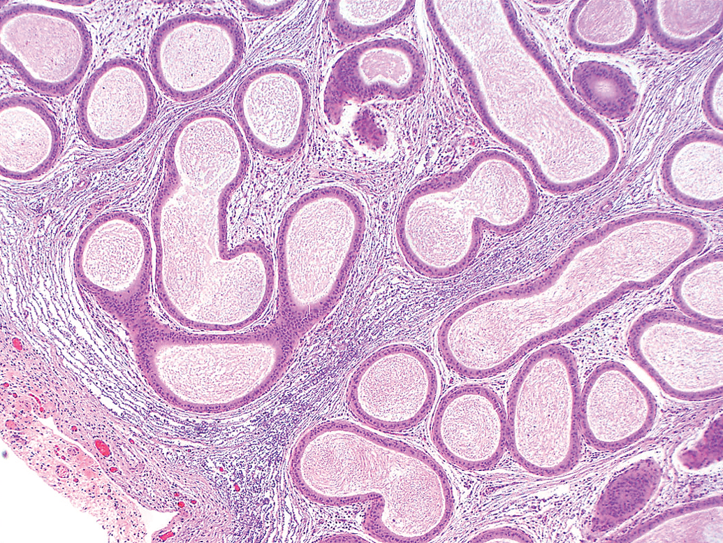
Expansion of the distal epididymal interstitium by mixed inflammatory cell infiltrates and myxoid edema (H&E, 4× objective).
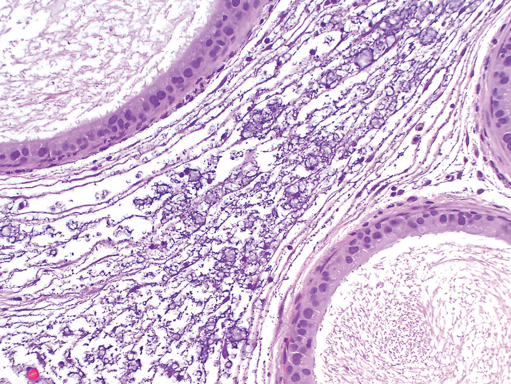
Myxoid edema characterized by granular to fibrillar basophilic material with clear spaces expanding the interstitium (H&E, 20× objective).
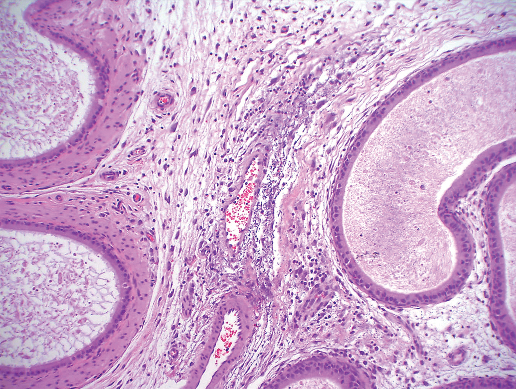
Locally acute inflammatory cell transmigration from vessels produced a vasculocentric appearance at low magnification; note the lack of vascular wall involvement (H&E, 20× objective).
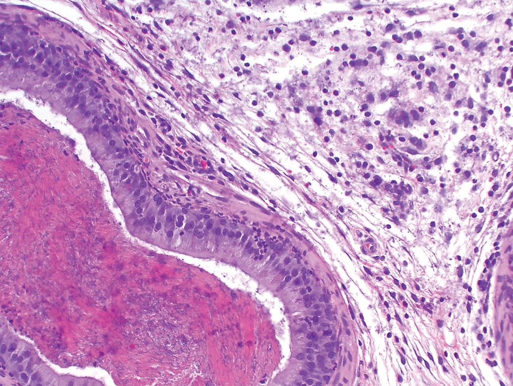
Predominantly mononuclear cell infiltrates accompanied by clusters of neutrophils that rarely penetrated tubules with resultant condensation of the luminal contents (H&E, 20× objective).
No macroscopic observations or notable organ weight differences (where available) were noted in the epididymis of any affected animal nor were clinical pathology correlates noted in routine hematology or chemistry parameters. No microscopic changes were noted in the testes of affected animals. The interstitial epididymal inflammation was morphologically distinct from and not associated with the more commonly recognized background focal lymphoid interstitial infiltrates or sperm granulomas present in isolated animals at subsequent necropsies of older cohorts from studies 2 and 3 (≥11 weeks of age) and interpreted as incidental background findings (Creasy et al. 2012; Yamasaki 1990).
Discussion
A search of historical control data from SD rats at another organization (Pfizer, Inc.) and extending back to 2008 identified 109 studies in which epididymides were examined and included a total of 792 control SD rats. Sections of epididymides from control rats with recorded findings consistent with epididymal mixed cell inflammation were reviewed, and 11 control rats from 8 studies had findings similar to those described; these 8 studies were conducted in 3 different test facilities (Groton, CT; Andover, MA; and San Diego, CA). When present, the incidence of the finding ranged from 9% (1/15 rats) to 44% (4/9 rats). All affected animals were 9 to 11 weeks old at the time of necropsy.
The overall low incidence of epididymal mixed cell inflammation (11/792 rats) noted in the historical data search suggests a rare finding. However, when present, the incidence in a given study can be high (up to 100% of control rats affected).
The young age of the affected rats and absence of the lesion in older cohorts suggested the likelihood of an age-related, possibly congenital change that is no longer present in the generally slightly older population examined in routine rat studies lasting ≥4 weeks. However, short-term exploratory studies of comparable duration to these studies are not uncommon, and similar mixed cell inflammatory lesions are not commonly noted at either of the 2 study facilities, suggesting this finding, if age related and/or congenital, is uncommon even in young rats. Although a sperm granuloma and focal lymphoid aggregates were noted in isolated older cohorts from studies 2 and 3, the low incidence and location differences suggested these commonly recognized background findings in SD rats were likely unrelated (Yamasaki 1990).
The lesions described were notable for the range of severity and chronicity observed at an early time point after arrival at the study facilities, suggesting a process that was ongoing, may have started before delivery, and potentially spread between animals before individual housing commenced after arrival at the facility. For 1 study where this was investigated (study 1), the distribution patterns of severity and/or chronicity of the described microscopic lesions did not correlate with the caging order within the affected animal room, suggesting that if any spread occurred, it would likely have taken place before arrival at the study facility. Therefore, an infectious cause remains possible; however, the lack of spread during the course of the studies and the sporadic occurrence in 3 studies across 2 facilities suggests if the condition had an infectious basis, it was not highly contagious and could be contained with routine disinfection procedures.
The location of the lesion in the cauda and distal corpus of the epididymis as well as the frequent severity differences between the generally bilaterally affected epididymides could suggest an ascending infection. In these studies, inflammation did not progress proximally and/or affect the testes of any animal. At the time of necropsy approximately 2 weeks after arrival in the study facility, no microorganisms were evident in Gram-stained sections for 1 study, even in locally acute lesions dominated by neutrophils, active transmigration of vessel walls, and myxoid edema. It has been noted that some organisms capable of inducing epididymitis may not be easily stained by Gram stains (Møller and Märdh 1980). However, the general lack of inflammation within the tubular lumina or involvement of the male accessory sex glands and concentration of the inflammation in the interstitium argues against an ascending infection (Møller and Märdh 1980). The absence of clinical or necropsy findings related to this microscopic lesion and clinical pathology findings that did not indicate a significant systemic inflammatory response underscored the localized nature of the change.
The vasculocentric appearance of the lesion in some of the more acute cases when viewed at low magnification was interpreted as a function of locally acute inflammation within the context of an ongoing process rather than inflammation focused on vessel walls. Any necrotic cellular debris was located in the interstitium outside of vessel walls, and edema and transmigrating inflammatory cells did not appear to disrupt the integrity of the vascular architecture.
An autoimmune process was considered unlikely, given the 100% incidence in 1 study and the morphologic appearance of the lesion with findings generally limited to the interstitium of the epididymis, a lack of apparent targeting of immunogenic material such as the luminal contents or epithelium in the epididymis, and the lack of changes in the testis, but could not be definitively excluded (Flickinger et al. 1990; Itoh, Hiramine, and Hojo 1991; Zhou et al. 1989).
The effects of toxicants targeting the epididymis to the exclusion of the testis can result in numerous changes, such as ductal occlusions, tubular dilatation, sperm granulomas, or primary effects in the epididymal tubular epithelium or may have effects on spermatozoa without notable changes in the epididymis. Interstitial findings for some of these compounds have included inflammation and/or fibrosis; however, they were accompanied by other epididymal changes not noted here. Among 20 reported epididymal toxicants, interstitial inflammation not accompanied by other lesions and similar to that noted in this article was not described (Hess 1998).
In the absence of a saline control group in all 3 studies, a relationship with some of the vehicle control article components could not be definitively excluded but appeared unlikely because similar findings have not been previously published or recorded at these facilities for any of the commonly used and commercially available constituents of these materials alone or in combination. Also, none of the components from the 3 vehicle control articles were included in all 3 studies.
Although an infectious cause, a relationship with vehicle control article components, or an autoimmune process could not be definitively excluded, a sporadic age-related change was considered most likely based on the occurrence of these lesions in animals of similar age from 3 separate studies but not in older cohorts from some of the same studies. A conclusive cause could not be demonstrated; consequently, a background finding in the epididymis of young mature male rats of unknown etiology is reported.
Footnotes
Acknowledgment
The authors would like to thank Steve van Adestine for assistance in the preparation of photomicrographs.
The author(s) declared no potential conflicts of interest with respect to the research, authorship, and/or publication of this article.
The author(s) received no financial support for the research, authorship, and/or publication of this article.