
Abstract
The incidence and range of spontaneous central nervous system tumors were determined in control Charles River rodents (Sprague-Dawley, Han-Wistar, Wistar rats, and CD-1 mice) from regulatory carcinogenicity studies carried out over the period 2002 to 2013 and were compared with the previously published data. In both species, the brain was notably more affected than the spinal cord. Incidences were comparable overall between rat strains (2.33%, 2.54%, and 2.89% in Wistar, Sprague-Dawley, and Han-Wistar strains, respectively) and were low in CD-1 mice (0.42% in 104-week studies and 0.2% in 80-week studies). Predominant tumor types were granular cell tumors in Wistar and Han-Wistar rats and malignant astrocytoma in Sprague-Dawley rats. Male rats were more frequently affected than females, but no sex predilection was apparent in CD-1 mice. Occasional early-onset tumors were diagnosed in rats from study week 23 onward. It is hoped that these results will provide the pathologist and the toxicologist with an up-to-date database of background neoplastic findings in widely used rodent strains.
Introduction
Rodent carcinogenicity studies are an important part of the drug development process. Of the different strains of rodents available, CD-1 mice and Sprague-Dawley, Han-Wistar, and Wistar rats are regularly used for this purpose in Europe. However, despite their common usage, specific and up-to-date information on the incidence of spontaneous neoplastic findings in the central nervous system (CNS) is not readily available in all strains of rodents. As some CNS tumors occur infrequently, marginal increases in incidence of a tumor type can be difficult to interpret, and historical control data are essential in assessing results of carcinogenicity bioassays.
The purpose of this study was to document the range and incidence of spontaneous brain and spinal cord neoplasms in the most frequently used rodent strains during routine carcinogenicity studies at Charles River Edinburgh over an 11-year period.
Materials and Methods
Animals
Brain and spinal cord samples from a total of 2,760 CD-1 mice (Crl:CD-1(ICR); 2,260 from 104-week studies and 500 from 80-week studies) and 2,562 rats (670 Sprague-Dawley [Crl:CD(SD)]; 1592 Han-Wistar [Crl:WI(Han)]; and 300 Wistar [Crl:WI(BR)]; all from 104-week studies) were obtained from control groups of Good Laboratory Practice-compliant preclinical carcinogenicity studies conducted between 2002 and 2013 at Charles River Edinburgh. The animals were purpose bred for laboratory use and were obtained from Charles River European suppliers (Charles River UK Ltd., Kent, United Kingdom). All control animals incorporated into the study were obtained from groups of animals that had been sham dosed with an appropriate vehicle.
Male mice were housed separately and female mice in groups of 3 animals per cage. Rats were housed in groups of 5 animals per cage by sex. Animal room temperature and humidity were automatically controlled at 19°C to 23°C and 40% to 70%, respectively, with a minimum of 15 air changes/hr. An automatic 12-hr light–dark cycle was maintained. Animals had free access to tap water in bottles with sipper tubes and were fed an ad libitum commercial rodent diet (Rat and Mouse [modified] No. 1 Diet SQC Expanded; Special Diet Service Ltd, Witham, Essex, England). Wooden chewsticks were also offered to all animals for environmental enrichment.
All studies were conducted in accordance with the UK Animals (Scientific Procedures) Act 1986, which conforms to the European Convention for the Protection of Vertebrate Animals Used for Experimental and Other Scientific Purposes (Strasbourg, Council of Europe).
Pathological Evaluation
Animals were humanely euthanized by a rising concentration of carbon dioxide and exsanguinated via femoral veins. Comprehensive necropsy was performed, and tissues were fixed by immersion in 10% neutral-buffered formalin, embedded in paraffin wax, sectioned to a 4- to 5-μm thickness, mounted onto glass slides, stained with hematoxylin and eosin (H&E), and coverslipped. Additional periodic acid-Schiff (PAS) staining was performed if deemed necessary by the study pathologist. Data from all studies were recorded by direct computer entry by the study pathologist using PLACES 2000 (Instem; Apoloco Limited Systems, Conshohocken, PA). Generally accepted terms were used in the diagnosis of proliferative and nonproliferative lesions (STP/ARP/AFIP SSNDC Guides for Toxicologic Pathology). Classification and nomenclature used in this project are those being considered in the recently published International Harmonization of Nomenclature and Diagnostic Criteria for lesions in rats and mice (Kaufmann et al. 2012). All neoplastic findings in each study underwent pathology peer review, and all data were reviewed by the Quality Assurance Department at Charles River’s Edinburgh facility prior to the release of the final pathology report.
Study Design
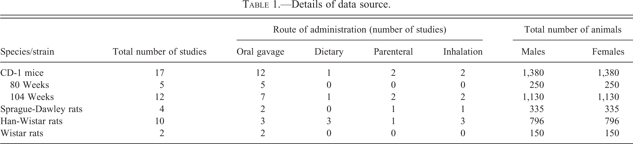
Information was gathered retrospectively from control groups of CD-1 mouse and Sprague-Dawley, Han-Wistar, and Wistar rat carcinogenicity studies evaluated during the period 2002 to 2013. Control groups varied in size, but at least 50 animals per sex were present in each study (Table 1).
Details of data source.
Study material including histological incidence tables, individual animal data listings, and selected glass slides (stained with H&E and, where applicable, PAS) were retrieved from the archives and analyzed for pathology findings.
Results
Tables 2 through 6 present a summary of brain and spinal cord tumors with their incidences.
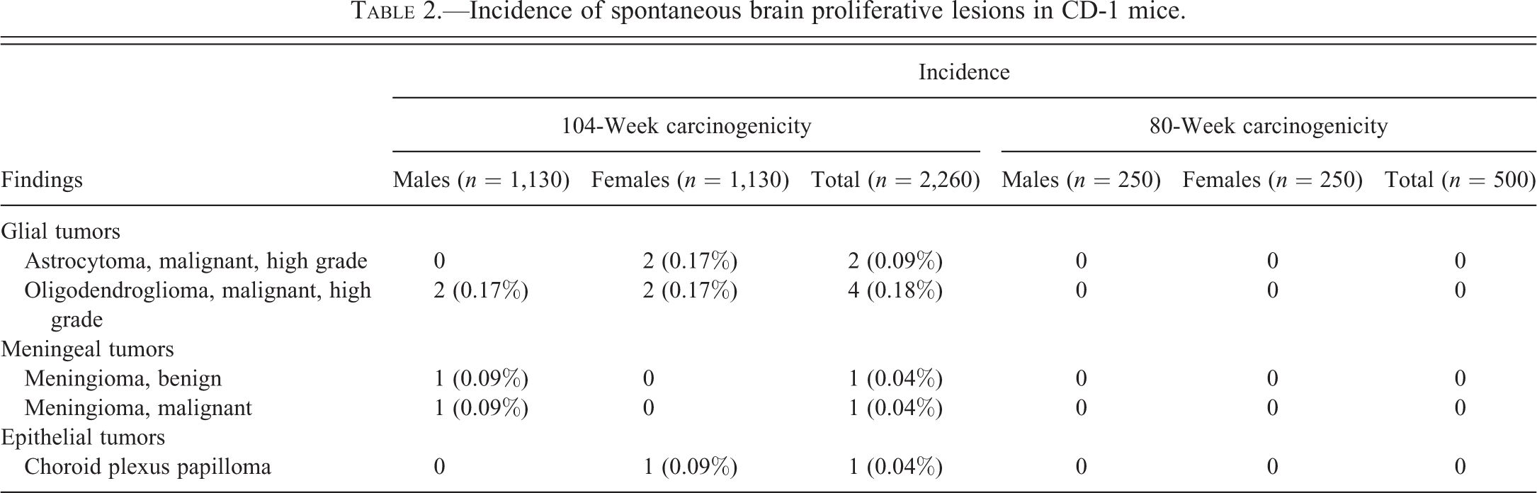
Incidence of spontaneous brain proliferative lesions in CD-1 mice.
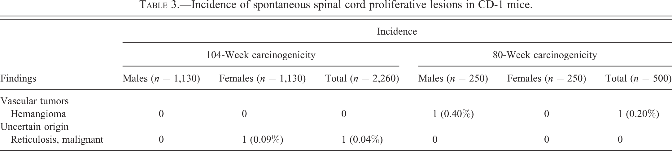
Incidence of spontaneous spinal cord proliferative lesions in CD-1 mice.
Incidence of spontaneous brain proliferative lesions in Sprague-Dawley, Han-Wistar and Wistar rats.
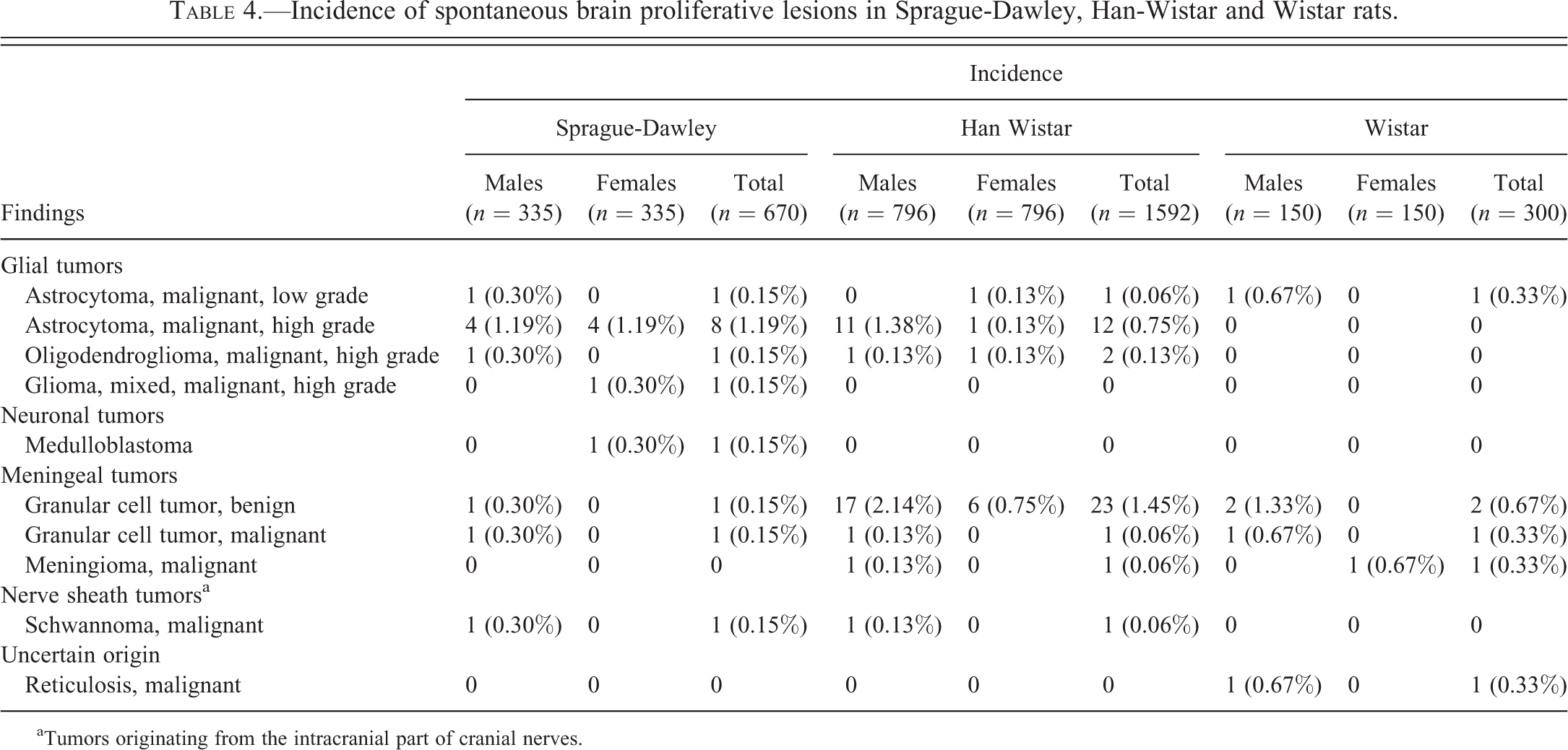
aTumors originating from the intracranial part of cranial nerves.
Incidence of spontaneous spinal cord proliferative lesions in Sprague-Dawley, Han-Wistar and Wistar rats.
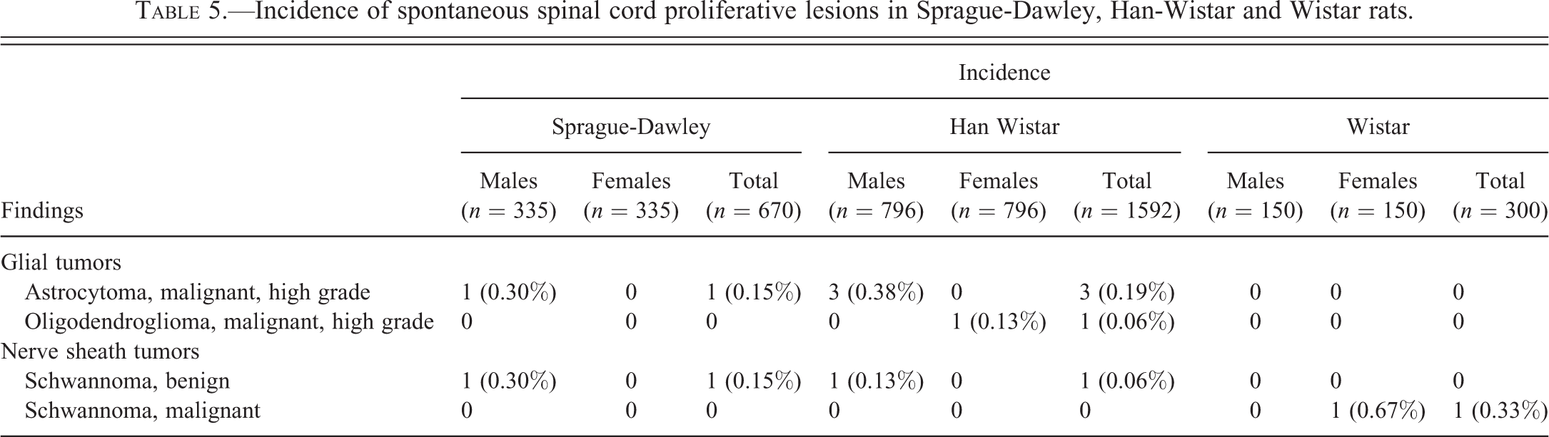
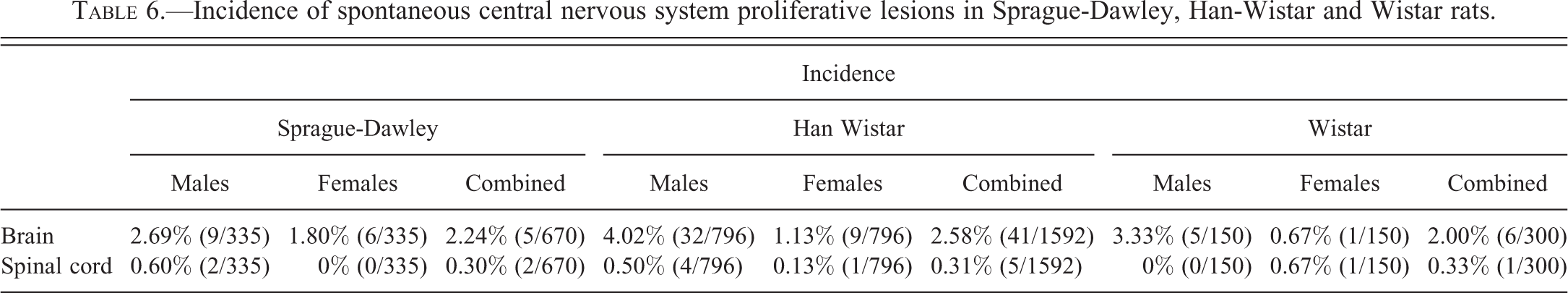
Incidence of spontaneous central nervous system proliferative lesions in Sprague-Dawley, Han-Wistar and Wistar rats.
CD-1 Mice
No brain tumors were observed in any of the 80-week studies. In 104-week studies, tumors of glial origin were the most common neoplastic finding. Due to their biologically aggressive behavior, they were all considered as malignant, and the differentiation between low- and high-grade tumors was based on cytological features (e.g., anisocytosis and anisokaryosis), mitotic index, and involvement of one or more areas of the CNS. High-grade oligodendroglioma was noted in 4 mice (0.18%) as early as 52 weeks. This tumor was composed of oligodendrocytes showing anaplastic features (high cellularity, atypia and pleomorphism, and increased mitotic index) and was frequently associated with necrotic and hemorrhagic foci, sometimes accompanied by “vascular garlands” at the periphery of the tumors (capillaries with atypical, proliferative endothelial cells). High-grade astrocytoma was observed in 2 animals (0.09%) and showed classic features such as high cellularity and invasiveness, with pleomorphic astrocytes forming perineuronal aggregates (satellitosis) or perivascular cuffing. A choroid plexus papilloma (intraventricular tumor composed of a thin fibrovascular stromal core lined by cuboidal, well-differentiated cells) was identified in 1 animal. A malignant and a benign meningioma (differentiated by the presence or absence of extension into the brain parenchyma, respectively) were diagnosed as incidental tumors on 1 occasion (0.04%) each.
There was a narrow spectrum of spinal cord neoplasms, with 1 hemangioma (0.20%) observed in an 80-week study and malignant reticulosis (composed of lymphocytic-to-histiocytic cells showing pleomorphism and atypia) occurring in 1 animal (0.04%) from a 104-week study.
Overall, 67% of CNS tumors diagnosed in premature decedents were considered to have contributed to the demise of the animal (data not shown).
Rats
Sprague-Dawley rats
A wide range of brain tumors were observed. Malignant high-grade astrocytoma was the most common (1.19%; Figure 1A), showing the same incidence in both sexes, and occurred as early as 64 weeks. Other malignant neoplasms were observed notably less frequently (0.15% each) and consisted of a high-grade oligodendroglioma (Figure 1B), a high-grade mixed glioma (composed of neoplastic oligodendrocytes and astrocytes, both populations forming at least 20% of the tumor; Figure 1C), a low-grade astrocytoma, a benign granular cell tumor (well-demarcated, noninvasive tumor that compressed the underlying brain parenchyma), a malignant granular cell tumor (differentiated from its benign counterpart by the invasion of the underlying nervous tissue), a malignant medulloblastoma (arising from the cerebellum, composed of neuroepithelial stem cells that showed atypical mitotic figures; Figure 1D), and a malignant schwannoma arising from cranial nerve roots. Males were more affected than females (2.69% and 1.80%, respectively).
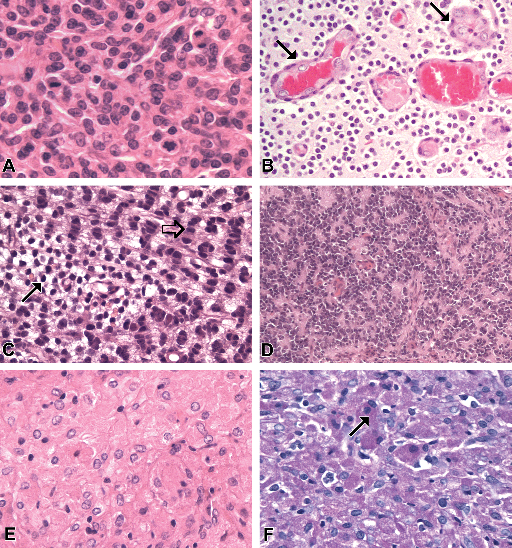
(A) Malignant high-grade astrocytoma, Sprague-Dawley rat. This tumor shows dense cellularity and is composed of cells with round-to-fusiform nuclei and indistinct borders. Several mitotic figures are present on this field. H&E. Original magnification: 400×. (B) Malignant high-grade oligodendroglioma, Sprague-Dawley rat. Neoplastic oligodendrocytes show the typical “fried-egg” appearance (round, hyperchromatic nucleus surrounded by a clear perinuclear halo and distinct cell borders). Note the atypical capillary endothelium hyperplasia (arrows). H&E. Original magnification: 200×. (C) Malignant low-grade-mixed glioma, Sprague-Dawley rat. This tumor is composed of 2 cell populations, arranged in adjacent areas of astrocytic (open arrow) and oligodendroglial (black arrow) origins. Each population provides more than 20% of the neoplasm. H&E. Original magnification: 400×. (D) Medulloblastoma, Sprague-Dawley rat. This densely cellular tumor is composed of small, round cells similar to granule cell layer neurons, arranged in sheets with occasional rosette formation. H&E. Original magnification: 200×. (E) Benign granular cell tumor, Wistar rat. Closely packed sheets of polygonal cells show an abundant cytoplasm and a central to eccentric nucleus. Note the eosinophilic, granular cytoplasm present in some neoplastic cells. Unlike granular cell hyperplastic foci, the tumor was associated with compression of the adjacent brain tissue. H&E. Original magnification: 400×. (F) Benign granular cell tumor, Wistar rat. Sparse to abundant PAS-positive granules are present in the cytoplasm of neoplastic cells. Original magnification: 400×. H&E = hematoxylin and eosin; PAS = periodic acid-Schiff.
Spinal cord tumors consisted of a malignant high-grade astrocytoma and a benign schwannoma (0.15% each) and were observed only in males. The latter tumor, originating from Schwann cells (myelinating cells of the peripheral nervous system) presumably located in adjacent cranial nerve roots, was composed of spindle cells with elongated, sharp-ended nuclei forming occasional nuclear palisades.
Han-Wistar rats
Benign granular cell tumor was the most frequent neoplasm observed in the brain (1.45%), the majority being diagnosed after study week 100, followed by high-grade malignant astrocytoma (0.75%). High-grade malignant oligodendroglioma was diagnosed in 2 animals (0.13%) as early as 32 weeks. Low-grade malignant astrocytoma, malignant granular cell tumor, malignant meningioma, and malignant schwannoma were observed on 1 (0.06%) occasion each. All tumor types were observed with higher incidences in males when compared with females (4.03% and 1.13%, respectively).
High-grade malignant astrocytoma (0.19%) and benign schwannoma (0.06%) were observed in the spinal cord from males, and a high-grade malignant oligodendroglioma (0.06%) was present in a female.
Wistar rats
Benign granular cell tumor (0.67%) was the most frequent tumor identified in the brain (Figure 1E and F), followed by malignant granular cell tumor, low-grade malignant astrocytoma, malignant reticulosis, and malignant meningioma (0.33% each). All were diagnosed in males, with the exception of the latter that was observed in a female on the 23rd week of study. The only tumor observed in the spinal cord was a benign schwannoma (0.33%).
Overall, 64% of CNS tumors diagnosed in premature decedents were considered to have contributed to the death of the animal (61.5% in Sprague-Dawley, 62.5% in Wistar, and 70% in Han-Wistar rats; data not shown).
Discussion
The aim of this study was to determine the incidences of spontaneous tumors of the CNS in rodents. Although such findings are known to be exceedingly rare in CD-1 mice and have seldom been reported, a variety of tumors of glial, epithelial, or meningeal origin were observed at Charles River Edinburgh with an overall incidence higher than in previous reports (Maita et al. 1988; Krinke et al. 2000; Giknis and Clifford 2010). Conversely, rat is a laboratory animal species that is more prone to developing CNS tumors. Overall there was minimal strain-specific difference in incidence, with 2.33%, 2.54%, and 2.89% of animals affected in Wistar, Sprague-Dawley, and Han-Wistar strains, respectively. If such incidences are in line with the literature in Sprague-Dawley and Han-Wistar rats (Zwicker et al. 1992; Giknis and Clifford 2011; Weber et al. 2011), they contrast with the low rate of spontaneous nervous system tumors that had been previously reported in the Wistar strain (Poteracki and Walsh 1998).
Wistar and Han-Wistar rats appeared more prone to developing meningeal tumors, mainly granular cell tumors. Neoplastic cells typically show PAS staining of lysosomal contents (Krinke et al. 2000) as well as positive immunostaining for Ricinus communis agglutinin 1 (RCA-1) and ionized calcium-binding adaptor molecule 1 (Iba-1), the 2 microglial markers. Granular cell tumors appear to be originating from a subset of macrophages associated with the meninges (Kolenda-Roberts et al. 2013).
The most frequent CNS tumor observed in Sprague-Dawley rats was malignant astrocytoma. Spontaneous astrocytomas are invariably negative to glial fibrillary acidic protein immunohistochemistry and have been thought to represent an immature astrocytic phenotype (Solleveld and Boorman 1990; Krinke et al. 2000; Weber et al. 2011). However, there is growing evidence that tumors previously diagnosed as astrocytic in origin may derive from another cell lineage. Recent investigations have shown that these tumors demonstrate positive immunoreactivity to anti-rat-activated macrophage/microglia antibody (ED-1), anti-rat macrophage/dendritic cell antibody (RM-4), lectin RCA-1, and protein Iba-1 (Nagatani et al. 2009; Kolenda-Roberts et al. 2013), which indicate that they could be of monocytic origin (macrophage or microglia). Some authors have suggested this tumor type be redesignated as malignant microglial tumors (Kolenda-Roberts et al. 2013).
Malignant reticulosis is a rare tumor, the existence of which has long been controversial (Cardesa et al. 1994; Krinke et al. 2000). It was observed in 1 Wistar rat and 1 CD-1 mouse across the 11-year period. The immunohistochemical profile of malignant reticulosis is similar to astrocytoma in many aspects, and both tumors are suspected to be derived from the same lineage (Nagatani et al. 2009).
Nervous system tumors were notably more common in the brain (approximately 90% in both species) than in the spinal cord, which is in accordance with previous reports (Zwicker et al. 1992; Weber et al. 2011). This might be attributable to a less thorough examination of the latter, which is limited to 3 sections across its 3 major segments and to gross abnormalities. Of interest is a recent change in routine brain trimming at Charles River Edinburgh, where the number of sections evaluated has been increased from 3 to 7 sections per brain, as recommended by the STP (Bolon et al. 2013). This might increase the detection of small-sized brain tumors in the long term.
Interestingly, the incidence of spontaneous nervous system tumors in CD-1 mice was comparable in males and females, whereas males were more affected in all strains of rats. Several reports have identified a comparable sex effect in rats (Solleveld and Boorman 1990; Zwicker et al. 1992; Weber et al. 2011), but to the authors’ knowledge, no such data have been published in mice.
Although the incidence of primary nervous system tumors is related to aging, early-onset tumors were diagnosed in Wistar and Han-Wistar rats after 23 and 32 weeks on study, respectively, and were considered to be the cause of death in both instances.
To the best of our knowledge, this is the most recent report of the incidences of background CNS tumors in control Charles River CD-1 mice and Sprague-Dawley, Han-Wistar, and Wistar rats used in chronic toxicity studies in recent years. Reference to the incidences reported should facilitate the differentiation of spontaneous from induced lesions in toxicological safety studies in these strains of rodents. Representative photomicrographs of the findings described are available on the goRENI (n.d.) website.
Footnotes
The author(s) declared no potential conflicts of interest with respect to the research, authorship, and/or publication of this article.
The author(s) received no financial support for the research, authorship, and/or publication of this article.