
Abstract
Thymomas from 277 Fischer 344/N (F344/N), 10 Sprague Dawley (HSD:Sprague Dawley SD) (SD), 129 Wistar Han [Crl:WI(Han)] (WH), and 4 Wistar Outbred (WO) rats were reviewed from long-term studies in the National Toxicology Program (NTP) database. The incidence of thymomas in F344/N rats was slightly higher in males than in females, while the incidences in SD and WH rats were higher in females than in males. Only male WO rats were used in NTP studies. Of the 277 thymomas in F344/N rats, 235 (84.8%) were benign and 42 (15.2%) malignant, 14 of which exhibited metastasis. Of the 10 thymomas in SD rats, 5 (50%) were benign and 5 (50%) were malignant, one of which exhibited metastasis. Of the 129 thymomas in WH rats, 126 (98%) were benign and 3 (2%) were malignant, 1 with metastasis. Of the 4 thymomas in WO rats, 3 (75%) were benign and 1 (25%) was malignant, with no metastases. Malignant thymomas in F344/N and WH rats showed a propensity to be the cause of death and to result in early mortality, whereas the benign thymomas were associated less often with decreased survival. No occurrences of this neoplasm were reported to be related to exposure to any test articles.
Introduction
Thymoma is defined as the neoplastic proliferation of thymic epithelial cells. By virtue of the cellular composition of the thymus, thymomas contain varying numbers of non-neoplastic lymphocytes. Various classification schemes have been described in veterinary and human medicine, and most are based on histomorphological features. One such scheme in the rat creates divisions based on the relative proportions of lymphocytes and neoplastic epithelial cells, thus specifying tumor types as epithelial type, mixed epithelial and lymphoid cell type, or “pure” lymphoid type (containing scattered epithelial cells to distinguish it from well-differentiated lymphoma). 1 Others suggest that lymphocyte-rich thymomas may be divided into 2 types, tumors with and without medullary differentiation. 2,3 Tumors exhibiting medullary differentiation have pale staining areas containing fewer lymphocytes and foci of large, pale epithelial cells and occasional Hassall’s corpuscles. These areas are subdivided into lobules by fibrous trabeculae, which may contain epithelial cells forming cords, tubules, and cysts. Thymomas without medullary differentiation consist of a mixture of small lymphocytes and epithelial cells of variable morphology. While epithelial cell morphology can be quite variable, cellular atypia has been found to be rare. 3 Epithelial cells of thymomas can form several morphological patterns, which have been described by one author as epidermoid (nonkeratinizing squamous epithelium); squamoid (with keratinization); papillary; ribbons, cords, or tubules; spindloid; endocrine-like (adenoid); neuroendocrine; and myoid. 4 According to the global open Registry Nomenclature Information System (goRENI), thymomas contain a mixture of epithelial cells and lymphocytes in varying proportions, ranging from predominantly epithelial cells to epithelial cells and lymphocytes with or without medullary differentiation. 5,6 Furthermore, the goRENI description separates thymomas into “epithelial” and “spindeloid” types based on cell morphology.
Thymoma is an uncommon tumor in most strains of rats and mice. Primary tumors of the thymus of Sprague Dawley (SD)–derived rats (strain: Tif: RAI) were reported from 7 different long-term or 2-year studies. 7 In that large review, 192 primary thymic tumors were identified (of 4,281 thymuses), of which 171 were classified as benign and 21 were called malignant. In the F344/N rat, spontaneously occurring thymomas are rare, and most of those have been reported to be benign. 8,9 A recent large study examined the incidences and histological features of thymomas and hyperplastic lesions in Wistar Hannover (WH) rats and found a high incidence of both lesions in this strain, with higher incidences observed in females than males. 10 Additionally, the authors found a higher incidence of these proliferative lesions in WH rats than in SD rats. The literature contains no reports of large studies of thymoma in Fischer 344 rats. The purpose of this study is to describe the morphological features, incidences, and behavior of thymomas in F344/N, SD, WH, and Wistar Outbred (WO) rats in National Toxicology Program (NTP) studies.
Materials and Methods
Animals and Study Design
The housing and care of the rats were based on the National Institutes of Health (NIH) Guidelines for the Care and Use of Laboratory Animals. 11 Each testing laboratory also had its own Institutional Animal Care and Use Committee that reviewed and approved the protocols prior to the initiation of the study. The NTP studies evaluated for this review were conducted from 1978 to 2011 and were chronic (2-year) studies. The exposures began at 7 to 8 weeks of age and continued for 104 to 105 weeks and generally involved groups of 50 rats of each sex (control and 2-3 treated groups).
For dosed feed and water studies, the animals were exposed ad libitum, while gavage exposures (generally corn oil vehicle) were dosed 5 times/wk with the controls receiving corn oil alone. For feed, water, and gavage studies, the rats were housed in polycarbonate cages on treated hardwood bedding. Water and feed were available ad libitum. For inhalation studies, the rats were housed individually in hanging wire cages and generally exposed approximately 6 h/d, 5 d/wk. Feed and water were available ad libitum; however, feed was not available during the exposure periods. Prior to 1980, available commercial diets were used for the studies. The NIH-07 open formula diet was used for most studies between 1980 and 1995. In 1995, the NTP studies switched to the NTP-2000 open formula diet that had a higher fiber and lower protein content in an effort to decrease the severity of chronic progressive nephropathy and increase survival. 12,13 The rats were housed under standard conditions with room temperatures generally between 21°C and 24°C with relative humidity between 35% and 65%. The animal rooms were on a 12-hour:12-hour light/dark schedule with a minimum of 10 air changes per hour. The animals were observed twice daily and weighed weekly for the first 13 weeks and monthly thereafter.
Complete necropsies were performed on all animals. All observed gross lesions and approximately 40 tissues were collected, fixed in 10% neutral-buffered formalin, stained with hematoxylin and eosin (H&E), and examined. All rats, including natural deaths and those that were moribund and killed during the course of the study, were necropsied. Thymic tissue and/or thymic masses were routinely collected from all rats on each study. The thymus was embedded flat in order to obtain a longitudinal section, when possible. If the thymus was grossly enlarged, it was trimmed at the largest dimension that would fit in a cassette.
Selection of Studies and Pathology Review
The NTP historical databases from 1978 to 2011 were queried for the diagnosis of benign thymoma, malignant thymoma, and thymoma. The diagnoses were confirmed by 5 pathologist coauthors (R.R.M., H.N., R.A.M., N.A., and D.E.M.). The databases contained data on 197,939 F344/N rats (175,136 rats from long-term studies and 22,803 rats used in short-term studies), 20,936 SD rats (13,525 rats from long-term studies and 7,411 rats from short-term studies), 3,624 WH rats (3,000 rats from long-term studies and 624 rats from short-term studies), and 260 WO rats (200 rats from long-term studies and 60 rats from short-term studies). All sections of thymomas were examined by light microscopy to confirm the presence of thymoma and to determine various morphologic features, patterns of growth, and the presence or absence of invasion and/or metastasis that could be used to evaluate whether the neoplasms might be categorized as benign or malignant. Tabulation of data was designed to determine thymoma occurrences between multiple strains of male and female rats, classification of tumors as benign or malignant, and to characterize general morphologic patterns to determine whether any were associated with any particular behavior propensities.
The classification of the neoplasms as benign or malignant was based on tumor expansion and invasion of surrounding tissues, the presence of metastatic lesions, and cytological features. Thymomas were distinguished from epithelial hyperplasias based on the size of the proliferation and the nodular and often compressive features of thymomas. Thymomas were classified as benign when they were confined to the mediastinal space, discrete, exhibited an expansile noninvasive growth pattern, and there was no evidence of metastasis. Extension into the mediastinal fat was considered a benign feature if there was connection to the main tumor mass and as long as the other benign tumor criteria were met. Malignant thymomas were characterized by implantation on serosal surfaces, metastasis to other organs, invasion of adjacent tissues (body wall, diaphragm, or thoracic organs), infiltration of mediastinal fat with nests of neoplastic cells but no apparent or suspected connection with the main tumor mass, and/or cytological features such as pleomorphism and atypia. To determine any relationship between biological behavior and malignancy of thymoma, individual necropsy record and/or pathology data of the NTP studies were reviewed and survival time, cause of death, and necropsy type were tabulated.
Results
Fischer 344/N Rats
Of 175,136 F344/N rats from long-term studies, 277 thymomas recorded in the NTP database were reviewed. No thymomas were reported in any of the 22,803 rats from short-term studies. Thymomas in F344/N rats in this survey were rare occurrences (277/175,136; 0.16% incidence). No occurrences of this tumor were found to be associated with treatment by test articles in these studies. More thymomas were recorded in male F344/N rats (56.3% [156/277]) than in females (43.7% [121/277]), and the overall incidence of thymomas remained slightly higher in males (0.18%) than females (0.14%), despite the higher number of males (88,414) than females (86,722) in long-term NTP studies (Table 1). Thymomas appeared grossly as firm, smooth, well-circumscribed masses within the anterior mediastinum (Figure 1). Benign thymomas in F344/N rats comprised 84.8% (235/277) of the thymomas reviewed, and the remaining 15.2% (42/277) were diagnosed as malignant thymomas based on metastasis, unequivocal tissue invasion, and/or cytological features (cellular pleomorphism, cellular atypia, karyomegaly, anisokaryosis, increased mitoses). In male rats 17.3% (27/156) of thymomas were malignant, and in female rats, 12.4% (15/121) were malignant. Metastases were observed in 14 (33.3%) of 42 animals with malignant thymomas, and in all 14 cases, metastatic lesions were observed in the lung. Metastases also were present in the lymph node of one animal and in the liver of another animal. All malignant thymomas that metastasized had features of the main tumor mass, and no other malignant epithelial neoplasms were present in other organs. Of the remaining 28 malignant thymomas that exhibited no metastases, 17 were diagnosed due to unequivocal tissue invasion, and 11 were diagnosed based on cytological features, such as pleomorphism, cellular atypia, karyomegaly, anisokaryosis, prominent nucleoli, and increased mitoses. These cytologic features typically associated with malignancy had little to no relationship to a propensity for invasion or metastasis.
Incidences of Thymomas in Male and Female Rats in NTP Long-Term Studies per Strain.
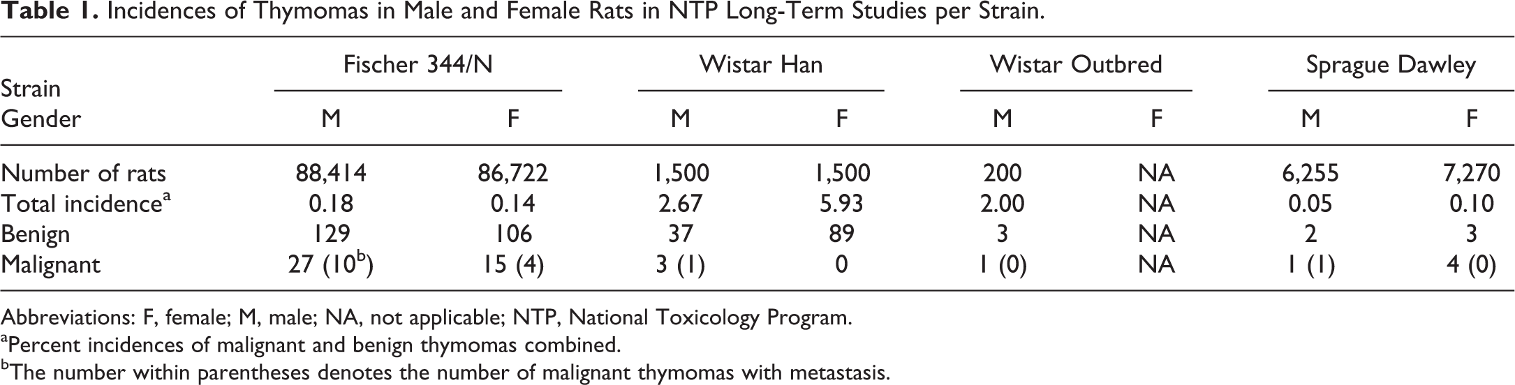
Abbreviations: F, female; M, male; NA, not applicable; NTP, National Toxicology Program.
aPercent incidences of malignant and benign thymomas combined.
bThe number within parentheses denotes the number of malignant thymomas with metastasis.
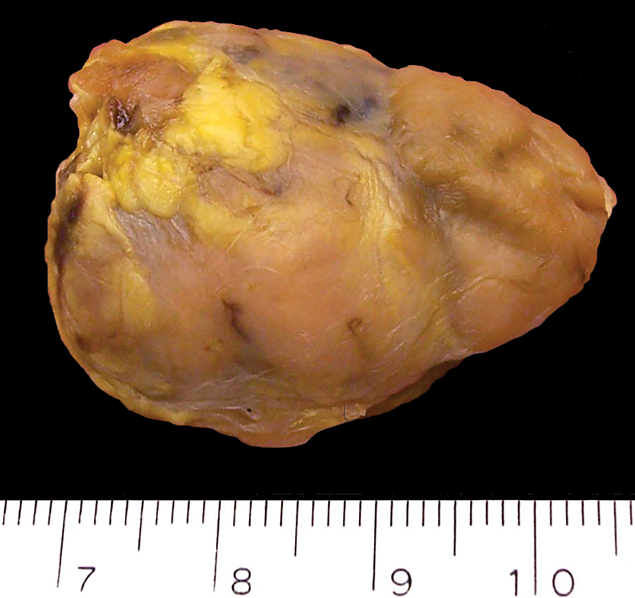
A benign thymoma measuring 3.7 cm × 2.8 cm was removed from the cranial mediastinum of a female F344/N rat from a 2-year study.
Thymomas in F344/N rats from our studies were morphologically heterogeneous but could be divided into 6 general categories according to morphologic features (Table 2). Most tumors displayed a mixture of more than 1 morphologic pattern, and these were categorized by the predominant pattern. The most common morphologic pattern consisted of epithelial cells arranged in cords and tubules and was present in 155 (56.0%) of 277 tumors in this review (Figures 2 and 3). This was followed by thymomas with a predominantly spindloid pattern, which occurred with a frequency of 79 (28.5%) of 277 (Figure 4). A papillary pattern was observed in 17 (6.1%) of 277 tumors and was frequently associated with cystic features (Figure 5). A squamous epithelial pattern was present in 12 (4.3%) of 277 tumors (Figure 6). A myoid pattern was present in 11 (4.0%) of 277 tumors (Figure 7). The myoid cell is a striated muscle cell that exhibits expression of desmin (Figure 8). Although it is possible that myoid cells within the thymomas in this series represented invasion of preexisting muscle, myoid differentiation of neoplastic cells could not be ruled out. A neuroendocrine pattern was observed in only 3 (1.1%) of 277 tumors (Figure 9). No particular pattern was unequivocally correlated with a metastatic or invasive propensity (data not shown).
Comparison of the Morphologic Patterns of Thymomas in Rats in NTP Studies.
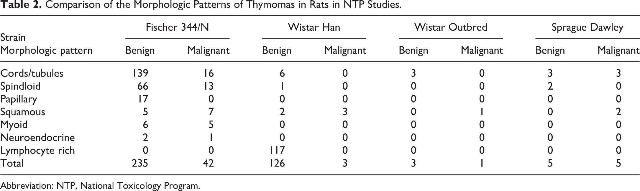
Abbreviation: NTP, National Toxicology Program.
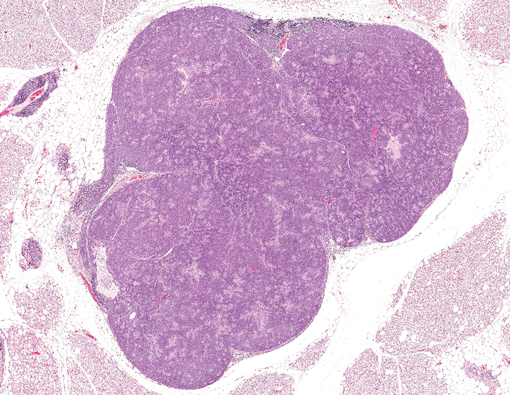
A well-circumscribed, nodular, benign thymoma from a male F344/N rat from a 2-year study. Hematoxylin and eosin.
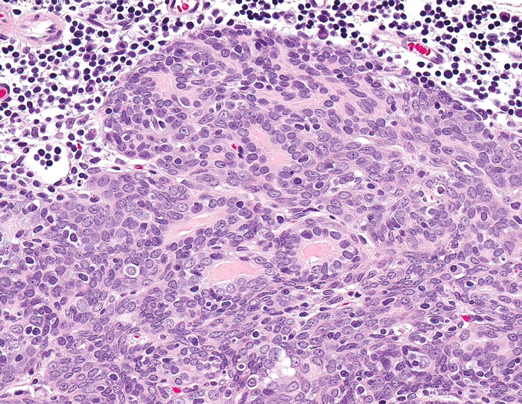
Higher magnification of Figure 2 showing the neoplastic epithelial cells forming cords and tubules. Hematoxylin and eosin.
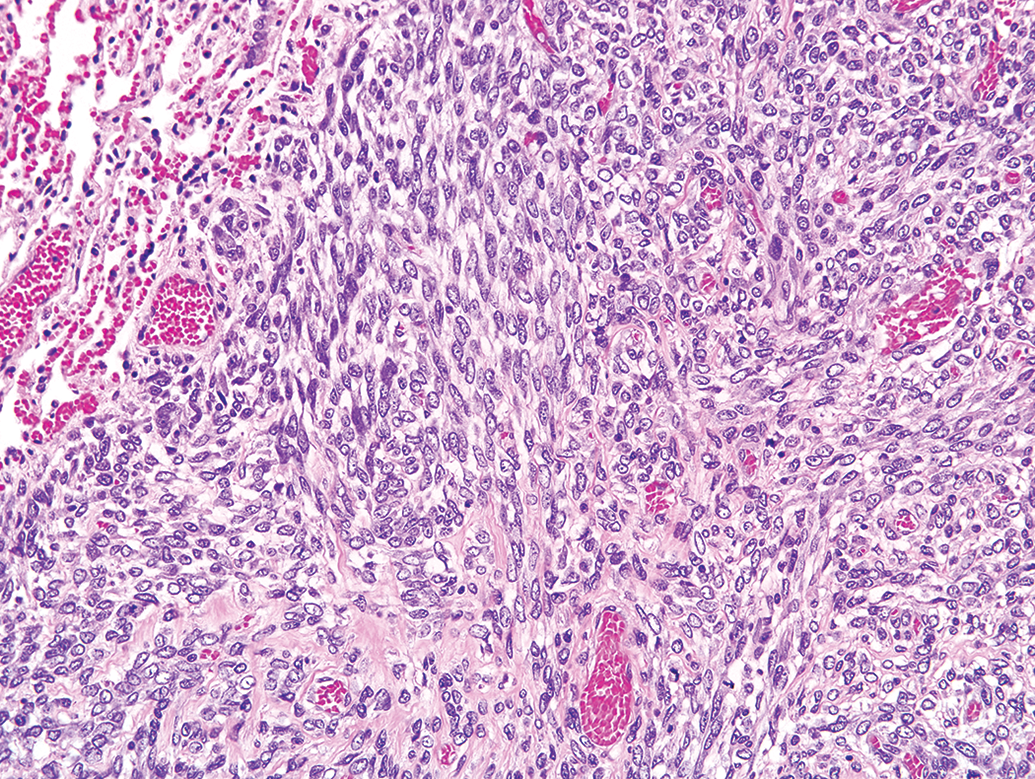
Lung metastasis from a malignant thymoma in a male F344/N rat from a 2-year study. Sheets of elongated neoplastic cells form a spindloid pattern. Hematoxylin and eosin.
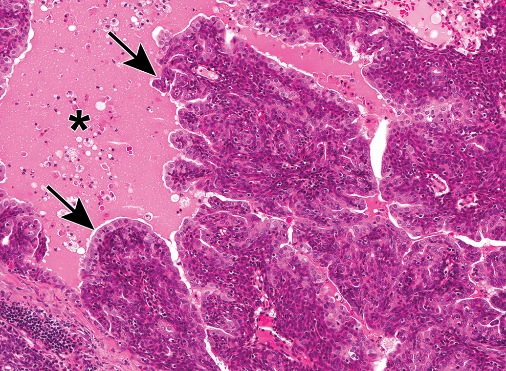
A benign thymoma from a male F344/N rat from a long-term study. The neoplastic epithelial cells form a papillary pattern. The papillary projections (arrows) protrude into a cystic space (asterisk). Hematoxylin and eosin.

A malignant thymoma from a male F344/N rat from a long-term study. The neoplastic epithelial cells form a squamous pattern. Hematoxylin and eosin.
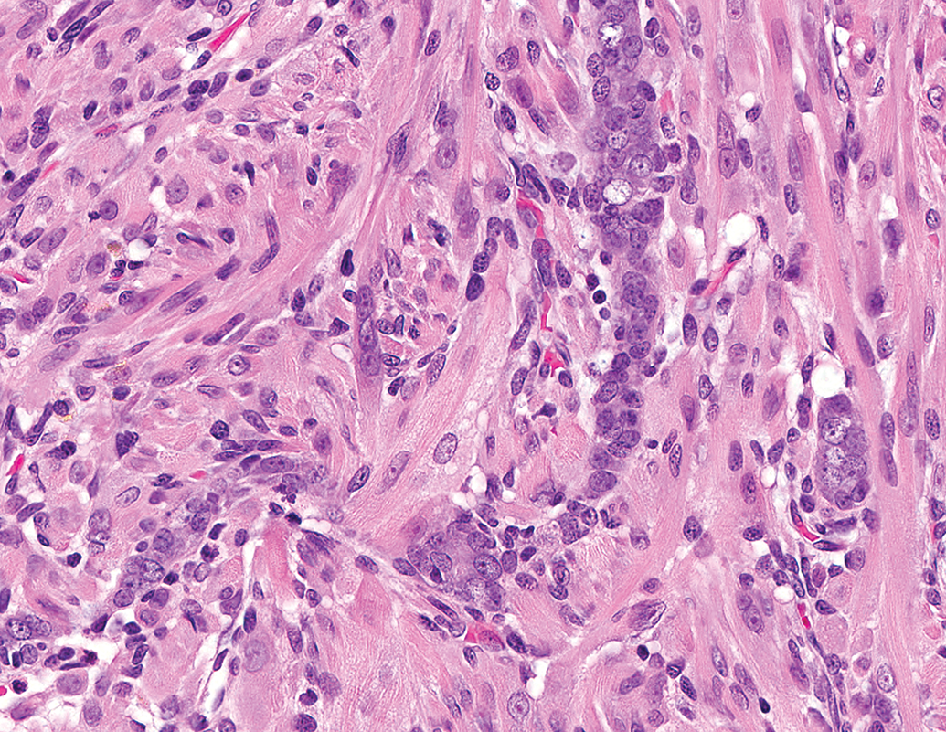
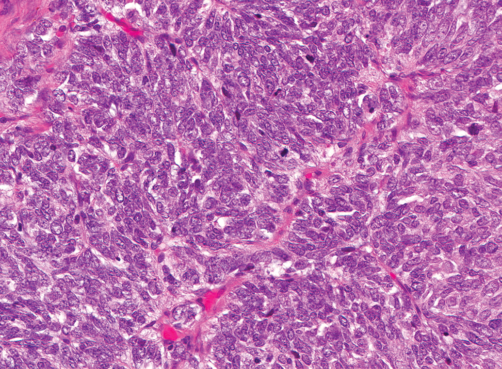
A malignant thymoma from a female F344/N rat from a long-term study. The neoplastic cells form a predominantly myoid pattern. Hematoxylin and eosin.
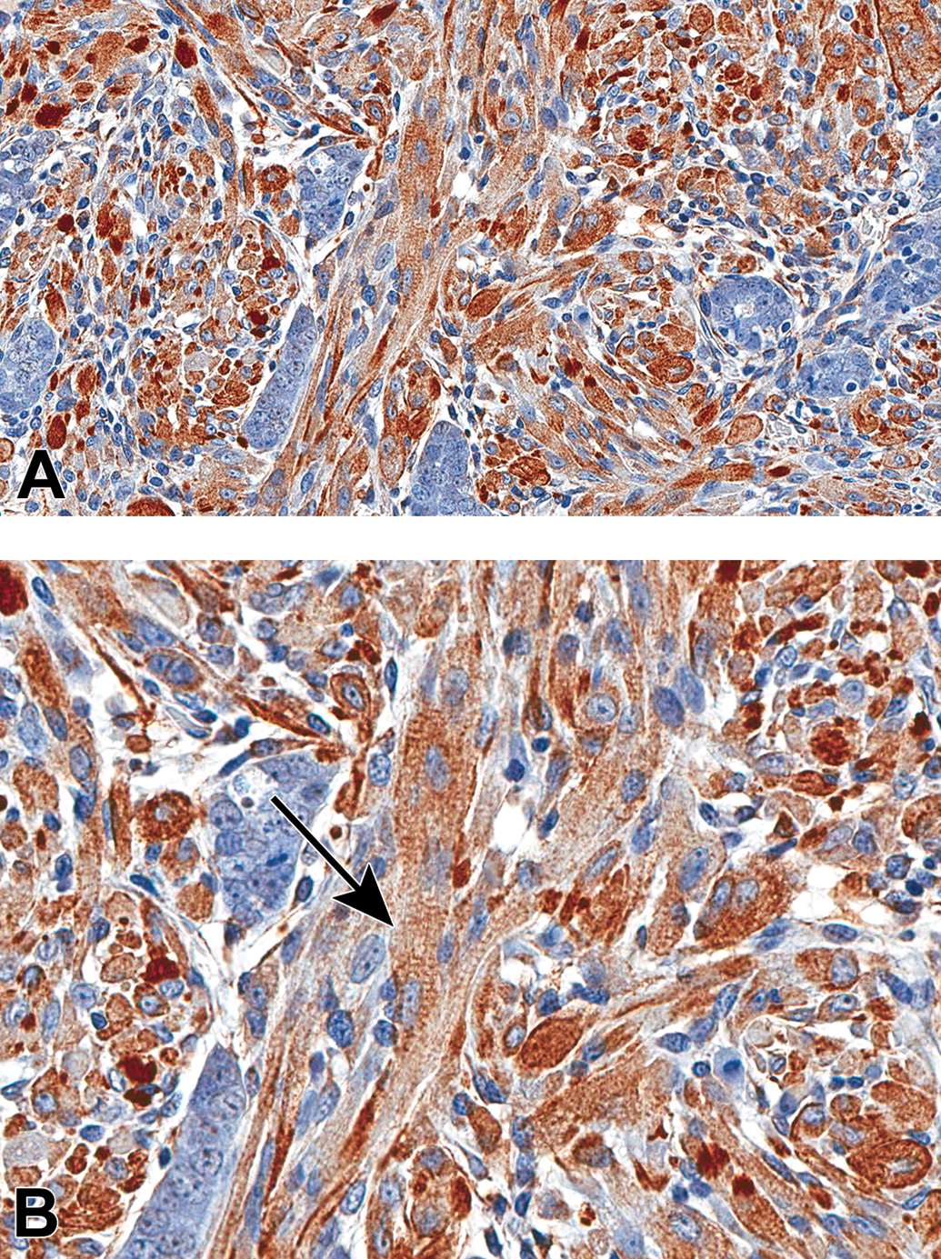
A, Immunohistochemical staining of neoplasm from Figure 7 showing expression of desmin. B, Higher magnification showing desmin-positive myoid cells with prominent cross striations (arrow).
A benign thymoma from a female F344/N rat from a long-term study. Packets of elongated neoplastic cells form a neuroendocrine pattern. Hematoxylin and eosin.
Four benign thymomas, 11 malignant thymomas without metastasis, and 11 malignant thymomas with metastasis were considered the primary cause of death. The survival days of animals with malignant thymomas were less than those of animals with benign thymomas (Table 3).
Correlation of the Malignancy of Thymoma With Survival Days, Cause of Death, and Termination Type in Fischer 344/N Rats.
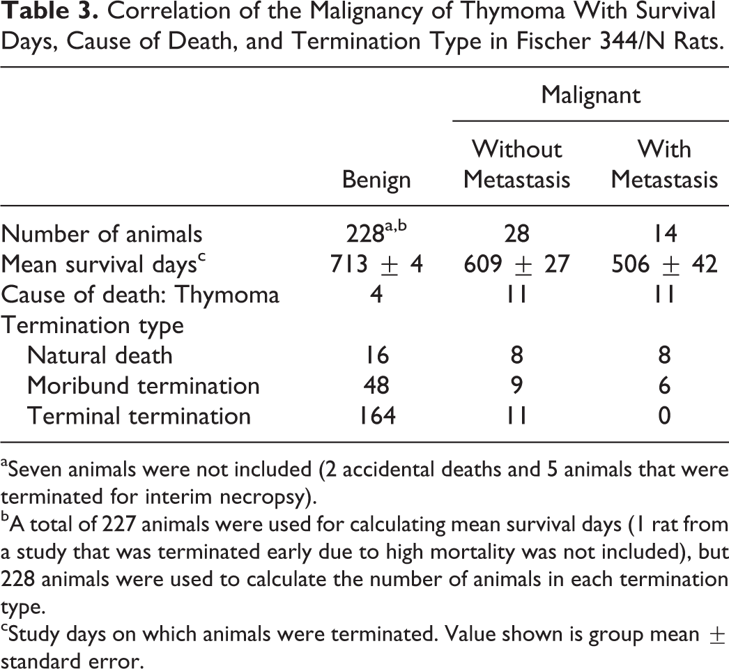
aSeven animals were not included (2 accidental deaths and 5 animals that were terminated for interim necropsy).
bA total of 227 animals were used for calculating mean survival days (1 rat from a study that was terminated early due to high mortality was not included), but 228 animals were used to calculate the number of animals in each termination type.
cStudy days on which animals were terminated. Value shown is group mean ± standard error.
Sprague Dawley Rats
Of 13,525 SD rats used in long-term studies, 10 thymomas recorded in the NTP database were reviewed. None were reported in any of the 7,411 SD rats from short-term studies. Thymomas in SD rats were rare occurrences (10/13,525; 0.07%) in this review, and no occurrences of this tumor in this strain were found to be associated with treatment by test articles in these studies. Of the 10 thymomas recorded in SD rats, 7 (70%) were recorded in females and 3 (30%) were recorded in males, but due to the higher number of females used in NTP studies, the overall incidence of thymomas in SD rats in long-term studies was 0.10% (7/7,270) in females and 0.05% (3/6,255) in males (Table 1). Benign and malignant thymomas each comprised 50% (5/10) of the thymomas reviewed in this strain. In male rats, 33% (1/3) were malignant, and in female rats, 57% (4/7) were malignant. Of the malignant thymomas, metastasis was observed in the mediastinal lymph node of 1 male, and the metastatic lesion had morphological features of the primary tumor. The 4 malignant thymomas that exhibited no metastases were diagnosed due to unequivocal tissue invasion.
Thymomas in the SD rats from our studies could be divided into 3 categories according to morphological features (Table 2). Most tumors exhibited features of more than 1 pattern, and these were categorized by the predominant pattern. Of the 10 thymomas, 6 (60%) had a predominant pattern of cords and tubules, while 2 (20%) displayed a squamous pattern, and 2 (20%) were categorized as having a spindloid pattern. The single metastatic thymoma exhibited a predominantly squamous pattern.
The single malignant thymoma with metastasis was recorded as the primary cause of death in that animal, and the number of survival days in this animal was less than those of animals with benign thymomas or malignant thymomas without metastasis. There was no significant difference between the survival days of animals with benign thymomas and the survival days of animals with malignant thymomas without metastasis (Table 4).
Correlation of the Malignancy of Thymoma With Survival Days, Cause of Death, and Termination Type in Sprague Dawley Rats.
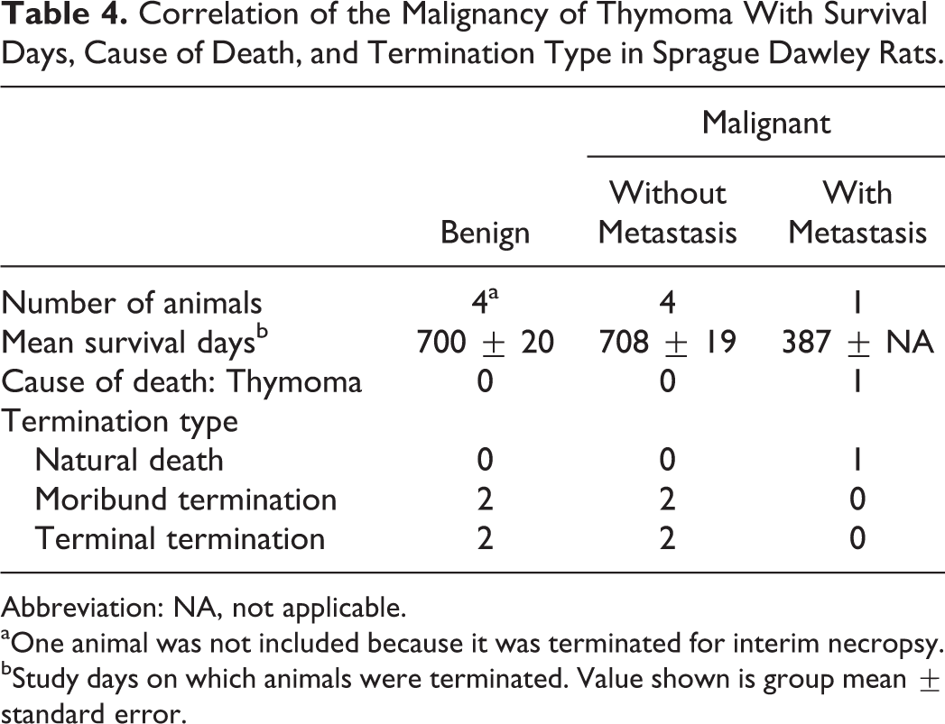
Abbreviation: NA, not applicable.
aOne animal was not included because it was terminated for interim necropsy.
bStudy days on which animals were terminated. Value shown is group mean ± standard error.
Wistar Han Rats
Of the 3,000 WH rats used in long-term studies, 129 thymomas recorded in the NTP database were reviewed. None were reported in any of the 624 WH rats from short-term studies. Thymomas in WH rats were uncommon occurrences (129/3,000; 4.30% incidence) in this review, and no occurrences of this tumor in this strain were found to be associated with treatment by test articles in these studies. Of the 129 thymomas recorded in WH rats, 89 (69%) were recorded in females and 40 (31%) were recorded in males, and the overall incidences of this tumor in WH rats in long-term studies were 2.67% (40/1,500) in males and 5.93% (89/1,500) in females (Table 1). Benign thymomas comprised 98% (126/129) of the thymomas reviewed, and the remaining 2% (3/129) were diagnosed as malignant thymomas. In males, 8% (3/40) of the thymomas were malignant, and 0% (0/89) were malignant in females. Of the malignant thymomas, metastasis was observed in the lung of 1 male, and the metastatic lesion had morphological features of the primary tumor. The 2 malignant thymomas with no metastasis were diagnosed due to unequivocal tissue invasion.
Thymomas in WH rats during this review could be divided into 4 categories based on morphological features (Table 2). Of the 129 thymomas in this strain, most displayed a lymphocyte-rich morphological pattern, with 117 (90.7%) falling into this category. Lymphocyte-rich thymomas were observed with and without a medullary pattern (Figures 10 and 11). Of the remaining thymomas, 6 (4.7%) had a predominant pattern of cords and tubules, 5 (3.9%) had a squamous pattern, and 1 (0.8%) was categorized as having a predominantly spindloid pattern. All 3 malignant thymomas in this strain exhibited a squamous pattern.
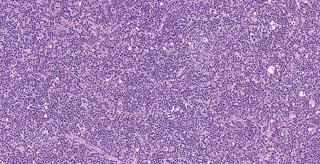
Benign thymoma from a female Wistar Han rat from a long-term study. There is a predominance of small lymphocytes, which partially obscure the underlying neoplastic epithelial cells. Hematoxylin and eosin.
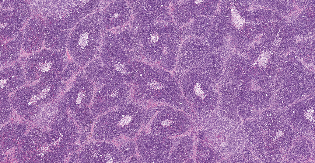
Benign thymoma from a female Wistar Han rat from a long-term study. This lymphocyte-rich thymoma has a medullary pattern, consisting of numerous lymphocytes separated into lobules by fibrous trabeculae and surrounding central foci of large pale-staining cells. Hematoxylin and eosin.
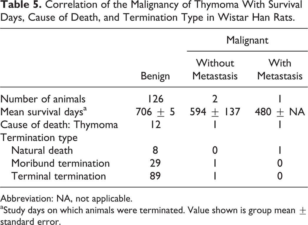
Thirteen benign thymomas, 1 malignant thymoma without metastasis, and 1 malignant thymoma with metastasis were recorded as the primary cause of death. The survival days of animals with malignant thymomas were less than those of animals with benign thymomas (Table 5).
Correlation of the Malignancy of Thymoma With Survival Days, Cause of Death, and Termination Type in Wistar Han Rats.
Abbreviation: NA, not applicable.
aStudy days on which animals were terminated. Value shown is group mean ± standard error.
Wistar Outbred Rats
Male WO rats were utilized for long- and short-term NTP studies associated with only 1 chemical. There were no female rats of this strain used in NTP studies. Of the 200 WO rats used in long-term studies, 4 thymomas recorded in the NTP database were reviewed. None were reported in any of the 60 WO rats used in short-term studies. Thymomas in WO rats were uncommon occurrences (4/200; 2.0% incidence) in this review (Table 1), and no occurrences of this tumor in this strain were found to be associated with treatment by test articles in these studies. Benign thymomas comprised 75% (3/4) of the thymomas reviewed, and the single malignant thymoma comprised 25% (1/4). Metastasis was not observed in the malignant thymoma, and a designation of malignancy was based on unequivocal tissue invasion.
The 4 thymomas in WO rats during this review could be divided into 2 categories based on morphological features, with 3 (75%) displaying a predominant pattern of cords and tubules and 1 (25%) exhibiting a squamous pattern, and this was the single malignant thymoma recorded in this strain (Table 2).
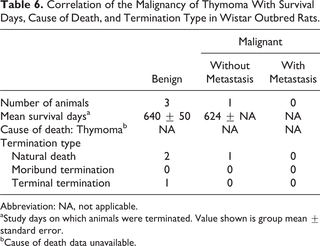
The primary cause of death data was unavailable for the 3 natural death WO rats in this review. The survival days of the single animal with a malignant thymoma were comparable to the survival days of those animals with benign thymomas (Table 6).
Correlation of the Malignancy of Thymoma With Survival Days, Cause of Death, and Termination Type in Wistar Outbred Rats.
Abbreviation: NA, not applicable.
aStudy days on which animals were terminated. Value shown is group mean ± standard error.
bCause of death data unavailable.
Discussion
Thymomas are neoplasms of thymic epithelial cell origin which contain variable numbers of non-neoplastic lymphocytes. Thymomas appear grossly as a firm, smooth mass in the anterior mediastinum, and compression of the surrounding tissues may or may not be present, depending on the size of the tumor. These tumors are rare in humans and most lab animals, with the exception of the Praomys (Mastomys) natalensis, 14 also known as the multimammate rat or common African rat; WH rats 10 ; several inbred strains of rats (BUF/Mna, 15 WAB, 16 and W/Nhg 17 ); European hamsters 18 ; and transgenic mice (SV40T 19 and E2F2 20 ). In the Fischer 344/N rat, thymomas are rare and are said to occur with no apparent sex difference, 8 and in this review, there were slightly more thymomas observed in male F344/N rats than in females. It has been previously reported that thymomas are relatively common in WH rats and occur more often in females, 21 and the finding of a higher incidence in females was supported in this review. Thymomas in SD rats have been found to occur more often in females, 21 and this finding was supported in this review.
There is no meaningful established classification system of thymomas in veterinary medicine, whereas in human medicine, there is the widely used classification system established by the World Health Organization (WHO). 22 Nevertheless, the WHO system has been adapted for use in one of the largest hamster thymoma papers to date. 18
Diagnostic paradigms have been described for rat thymomas, including those dividing them according to the proportions of epithelial and lymphoid cells, 1 as well as divisions based on thymomas with and without medullary differentiation. 2,3 Although understandable and usable, these paradigms do not seem to add significantly to our ability to interpret the rat thymoma in relation to toxicologic or biological significance.
The thymomas in F344/N, SD, WH, and WO rats in this review were variable in microscopic appearance and were divided into 6, 3, 4, and 2 categories, respectively, based on morphological patterns (see Table 2). Although many tumors displayed a mixture of more than 1 morphologic pattern, they were categorized by the predominant pattern. A small number of thymomas in F344/N rats were categorized as having a myoid or neuroendocrine pattern. The thymic medulla normally contains a variety of cell types, including myoid cells and neuroendocrine cells, 23 and it is possible that these cell types were present in thymomas categorized as having a predominantly myoid or neuroendocrine pattern. Thymomas with cords/tubules, spindloid, and papillary patterns were relatively common in most strains and tended to be classified as benign. While thymomas in WH rats could be separated into 4 patterns, the majority were categorized as lymphocyte rich, a pattern that was not observed in the other strains. Despite the presence of large numbers of lymphocytes that often obscured the underlying neoplastic epithelial cells, these were nevertheless considered epithelial cell neoplasms. The immunohistochemical epithelial cell marker, cytokeratin-18, has been useful in demonstrating the large number of epithelial cells that can be difficult to appreciate amid the dense non-neoplastic lymphocyte population when relying solely upon an H&E stain. 24 The lymphocyte-rich thymomas were present with and without medullary differentiation, and all were considered benign. All 3 malignant thymomas in WH rats and the 1 malignant thymoma in a WO rat were categorized as having a squamous pattern, and thymomas with a squamous pattern also tended to be malignant in F344/N and SD rats. Although a thymic origin was considered likely for the tumors with a squamous pattern, an ultimobranchial tissue origin could not be ruled out. Despite these tendencies, separating thymomas into several subclassifications appears to have limited scientific relevance in terms of biological behavior, and it seems adequate to designate them only as benign or malignant until new information is obtained.
Classifying thymomas as benign or malignant traditionally has been based on cytologic features and biological behavior such as invasion and metastasis, as discussed in the 2 volumes of International Classification of Rodent Tumors. 25,26 Although the recommendations from the International Harmonization of Nomenclature and Diagnostic Criteria are not yet published, the information accessible through goRENI suggests that the primary means of differentiating malignant from benign thymomas is by the presence of marked invasion of adjacent tissues and/or metastases in malignant thymomas. 5,6 During this review, there was little evidence that cytologic features typically associated with malignancy were necessarily correlated with invasion or metastasis; however, there was some indication that morphology influenced behavior.
In conclusion, no occurrences of this neoplasm were reported to be related to exposure to any test articles. Thymomas with cords/tubules, spindloid, and papillary patterns were more common and more likely to be benign. Lymphocyte-rich thymomas were common and were only observed in WH rats, and all were considered benign. Thymomas with a squamous pattern had a slight tendency to be malignant. Morphological patterns could be more relevant to biological behavior than cytological features.
Footnotes
Acknowledgments
The authors would like to thank Beth Mahler, Eli Ney, Maureen Puccini, and Emily Singletary for their expert technical assistance with the acquisition of images and the compilation of figures and image plates. The authors would also like to thank Julie Foley and Natasha Clayton for their assistance with immunohistochemistry.
Declaration of Conflicting Interests
The author(s) declared no potential, real, or perceived conflicts of interest with respect to the research, authorship, and/or publication of this article.
Funding
The author(s) disclosed receipt of the following financial support for the research, authorship, and/or publication of this article: This work was supported by the Intramural Research Program of the NIH, National Institute of Environmental Health Sciences, Intramural Research project ZIA ES103319-0, and Contract HHSN273201500013C, performed for the National Toxicology Program, National Institute of Environmental Health Sciences, and National Institutes of Health.