
Abstract
The authors compared the mortality and cardiac biomarker responses in three outbred stocks of Sprague Dawley rats (CD/IGS, Sasco, Harlan) treated with isoproterenol hydrochloride. Cardiac injury was confirmed by histologic evaluation, and increases in cardiac troponin I concentration in serum were measured by two methods. CD/IGS rats had a higher incidence and earlier mortality compared with Sasco or Harlan rats. Harlan rats had lower severity scores for cardiomyocyte degeneration/necrosis compared with the other stocks. Post–isoproterenol treatment cardiac troponin I concentrations were greater in CD/IGS and Sasco rats compared with Harlan rats. Concentrations of cardiac troponin T followed a similar pattern to that of cardiac troponin I in rats treated with isoproterenol. Myosin, light chain 3 concentrations increased in all rats treated with isoproterenol, but there was no difference between the three stocks in the magnitude or pattern of the dose response. Increases in fatty acid binding protein 3 concentrations were detected in only the highest dose group at the earliest timepoint postdose for all three stocks of rats. Results of these studies illustrate the need for investigators to recognize the potential differences in response between stocks of Sprague Dawley rats treated with cardiotoxicants or novel chemical entities.
Keywords
Introduction
Measurement of cardiac troponin (cTn) concentration in serum or plasma to diagnose and monitor cardiac injury is an accepted practice in human and veterinary medicine (Adin et al. 2006; Burgener et al. 2006; Collinson, Boa, and Gaze 2001; Feng et al. 2005; Gaze and Collinson 2005; Herman et al. 2006; O’Brien 2006; O’Brien et al. 2006). Although initially employed as the gold standard for the diagnosis of acute coronary syndrome and myocardial infarction in people (Antman et al. 2000), quantitation of this intracellular globular protein released from injured cardiac myocytes has proven helpful in the diagnosis of cardiac injury due to a variety of causes (NACB Writing Group et al. 2007; O’Brien 2006). Cardiac troponin concentration has been termed the most effective translational safety biomarker for cardiotoxic injury that results in disruption of cardiac cell membrane integrity (O’Brien 2008), and inclusion of cTn concentration as an indication of cardiac injury in human clinical trials is supported by the European Society/American College of Cardiology (Thygesan et al. 2007).
The complete analytical and biologic validation of a new biomarker for cardiac injury is an arduous task that may require years of work and the cumulative input of many investigators. Members of the nonprofit Health and Environmental Sciences Institute (HESI) Cardiac Troponins Biomarker Working Group reviewed nine commercially available assays used to measure cTn concentration, identified differences in species cross-reactivity for antibodies used in cTn assays, and noted that there were important differences in precision and dynamic range of the assays. Several of these assays worked very well in people, monkeys, dogs, and rats, while a few assays were viewed as having unsatisfactory performance, particularly in rats (Apple et al. 2005, 2007, 2008; Pettit et al. 2007).
The HESI group also engages in studies of the biologic validation or performance of cTn assays in various animal models (Clements et al. 2010) and gathers scientists of differing focus and expertise to engage in discussions of issue resolution regarding cardiac safety (Berridge et al. 2009; Pettit, Berridge, and Sarazan 2010). In a recent workshop held in Washington, D.C. (Berridge et al. 2009), cardiologists, clinical chemists, veterinary clinical and anatomic pathologists, physiologists, toxicologists, and regulatory scientists agreed on the need for more information describing the differences in cTn concentrations in various demographic and ethnic groups of people and strains and stocks of laboratory animals. This request, coupled with our observations of differences in response of certain rats to various cardiotoxicants used in internal studies, compelled us to explore differences in mortality and cTn response among three stocks of Sprague Dawley (SD) rats to isoproterenol (iso).
The best approach for designing and implementing cardiac biomarkers in efficacy and safety studies in drug development is controversial and varies greatly between research organizations. Some of the current trends in cardiac biomarker research, as evidenced by changes in cTn assays, include lowering the limit of sensitivity of the assays and decreasing the volume of sample required for cTn assays used in both people and laboratory animals (Schultze et al. 2008, 2009; Todd et al. 2007; Wu et al. 2006, 2009). Another helpful approach some groups advocate is the inclusion of a panel of organ-specific biomarkers (Rebar and Boon 1983) determined at various times throughout the length of a study. Our planned study of the cTn response in three stocks of SD rats afforded us an additional opportunity to evaluate the clinical utility of an investigational panel of cardiac biomarkers under development within our organization. This panel consisting of cardiac troponin I (cTnI; TnnI3), cardiac troponin T (cTnT; TnnT2), fatty acid binding protein 3 (Fabp3), and myosin, light chain 3 (My13) is measured using a MesoScale Discovery (MSD) electrochemiluminescent platform. This panel of cardiac biomarkers has already passed a rigorous paradigm of analytical validation and awaits clinical evaluation of its usefulness in drug development. Therefore, the objectives of this study were to evaluate the effects of a single subcutaneous dose of isoproterenol hydrochloride (iso) on three different stocks of SD rats through the assessment of mortality and microscopic evaluation of the hearts and to correlate these findings with the concentrations of cTnI in serum using a well-validated, commercially available method of cTn quantitation (Access Immunoassay System; Beckman Coulter Inc., Brea, CA; Apple et al. 2008). In addition, we sought to evaluate the utility of an investigational MSD panel of cardiac biomarkers (cTnI, cTnT, Fabp3, and My13 concentrations) by comparing the results of this panel of serum biomarkers with the Access cTnI concentration and microscopic heart lesions reported in the different stocks of SD rats.
Materials and Methods
Institutional Compliance Statement
All animal studies described in this publication were approved by the Eli Lilly and Company Study Design and Approval Committee. Studies were contracted to MPI Research, Inc. (Mattawan, MI). MPI Research, Inc. is an animal facility accredited by the Association for Assessment and Accreditation of Laboratory Animal Care International, and these studies were approved by the Institutional Animal Care and Use Committee at MPI Research, Inc.
Animals
Male CD/IGS Sprague Dawley rats (CD/IGS SD; n = 100) and male Sasco Sprague Dawley rats (Sasco SD; n = 100), approximately 8.5 to 9 weeks of age and approximately 250 to 350 g body weight, were obtained from Charles River Laboratories (Portage, MI). Male Hsd Sprague Dawley rats (Hsd SD; n = 100) approximately 8.5 weeks of age and approximately 250 to 350 g body weight were obtained from Harlan Laboratories (Indianapolis, IN).
Rats were individually housed in suspended, stainless-steel, wire-mesh–type cages in an environmentally controlled room maintained between 64°F and 79°F and between 30 and 70% humidity with a 12-hr light-dark cycle. Food (Lab Diet Certified Rodent Diet #5002; PMI Nutrition International, Inc., Henderson, Co) and drinking water were supplied ad libitum. Studies began after an acclimation period of at least 1 week during which the animals were observed daily for clinical signs of disease or poor health.
Isoproterenol Formulation and Administration
Isoproterenol HCl (iso; catalog #16504; Sigma-Aldrich, St. Louis, MO) was provided at a theoretical purity of 85.3% and stored at –10 to –30°C dessicated. Test article formulation concentrations were calculated as the free base using a correction factor of 1.1723%. No adjustments were made for purity. The iso was mixed with saline (0.9% sodium chloride for injection, USP), and preparations were made as a serial dilution from a single stock solution to achieve the desired concentrations. Fresh formulations were prepared for each concentration on the day of dosing.
The iso preparations were administered once as a subcutaneous bolus injection, between the skin and underlying layers of tissue in the scapular region on the back of each rat. Individual doses were based on body weights measured on the day prior to dosing and administered in a 1-ml/kg dose volume.
Selection of Animals
All rats with body weights that fell within 20% of the mean for the entire group were assigned to control or treatment groups using a simple randomization procedure to minimize between-group differences in body weight. Body weights ranged from 286 to 409 g at randomization. Rats were implanted with a microchip bearing a unique identification number.
Experimental Design
Groups of 20 rats (CD/IGS SD, Sasco SD, or Hsd SD) were given a single subcutaneous dose of 0.00, 0.01, 0.03, 0.10, or 0.30 mg iso/kg. Groups of 5 to 10 rats per stock per time point were euthanized at 4, 24, or 48 hr following dose administration.
Blood Collection and Processing
Immediately preceding euthanasia and necropsy, blood samples were obtained via the retro-orbital sinus under isoflurane anesthesia. Whole blood (2 ml) was collected and allowed to clot at room temperature for approximately 45 min before being spun in a centrifuge to obtain serum. Serum samples were stored frozen at –70°C in multiple polypropylene tubes until thawed for biomarker analysis.
Mortality, In-Life (Cageside) Observations, and Body Weights
All rats were observed twice daily for morbidity, mortality, signs of injury, and access to food and water. Body weights were recorded upon arrival and 1 day prior to dosing.
Necropsy and Histopathologic Examination
Rats were anesthetized by isoflurane inhalation and euthanized by exsanguination. Hearts were excised and separated from the pericardium, and the chambers were left unopened prior to immersion in 10% neutral buffered formalin for a minimum of 48 hr. Hearts were trimmed in the longitudinal plane (apex to base) to open all chambers. Both halves of each heart were processed routinely and embedded in paraffin for histopathologic examination. Sections were cut at 5 µm and stained with hematoxylin and eosin. The resulting histologic slide contained two near-midline longitudinal sections of heart, providing representation of all four chambers, the ventricular septum, both ventricular free walls, and both atria. Tissues were evaluated by a single board-certified veterinary pathologist at Eli Lilly and Company (Indianapolis, IN).
Two longitudinal sections were examined from each heart. Severity grading of the cardiac lesions in this study was based on a subjective assessment of the approximate percentage of the total myocardial area within the distal half of the ventricles that was occupied by changes related to cardiomyocyte degeneration/necrosis. The longitudinal sections obtained from both halves of the bisected hearts were considered in determining the severity grade for each rat. The following grading scale was used: minimal = <10%, slight = 10 to <25%, moderate = 25 to <50%, marked = 50 to <75%, and severe = >75% of distal ventricular myocardium altered by changes related to cardiomyocyte degeneration/necrosis. These changes included degenerate/necrotic cardiomyocytes, interstitial and/or intramyocellular inflammatory infiltrates, and/or interstitial fibroplasia.
cTnI Concentration
Sera from control vehicle- and iso-treated rats were analyzed for the concentration of cTnI using the Access Immunoassay System (Beckman Coulter Inc., Brea, CA). Precision, accuracy, and species cross-reactivity of the Beckman Access Immunoassay have been described, and the performance of this assay was compared with several other commercially available assays for cTn quantitation (Apple et al. 2008). The lower limit of quantitation (LLOQ) and upper limit of quantitation (ULOQ) of this assay in our laboratory were 0.01 and 90.00 ng/ml, respectively.
Muscle Injury Panel Analysis
Serum samples from vehicle control– and iso-treated rats were analyzed for concentrations of cTnI, cTnT, Fabp3, and My13 using an MSD (Gaithersburg, MD) electrochemiluminescent multiplex immunoassay (#K15181). The assay was performed according to the manufacturer’s instructions and validated in accordance with “fit-for-purpose” biomarker validation recommendations of Lee and colleagues (2006) for precision, accuracy, and recovery. The LLOQ and ULOQ of the assays were, respectively, as follows: cTnI, 0.24 and 250.00 ng/ml; cTnT, 1.24 and 312.4 ng/ml; Fabp3, 0.48 and 62.40 ng/ml; and My13, 0.28 and 550.00 ng/ml. Serum samples were diluted 1:4 in diluent provided by the manufacturer, and 0.025 ml of serum was added to the plate in duplicate wells.
Bioanalytical Determination of Iso Concentration
Serum samples from vehicle control– and iso-treated rats were analyzed for iso concentrations by liquid chromatography–tandem mass spectrometry (LC/MS/MS). Serum samples (50 µl) were processed by protein precipitation with 100 µl acetonitrile using metoprolol (Sigma Aldrich, cat #M5391) added to each sample prior to extraction as an internal standard. After centrifugation, a 100-µl aliquot of the supernatant was dried by vacuum centrifugation and reconstituted in initial mobile phase (0.1% formic acid) for injection. The liquid chromatography system consisted of a CTC Analytics HTS Pal autosampler (Leap Technologies, Carrboro, NC) and a Shimadzu liquid chromatograph equipped with LC-20AD pumps (Shimadzu Scientific Instruments, Columbia, MD). Gradient elution involving a binary mixture of 0.1% formic acid and acetonitrile occurred at a flow rate of 0.3 ml/min using a Varian Monochrome C18 column (5 µm, 100 × 2.0 mm). Online mass spectrometric detection was conducted by a Quantum Ultra triple quadrupole (ThermoFisher Scientific, San Jose, CA) in the selected reaction monitoring mode (SRM) under positive ion electrospray ionization. The SRM transitions monitored for iso and metoprolol were m/z 212.1 → 107.1 and m/z 268.1 → 133.1, respectively. Calibration curves were prepared in control rat serum using iso. Spiking solutions were prepared in 0.1% formic acid without correcting for compound potency. During analysis, all study samples were bracketed by duplicate 8-point standard curves covering a range of 31 to 8000 pg/ml. Peak integration and linear regression (1/X2 weighting) were performed using Xcalibur software v1.
Statistics
Design
This study included 15 groups of rats (5 dose levels of iso at 0 [vehicle control], 0.01, 0.03, 0.1, and 0.3 mg iso/kg used in three stocks of SD rats). Microscopic evaluation was performed on hearts collected from rats that survived to study termination as described previously. For statistical analysis, a numeric score was assigned to each histologic grade of the cardiac lesion severity: 0 for none, 1 for minimal, 2 for slight, 3 for moderate, 4 for marked, and 5 for severe. Since all animals survived to at least 4 hr after iso dose but not necessarily to 24 and 48 hr after iso dose, no statistical comparisons were attempted to evaluate the time effect on the results.
Analysis
For histologic scores of cardiac lesion severity, many identical scores were observed within each combination of rat stock and dose of iso. The assumptions needed to apply normal analysis of variance models could not be confirmed. Therefore, a one-factor analysis was performed for each dose level at each time point on the rank-transformed scores. These analyses were followed by pairwise comparisons among the stocks of SD rats.
For biomarkers measured in serum, statistical analyses were performed on 4-hr post–iso dose data for each parameter separately. The cTnI, cTnT, Fabp3, and My13 concentrations from each animal were fitted to a two-factor analysis of variance model using the MIXED procedure in SAS (SAS Institute Inc. 2004). The two factors were stock and dose level. This model included an interaction term between the two main factors and was followed up with pairwise comparisons among stocks or dose levels to help identify the source of significant effects. The Shapiro-Wilk test (Shapiro and Wilk 1965) for normality and Levene’s test (Levene 1960) for homogeneity of variance were conducted to check the validity of assumptions. As a result of these checks, all biomarker data were subjected to a base 10 logarithmic transformation prior to statistical analyses. Descriptive statistics including mean, standard deviation, and sample size for each stock by group combination were calculated for each 4-, 24-, and 48-hr post-iso dose. For statistical analysis and calculation of fold-change from vehicle control, a numerical value defined as 0.01 subtracted from the assay LLOQ value for the analyte was substituted for biomarker results that were reported as below the lower quantifiable limit. For example, if the reported result for MSD cTnI was below the limit of quantitation, and the LLOQ for the assay is 0.24, the value used for statistical evaluation would be 0.24 – 0.01 = 0.23. Shapiro-Wilk’s test and Levene’s test were performed at the significance level of .01. All other tests were performed at the significance level of .05.
Results
In-Life (Cageside) Observations
Although several rats were found dead acutely posttreatment with iso, no obvious signs of ill-thrift or distress were reported at any time prior to death.
Mortality
All three rat stocks had treatment-related early deaths that occurred between 24 and 48 hr following doses of ≥0.1 mg iso/kg. The CD/IGS SD had a greater incidence of mortality (4 rats given 0.3 mg iso/kg and scheduled to be killed at 24 hr, 6 rats given 0.1 mg iso/kg and scheduled to be killed at 48 hr, and all 10 rats given 0.3 mg/kg and scheduled to be killed at 48 hr) compared with the Hsd SD rats (2 rats given 0.1 mg iso/kg and scheduled to be killed at 48 hr and 8 rats given 0.3 mg iso/kg and scheduled to be killed at 48 hr) or the Sasco SD (2 rats given 0.1 or 0.3 mg iso/kg and scheduled to be killed at 48 hr) given the same dose of compound. The cumulative incidence of treatment-related early deaths was highest for CD/IGS SD (20), followed by Hsd SD (10) and Sasco SD (4). The CD/IGS SD stock had the earliest occurrence of unscheduled deaths, with mortality occurring between 4 and 24 hr in the group given 0.3 mg iso/kg.
Microscopic Findings
For all three stocks of SD rats, iso-induced cardiac lesions were characterized by cardiomyocyte degeneration/necrosis oriented primarily within the distal apex of the left ventricle, with less frequent involvement of the distal right ventricular free wall and infrequent involvement of the proximal ventricles toward the heart base. In addition, within the distal left ventricle, lesions tended to occur around the ventricular chamber.
The microscopic features of cardiomyocyte degeneration/necrosis varied in relation to the time of death or sacrifice after dosing (Table 1 ). At 4 hr, cardiomyocyte changes were peracute, characterized by cellular swelling with sarcoplasmic hypereosinophilia, hyalinization, and nuclear pyknosis or fragmentation. An associated leukocyte response was either absent or characterized by only minimal interstitial (pericellular) or intramyocellular neutrophil infiltrates. At 24 hr, changes were acute, characterized by more extensive sarcoplasmic fragmentation and increasingly intense interstitial and intramyocellular mixed leukocyte infiltrates dominated by macrophages. At 48 hr, changes were usually subacute, characterized by intense intramyocellular and interstitial macrophage infiltrates and an increasingly prominent stromal cell response including cellular hypertrophy and increased numbers of mitotic figures.
Compound-Related Changes in Three Stocks of Sprague Dawley Rats following a Single Dose of Isoproterenol Hydrochloride
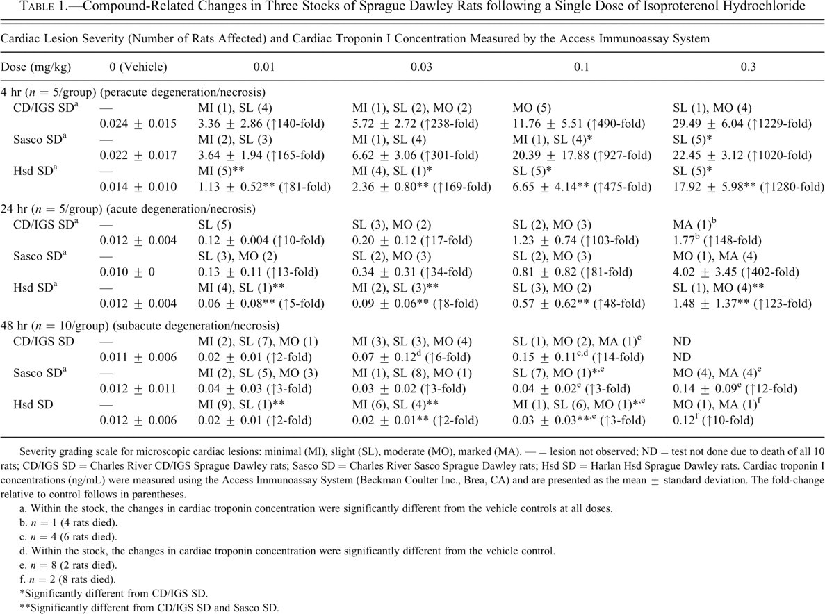
Severity grading scale for microscopic cardiac lesions: minimal (MI), slight (SL), moderate (MO), marked (MA). — = lesion not observed; ND = test not done due to death of all 10 rats; CD/IGS SD = Charles River CD/IGS Sprague Dawley rats; Sasco SD = Charles River Sasco Sprague Dawley rats; Hsd SD = Harlan Hsd Sprague Dawley rats. Cardiac troponin I concentrations (ng/mL) were measured using the Access Immunoassay System (Beckman Coulter Inc., Brea, CA) and are presented as the mean ± standard deviation. The fold-change relative to control follows in parentheses.
a. Within the stock, the changes in cardiac troponin concentration were significantly different from the vehicle controls at all doses.
b. n = 1 (4 rats died).
c. n = 4 (6 rats died).
d. Within the stock, the changes in cardiac troponin concentration were significantly different from the vehicle control.
e. n = 8 (2 rats died).
f. n = 2 (8 rats died).
*Significantly different from CD/IGS SD.
**Significantly different from CD/IGS SD and Sasco SD.
Although of similar microscopic character, the severity of cardiac lesions varied between stocks of SD rats (Fig. 1 ). At 4 hr, Hsd SD rats had significantly lower microscopic severity scores for cardiac lesions than CD/IGS SD rats given all doses of iso. In addition, Hsd SD rats given 0.01 mg iso/kg had lower severity scores than the Sasco SD rats. Sasco SD rats given 0.1 or 0.3 mg iso/kg also had significantly lower severity scores than the CD/IGS SD rats given the same dose of compound. At 24 hr after iso dose, Hsd SD rats had significantly lower cardiac lesion severity scores than the other two stocks for all but the 0.1 mg iso/kg groups. At 48 hr, Hsd SD rats again had significantly lower severity scores than the other two stocks given 0.01 or 0.03 mg iso/kg. Both Hsd SD and Sasco SD rats given 0.1 mg iso/kg had significantly lower severity scores than CD/IGS SD. No statistical comparison was made for the three stocks of SD rats given 0.3 mg iso/kg and killed at 48 hr postdose due to the death of all CD/IGS SD rats given this dose.
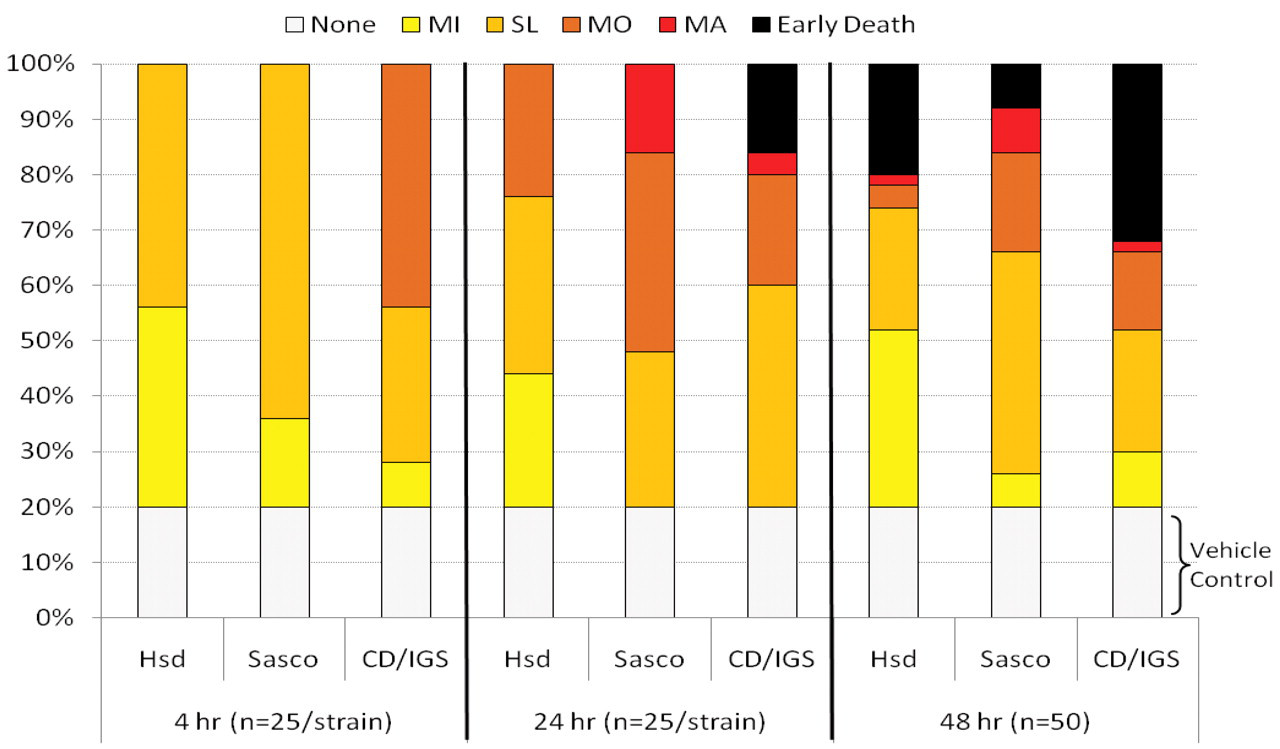
Distribution of histologic cardiac lesions. Numbers of animals with histologic cardiac lesions (degeneration/necrosis) or early death reported as a percentage of total treated animals for each time point (4, 24, and 48 hr) and each rat stock after a single dose of isoproterenol hydrochloride in Sprague Dawley rats. There were no histologic findings (None) in the vehicle control groups of rats, which represented 20% of each group at each time point. Score abbreviations: MI = minimal (yellow); SL = slight (gold); MO = moderate (orange); MA = marked (red); Early Death (black).
Cardiac Troponin I Concentration (Beckman Access)
The concentrations of the cTnI in serum were extremely low in the vehicle control groups in all three stocks of SD rats and well within the reference intervals for CD/IGS SD rats (0.01-0.07 ng/ml) established in our laboratory. Thus, no statistical evaluation was performed for the stock difference in the vehicle control rats. Compound-related findings consisted of dose-dependent increases in the concentration of cTnI in the CD/IGS SD, Sasco SD, and Hsd SD rats (Table 1). For all three stocks of rats at each dose, cardiac injury as measured by increases in the concentration of cTnI was greatest at 4-hr post-iso administration and progressively decreased at 24 and 48 hr.
For all iso dose levels at 4 and 24 hr, Hsd SD rats had significantly lower cTnI concentrations (p < .05) than the other two stocks of SD rats. At 48 hr after iso administration, most of the cTnI concentrations in all three stocks of SD rats given up to 0.1 mg iso/kg were low and similar to those of vehicle-treated controls. No statistical comparisons of stock differences were made at the 48-hr time point.
MSD Muscle Injury Panel Results
cTnI Concentration (Figs. 2A and 2B)
The concentrations of the cTnI in serum were low in the vehicle control groups in all three stocks of SD rats. Dose-related increases in cTnI concentrations measured using the MSD assay were observed in the serum of all three rat stocks at 4 hr after the single dose of iso, with the Sasco SD rats often having the highest cTnI concentrations. However, iso treatment–related changes in cTnI concentrations were more pronounced in the CD/IGS SD rat stock with mean fold-change from control values increasing from threefold at 0.01 mg/kg to 34-fold in the 0.3-mg/kg group at 4 hr postdose.
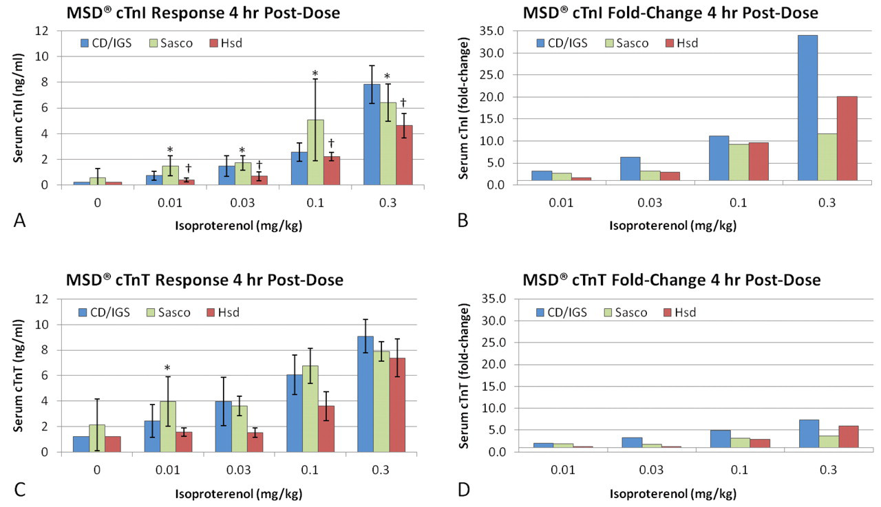
Rat stock comparison of MesoScale Discovery (MSD) biomarker concentration and fold-change results. Data are presented as mean ± standard deviation. n = 5/group. The most pronounced biomarker responses on the MSD multiplex panel were observed at 4 hr after isoproterenol hydrochloride (iso) dose. Data from 24 and 48 hr are not shown. Statistical evaluations were not performed on the fold-change data. MSD biomarker fold-change responses were reported as group mean fold-change from group mean vehicle control at 4 hr postdose. (A) Iso induced a generally dose-responsive release of cardiac troponin I (cTnI) in the serum of all three rat stocks, with Harlan Hsd Sprague Dawley rats (Hsd SD) responding with the lowest concentrations of cTnI. (B) The Charles River CD/IGS Sprague Dawley (CD/IGS SD) rat stock responded with the greatest magnitude of cTnI change when evaluated as mean fold-change from vehicle controls. (C) Iso induced a generally dose-responsive release of cardiac troponin T (cTnT) in the serum of all three rat stocks. Similar responses were observed in the Charles River Sasco Sprague Dawley (Sasco SD) and CD/IGS SD rats, whereas the Hsd SD rats had lower cTnT concentrations than the other two stocks. (D) The CD/IGS SD stock produced a larger fold-change response as compared with Sasco SD and Hsd SD rats. The magnitude of cTnT fold-change was markedly lower than the cTnI fold-change response in all three stocks. *Significantly different from CD/IGS SD (p < .05). †Significantly different from CD/IGS SD and Sasco SD (p < .05).
Statistical comparisons of the cTnI values obtained using the MSD assay among the stocks were performed only for the 0.01- to 0.3-mg/kg groups at the 4-hr time point because many values were below the LLOQ for each group at the 24- and 48-hr time points. Values from the vehicle control animals at each time point were not used in the comparisons because they also were below the LLOQ. In general, the Hsd SD rats had lower cTnI concentrations than the CD/IGS SD and Sasco SD rats (p < .05).
cTnT Concentration (Figs. 2C and 2D)
The concentrations of cTnT in serum were low in the vehicle control groups in all three stocks of SD rats. Variability in concentrations of cTnT in the vehicle controls was relatively greater in Sasco SD rats compared with CD/IGS SD or Harlan SD rats. When administered 0.01, 0.03, 0.1, or 0.3 mg/kg iso, the Hsd SD rats generated the lowest cTnT concentrations in serum as compared with Sasco SD or CD/IGS SD rats (p < .05) at 4 hr postdose, with some values falling below the LLOQ. Generally, dose-related increases in cTnT concentrations were observed at all doses for all rat stocks. Sasco SD rat cTnT concentrations were significantly greater (p < .05) than, or similar to, the CD/IGS SD at 0.01, 0.03, and 0.1 mg iso/kg. However, the dose-related fold-change increase was actually greater for the CD/IGS SD (7.4-fold) than the Sasco SD (3.7-fold) rats. cTnT concentrations were similar across all stocks administered 0.3 mg/kg iso, with the magnitude of fold-change from control slightly higher in the CD/IGS SD stock.
Statistical comparisons of the MSD cTnT values among the stocks were performed only for the 0.01- to 0.3-mg/kg groups at the 4-hr time point because many values were below the LLOQ for each group at the 24- and 48-hr time points. Values from the vehicle control animals at each time point were not used in the comparisons because they also were below the LLOQ.
Fabp3 Concentration (Figs. 3A and 3B)
Fabp3 concentrations in the serum from the vehicle control groups in all three stocks of SD rats were consistently low and similar to concentrations in the serum of rats treated with 0.01 to 0.1 mg iso/kg. Significant increases in Fabp3 concentrations compared with vehicle controls were detected in the 0.3-mg/kg dose group at 4 hr postdose for all three stocks of SD rats (p < .05). However, no statistically significant stock differences were detected at 4 hr postdose for Fabp3 concentrations. Fold-changes in Fabp3 concentrations in SD rats given iso compared with the respective vehicle control groups were minimal (less than threefold) at all doses and similar between stocks at all time points.
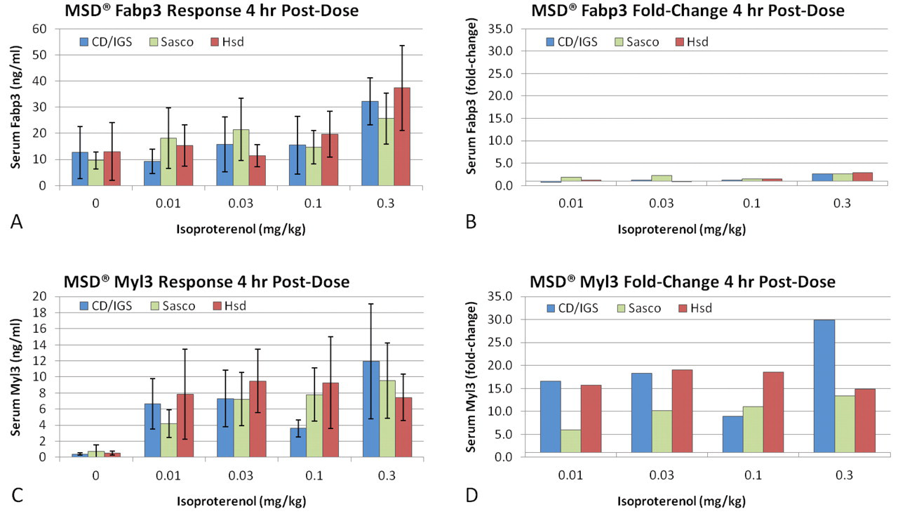
Rat stock comparison of MesoScale Discovery (MSD) biomarker concentration and fold-change results. Data are presented as mean ± standard deviation. n = 5/group. The most pronounced biomarker responses on the MSD multiplex panel were observed at 4 hr after isoproterenol hydrochloride (iso) dose. Data from 24 and 48 hr are not shown. Statistical evaluations were not performed on the fold-change data. MSD biomarker fold-change responses were reported as group mean fold-change from group mean vehicle control at 4 hr postdose. (A) Increases in concentrations of fatty acid binding protein 3 (Fabp3) were detected at 4 hr postdose in the serum of all three stocks of rats at the 0.3-mg/kg dose only (p < .05). However, no statistically significant interstock differences were detected at 4 hr postdose for Fabp3 concentrations. (B) Treatment-related fold changes in Fabp3 compared with the respective vehicle control groups were minimal (less than threefold) and similar between stocks. (C) At 4 hr, iso treatment induced the release of myosin light chain 3 (My13) in all three rat stocks but not generally in a dose-responsive manner. (D) The My13 concentrations were less than fivefold above vehicle control. *Significantly different from Charles River CD/IGS Sprague Dawley (CD/IGS SD) rats (p < .05). †Significantly different from CD/IGS SD and Charles River Sasco Sprague Dawley rats (p < .05).
At 24 hr postdose, no obvious trends were identified for dose effect or stock differences. At 48 hr postdose, Hsd SD rats had greater Fabp3 concentrations (p < .05) than Sasco SD rats, and values were greater than those of the surviving CD/IGS SD rats given up to 0.1 mg/kg iso.
My13 Concentration (Figs. 3C and 3D)
The concentrations of My13 in the serum of vehicle control rats were consistently low among rats within stock groups. At 4 hr after iso dose, all three rat stocks responded to iso treatment with greater than fivefold My13 concentrations above vehicle control in all iso-dosed groups. The 4-hr My13 concentrations were the higher responses compared with 24 or 48 hr posttreatment. No stock effects were detected for My13 concentrations at 4 hr after iso dose. The magnitude of the fold-changes was large but not dose-responsive across stocks. At 24 hr, My13 concentrations in the Hsd SD rats were lower than in the CD/IGS SD rats (p < .05) and Sasco SD (p < .05) rats. However, the magnitude of change from control was generally smaller in the Sasco SD than the Hsd SD or CD/IGS SD rats. Isoproterenol treatment–related increases in My13 concentrations (p < .05) were observed across stocks at 0.01 through 0.3 mg/kg at 4, 24, and 48 hr after iso dose. In contrast to cTnI, cTnT, and Fabp3 concentrations, which were generally similar to the concurrent vehicle controls (Fig. 4 ), serum My13 concentrations were sustained greater than twofold from vehicle control at 24 hr in all stocks.
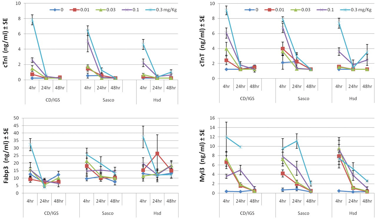
MSD biomarker kinetic response following a single dose of isoproterenol hydrochloride (iso). Samples were collected from individual rats at 4, 24, or 48 hr after a single dose of vehicle (control; 0 mg/kg) or iso (0.01, 0.03, 0.1, or 0.3 mg/kg). Data are presented as mean ± standard error. n = 5/group. The 48-hr time point was not available from the 0.3-mg/kg dose group of Charles River CD/IGS Sprague Dawley rats due to early deaths.
Bioanalytical Determination of Iso Concentration
LC/MS/MS quantitation of iso was conducted on the serum samples collected from the three rat stocks to ascertain whether the observed differences in cardiotoxicity findings could be attributed to differences in drug exposure. Despite the sensitivity of the method used (lower limit of detection ~10 pg/ml), measurable levels of iso were observed only for the highest dose (0.3 mg/kg) at 4 hr. Because of the rapid clearance of iso, which has a reported elimination half-life of less than 10 min in SD rats (Hadwiger et al. 1997), this observation was not unexpected, particularly since the time points and doses were selected to facilitate cardiotoxicity assessment. Table 2 lists the iso concentrations measured in the three SD stocks at 4 hr (0.3 mg/kg). The range in reported concentrations spanned from 119.1 to 548.4 pg/ml, all values readily detected by the method used. According to the mean values reported in Table 2, the CD/IGS SD stock had the lowest exposure and Sasco SD had the highest. Different levels of within-stock variation were also observed for each stock, with Sasco SD exhibiting the highest variation and Hsd SD having the lowest.
Concentrations of Isoproterenol Hydrochloride (Iso) in Serum Determined Using LC/MS/MS
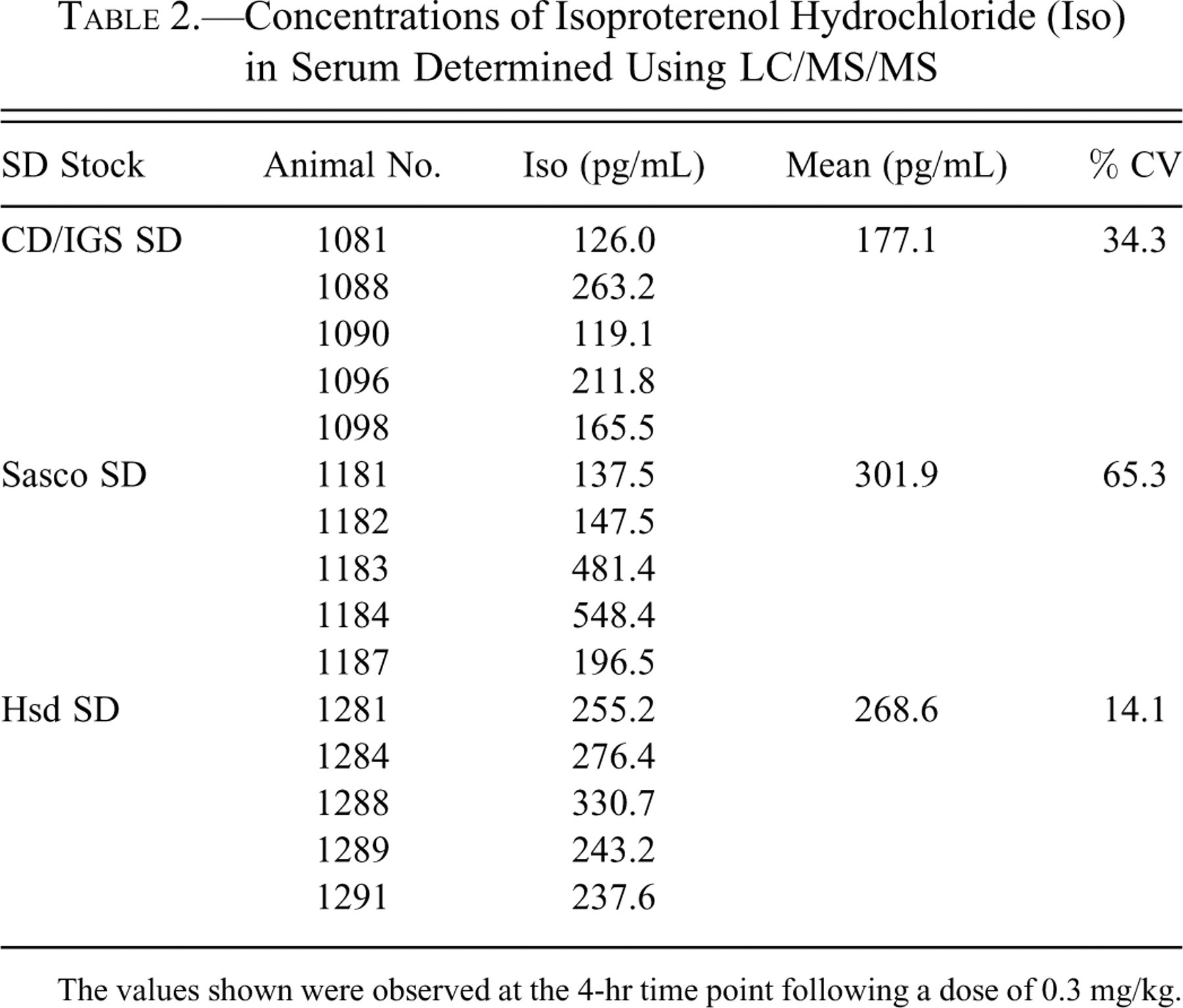
The values shown were observed at the 4-hr time point following a dose of 0.3 mg/kg.
Discussion
The appropriate evaluation and effective use of safety biomarkers in preclinical toxicology studies require consideration of many factors: the specificity and robustness of the biomarker for the indicated toxicity, the analytical specificity and sensitivity of the biomarker assay, and the translation of the biomarker across species, strains, and stocks. Our study employed a combination of well-characterized and investigative biomarkers to monitor cardiac injury. The measurement of the cTnI and cTnT, as specific indicators of myocardial injury is well documented, and increases in the concentration of cTn have been correlated with histopathological findings of cardiac injury in rats (O’Brien 2006, 2008).
Fabp3 is a cytosolic protein that is present in plasma of healthy humans and rodents (Sorichter et al. 1998; Pritt et al. 2008). The concentration of Fabp3 increases in serum or plasma following cardiac damage and is reported to have a kinetic profile similar to that of cTnI, with an early plasma response approximately 0.5 to 2 hr after iso treatment in rats given a single dose of >1 mg isoproterenol/kg (Clements et al. 2010; Tonomura et al. 2009). In some studies, Fabp3 has been reported to be more sensitive than cTns for detection of ischemic injury (Mion et al. 2007; Niizeki et al. 2007). However, Fabp3 is acknowledged as a less specific marker of cardiotoxicity due to the presence of the protein in many other tissues such as skeletal muscle, brain, and kidney (Glatz and van der Vusse 1996; Zhen et al. 2007). Tonomura et al. (2009) hypothesized that the observed increase in Fabp3 concentration with iso treatment is probably related to slow-twitch skeletal muscle toxicity.
My13, also known as myosin light chain 1 (MLC1), is a 23-kDa isoform of one of the subunits of myosin expressed in ventricular and slow-twitch skeletal muscles and is not a specific biomarker for cardiac injury. My13 has an extended kinetic profile compared with cTns (Berna et al. 2007). However, My13 is also less selective than cTnI, and measurement can be affected by skeletal muscle injury (Pritt et al. 2008). When applying a strategy of combining multiple biomarkers into a diagnostic panel, My13, when paired with cTnI and skeletal TnI, assists in the elucidation of the tissue origination of increased My13 concentration.
Iso was chosen for induction of cardiac lesions based on its well-characterized cardiotoxicity. Iso is a potent β-agonist that does not distinguish between β1 and β2 receptors. In the rat, high doses of iso rapidly stimulate β1 and β2 receptors in the heart, inducing an abnormally rapid heart rate, attributed to the β1 activity, and a decreased blood pressure caused by the β2 activity. This results in cardiac tissue anoxia/hypoxia and myocardial necrosis (Rona et al. 1959; York et al. 2007).
The iso-treated rat has been used in cardiovascular research for many years and is an accepted model for cardiac injury (Herman et al. 2006; York et al. 2007; Schultze et al. 2008; Clements et al. 2010). Serum cTnI and cTnT concentrations have been reported to be significantly increased with iso-induced cardiac injury in rats (Bleuel et al. 1995; Bertinchant et al. 2000; Wallace et al. 2004; Schultze et al. 2008). Although a variety of rat strains and stocks are used frequently in cardiovascular toxicity studies, few studies have compared the responses of these various rat strains and stocks to standard doses of cardiotoxicants.
In our study, important differences in mortality, cardiac lesion severity, and cardiac biomarker responses were observed between the three stocks of SD rats treated with iso. Doses of iso were selected to span a range of exposures that would induce no or little toxicity to obvious toxicity with the possibility of mortality and allow a comparative assessment of the response curves for each rat stock. This resulted in mortality at the high doses with variable incidence across stocks. Higher mortality in the CD/IGS SD stock compared with the Sasco SD and Hsd SD stocks was one manifestation of differing sensitivities to iso cardiotoxicity. The exact causes for the differences in mortality and magnitudes of cardiac injury observed between the three stocks of SD rats used in these investigations are not known completely. To test for the possibility that the different stocks exhibited differential exposure to iso, perhaps due to differential dosing, absorption, or metabolism, an iso-selective LC/MS/MS assay was developed (Table 2). Despite the known rapid clearance of iso (Hadwiger et al. 1997), this effort was undertaken to see if an indication of differential exposure could be observed at the earliest time point (4 hr). For the highest dose of iso (0.3 mg/kg), levels well above the lower quantitation limit of the assay (31 pg/ml) were detected at 4 hr. Because the iso concentrations in serum bear no apparent correlation to the observed toxicity findings and biomarker data, our tentative conclusion is that differential exposure was not responsible for the observed toxicity differences between stocks. Additional studies designed using earlier sample collection time points would be needed to definitively explore the relationship between iso toxicokinetics and cardiotoxicity for the SD rat stocks investigated.
We considered other factors that may have affected the rats in the current study and caused the differences in cardiac biomarker results and mortality that we observed. O’Brien et al. (2006) detected age-dependent increases in cTnI concentrations in SD rats. Specifically, 6- and 8-mo-old male rats had a 10-fold increase in cTnI concentrations compared with 3-mo-old male rats. In our study, SD rats (CD/IGS SD, Sasco SD, and Hsd SD) were approximately 8.5 to 9 weeks of age at time of purchase. After the acclimation period, test rats weighed 286 to 409 g. To decrease the probability of between-group differences in body weight (an indirect marker of age) affecting our data, rats were randomized to test groups according to body weight. A detailed review of individual rat biomarker and mortality data indicated that there was no direct relationship between body weight and increases or decreases in concentrations of cardiac biomarkers including cTnI, cTnT, Fabp3, or My13 or severity of histologic scores of cardiac injury in this study of SD rats given low doses of iso. In addition, our research group has observed very similar patterns of changes in these cardiac biomarkers and mortality in previous sham studies that used these three stocks of SD rats that were of closer ages and weights than those described in this article (data not shown). Therefore, we felt that the effects of age and/or weight were not major factors in this particular study of stock-related differences in SD rats treated with low doses of iso.
Hasic and colleagues (2007) treated Wistar rats with a high concentration of iso (250 mg iso/kg ip). At 4 hr after iso administration, rats that weighed 250 to 400 g had increased cTnT concentrations and more extensive cardiac histological changes than did rats that weighed 260 to 280 g. In contrast, the SD rats that we used in our studies received low doses of iso (0.00, 0.01, 0.03, 0.10, or 0.30 mg iso/kg), and no age-related alterations in cardiac biomarker results or mortality were observed between rats that weighed 286 to 409 g at randomization.
Iso-related cardiomyocyte degeneration/necrosis occurred at all doses in each of the three rat stocks. Although differences between stocks were incremental, the Hsd SD stock had more rats with lower severity grades for cardiomyocyte degeneration/necrosis at most doses and time points. Cardiovascular-related strain and stock differences in rodents are not an uncommon observation. Van Den Brandt, Kovácks, and Klöting (1999) compared blood pressure, heart rate, and motor activity in six strains of inbred rats and wild rats housed under standard laboratory conditions. Using surgically implanted telemetry devices, the investigators concluded that inbred rat strains have disturbances in blood pressure that may contribute to their morbidity and mortality. Eriksson, Kerecsen, and Bunag (1991) found important differences in central cardiovascular regulation between SD and Fischer 344 rats. Infusion of enalapril lowered blood pressure in SD rats but not in Fischer rats.
Because we did not evaluate heart rate or blood pressure in these studies, we are not able to comment on the potential for differences in the pharmacodynamic responses among these three stocks. We were unable to locate specific literature that described any inherent differences in cardiac vasculature in these stocks of SD rats. It is possible that the stocks may differ with regard to density of collateral vessels to the heart and capacity to maintain oxygen tension during iso treatment. Osadchii and colleagues (2007) evaluated SD rats (genetic line not specified) and Wistar-Kyoto rats for differences in cardiac adrenergic tone. Although SD rats had higher sympathetic activity, the investigators concluded that the detected differences did not contribute to phenotypes associated with progressive cardiac disease. Parini and associates (1988) identified age-related changes in the density of adrenergic receptors in heart and kidney that may have contributed to hypertension in the Lyon hypertensive rat. Using Fischer 344 and Brown-Norway rats, Snyder et al. (1998) concluded that age-related changes in the capacity of cardiac adrenergic nerve terminals to release norepinephrine are not strain specific. It is possible that the density of β-adrenergic receptors may vary between the stocks of SD rats.
In our study of SD rat stocks, serum cTnI concentration, a biomarker for cardiac muscle injury, was measured by two different methods (Beckman Access and MSD). The Beckman Access assay is analytically more sensitive and detects lower concentrations of cTnI. A small number of samples measured using the MSD cTnI assay had results below the LLOQ. However, results from both test systems indicated that cTnI concentrations were generally lower in iso-treated Hsd SD rats compared with Sasco SD or CD/IGS SD rats treated similarly. The magnitude of response (fold-change from control) was lower for Sasco and Hsd SD rats as compared with the CD/IGS stock. In addition, the results of the MSD Muscle Injury Panel indicated time- and dose-dependent increases in cTnT concentrations in all three stocks, with lower cTnT concentrations in the Hsd SD stock compared with the Sasco SD or CD/IGS SD rats. Although cTnT fold-change responses were of smaller magnitude than the cTnI responses, the treatment-related responses of both cTnI and cTnT were generally of higher magnitudes in the CD/IGS SD rat stock at 4 hr postdose for all iso doses. The difference in magnitude of cTnI and cTnT responses at 4 hr after 0.01-mg/kg iso dosing, in which myocyte degeneration/necrosis was observed in 80% of animals in this study, suggested less analytical sensitivity of the cTnT biomarker to detect cardiac injury. This may be an assay sensitivity issue as the cTnT assay has a reported LLOQ that is approximately fivefold higher than cTnI assay.
The concentrations of Fabp3 and My13 in serum were also evaluated for correlation with the incidence and severity of cardiac lesions in the iso-treated SD rat stocks. Fabp3 appeared to be the least sensitive indicator of cardiac injury, with significant increases (p < .05) occurring only at the highest dose (0.3 mg/kg) in all three SD rat stocks at 4 hr postdose. My13 concentration in serum increased markedly in all rat stocks at all doses with iso treatment. Although the increased My13 concentrations and the calculated fold-change from control did not trend with dose, My13 appeared to be a very sensitive indicator of cardiac injury, with significant increases occurring at the lowest dose of 0.01 mg/kg iso, where minimal to moderate myocyte degeneration/necrosis was reported to occur in the majority of rats. Increased My13 concentrations were still detectable 48 hr after iso treatment, confirming that My13 is a kinetically robust marker, as previously reported by Berna and colleagues (2007).
Our group found several advantages in using the MSD panel of cardiac biomarkers in these investigative studies: small sample volume requirement and different kinetic profiles of response in some of the biomarkers that in turn allowed ease in following the pattern of cardiac injury. Using the MSD panel, we were able to obtain information on four cardiac biomarkers (cTnI, cTnT, Fabp3, and My13) using only 55 µl of serum. This conservation of serum is a distinct advantage in rodent toxicity studies, when blood volumes may be limited. A second advantage is the unique combination of biomarkers in the panel that permit the user to follow the pattern of response to cardiac injury. In the iso-treated rat model, cTnI, cTnT, and Fabp3 responded relatively quickly, and values returned to concurrent vehicle control values soon after the cardiac insult. In contrast, My13 concentrations remained above those of vehicle controls for a longer period of time, providing a bigger window for detection of cardiac injury. Disadvantages of using the MSD panel were few and were centered on the decreased sensitivity of each individual assay, which often occurs when multiple biomarker assays are assembled into a single panel.
The results obtained in this study provided important information regarding the differences between three SD stocks exposed to a compound that produced cardiac lesions and compared the serum biomarkers that are useful in detecting the injury. Increasing doses of iso produced cardiac damage in all three stocks of SD rats, with the Hsd SD stock exhibiting less severe cardiac damage and the CD/IGS SD exhibiting more severe cardiac damage than Sasco SD stock, as assessed by mortality, histologic examination of cardiac tissue, and changes in the concentrations of serum biomarkers. These findings demonstrate that the incidence of mortality and magnitudes of cardiac biomarker responses to cardiotoxic molecules can be influenced by the individual stock of SD rat. The implications of this observation may be influential to the selection of rats for cardiovascular safety, physiology, and pathobiology studies within the pharmaceutical industry, various academic institutions, and regulatory agencies. It is critical that investigators understand variations in rodent models applied to drug safety assessment and consider stock differences in sensitivity and biomarker responses when interpreting and comparing results of studies. Our data emphasize the need for consistency in rat selection when comparing results across numerous molecules within an individual drug platform or comparing results of safety assessment studies performed at different companies or different sites within global research organizations.
Footnotes
The authors declared no potential conflicts of interests with respect to the authorship and/or publication of this article. The authors received no financial support for the research and/or authorship of this article.