
Abstract

Significance Statement
Oncocytomas are rare benign salivary gland tumors composed predominantly of oncocytes, which are large epithelial cells with eosinophilic granular cytoplasm resulting from an accumulation of mitochondria. We present a submandibular gland (SMG) oncocytoma with difficulties in radiological diagnosis. Magnetic resonance imaging (MRI) findings of intermediate signal intensity similar to native SMG may be able to radiologically differentiate oncocytomas from other salivary gland tumors.
An 84-year-old man was referred to our department for a clinically-palpable left submandibular mass and was undergoing anticoagulant therapy for nonvalvular atrial fibrillation and old cerebral embolism. The patient had no facial nerve paralysis. Upon clinical palpation, the painless mass was elastic. The left oral floor was intact, and salivary flow from the Wharton’s duct at digital submandibular pressure was normal. Contrast-enhanced computed tomography (CT) showed a poor-circumscribed, homogeneous lesion (21 × 18 × 18 mm) with a density similar to normal SMG in the left SMG (Figure 1A, B). Sequential series from MRI (Figure 1C-1F) revealed a poorly-defined, heterogeneous lesion (18 × 17 × 17 mm) with intermediate signal intensity similar to normal SMG in the T1-weighted image, T2-weighted image, fat-suppressed T2-weighted image, and gadolinium-enhanced fat-suppressed T1-weighted image. Ultrasonography showed a well-circumscribed 16 × 15 mm heterogeneous echogenicity lesion with abundant blood flow by color Doppler (Figure 1G, 1H). We recommended fine-needle aspiration (FNA), but the anticoagulated patient refused FNA due to the risk of bleeding. The clinical and radiological diagnosis was SMG tumor. The patient underwent extraoral removal of the SMG tumor under general anesthesia. Because the rapid intraoperative pathological diagnosis was oncocytoma, the wound was closed after complete removal of the SMG, adequate hemostasis and placement of a suction drain. The pathological examination showed that the tumor was circumscribed with a fibrous capsule and was composed of monotonous, polygonal, and eosinophilic epithelial cells with a low N/C ratio (Figure 2), and the final pathological diagnosis was oncocytoma. The postoperative course was uneventful, and there was no recurrence 1.5 years after surgery.
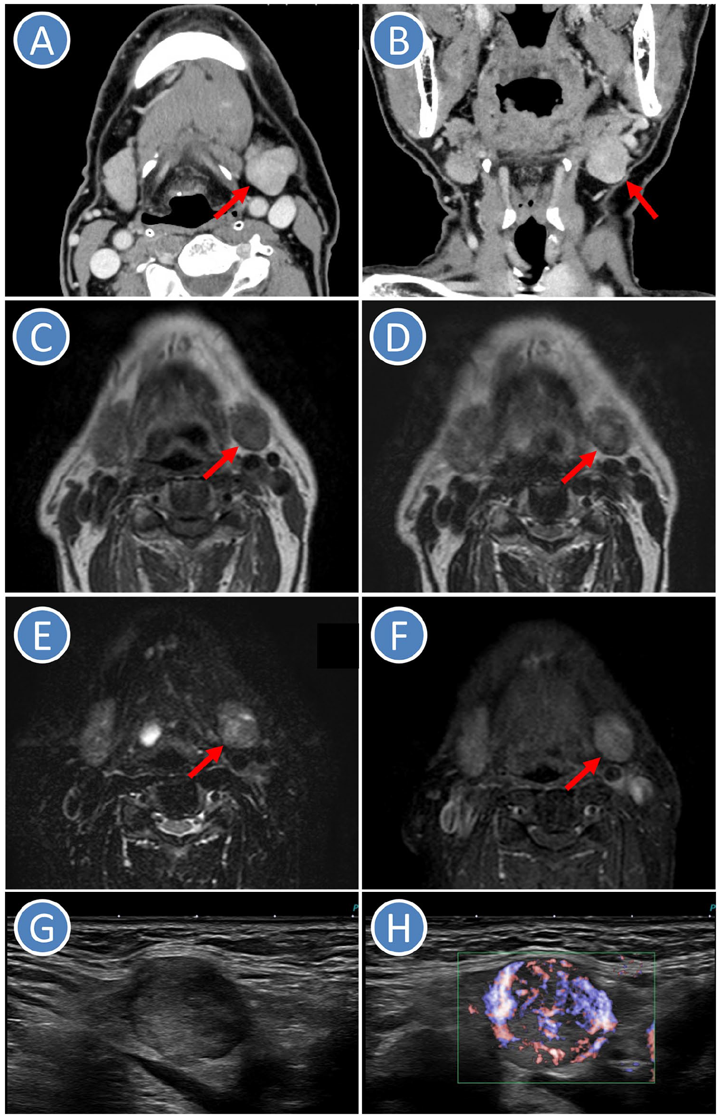
Radiological images. Arrow indicates the tumor. (A) CT (Axial image). (B) CT (Coronal image). (C) MRI (T1-weighted image). (D) MRI (T2-weighted image). (E) MRI (fat-suppressed T2-weighted image). (F) MRI (gadolinium-enhanced fat-suppressed T1-weighted image). (G) US (B-mode image). (H) US (color Doppler image). CT, computed tomography; MRI, magnetic resonance imaging; US, ultrasonography.
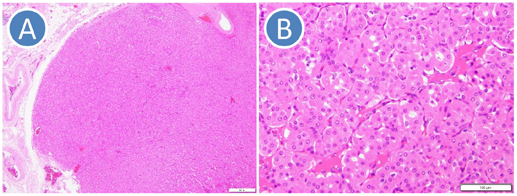
Pathological images (hematoxylin and eosin staining). (A) Low-power field. (B) High-power field.
Oncocytomas are rare benign salivary gland tumors composed predominantly of oncocytes, which are large epithelial cells with eosinophilic granular cytoplasm resulting from an accumulation of mitochondria. Oncocytomas generally occur in the parotid glands, while SMG oncocytomas have been reported to account for 0.2% to 0.9% of all SMG tumors in a large number of studies.1 –3 SMG oncocytomas clinically present as unilateral painless masses, but bilateral SMG oncocytomas have been reported as extremely-rare cases. 4
Although CT is used to evaluate salivary gland lesions, even contrast-enhanced images may sometimes not show lesions despite high clinical suspicion. 5 There are no reliable distinguishing features of oncocytomas on CT or ultrasonography, while MRI may be able to characterize oncocytomas preoperatively. 6 Although there are few studies of characteristic MRI findings of oncocytomas, Patel et al. 6 showed that parotid gland oncocytomas were hypointense on T1-weighted images but isointense to the native parotid gland on fat-saturated T2-weighted and postcontrast T1-weighted images. In the present case, the oncocytoma had a density similar to normal SMG in contrast-enhanced CT images. MRI showed intermediate signal intensity similar to native SMG on the T1-weighted image, the T2-weighted image, the fat-suppressed T2-weighted image and the gadolinium-enhanced fat-suppressed T1-weighted image. Therefore, our oncocytoma was considered a so-called invisible CT and MRI lesion.
Footnotes
Ethical Considerations
Our institution does not require ethics approval for reporting individual cases. A written informed consent form was obtained from the patient.
Funding
The author(s) received no financial support for the research, authorship, and/or publication of this article.
Declaration of Conflicting Interests
The author(s) declared no potential conflicts of interest with respect to the research, authorship, and/or publication of this article.
Data Availability Statement
The datasets used and/or analyzed during the current study are available from the corresponding author on reasonable request.