
Abstract
Calcification of auricular cartilage is a rare condition. This phenomenon might be associated with frostbite, local trauma, inflammation, or systemic diseases. Calcification that progresses to the external ear canal cartilage is even rarer. We present an extremely rare case of bilateral auricular and external ear canal cartilage calcification in a patient with acromegaly. Clinicians should be aware that auricular and external ear canal cartilage calcification can occur in a patient with acromegaly.
Introduction
Calcification of auricular cartilage is a rare condition. This phenomenon might be associated with frostbite, local trauma, inflammation, or systemic diseases.1-7 Among patients with calcification of auricular cartilage, it has been reported that calcification can progress in the external ear canal cartilage.1,5 Herein, we present a case with bilateral auricular and external ear canal cartilage calcification.
Case report
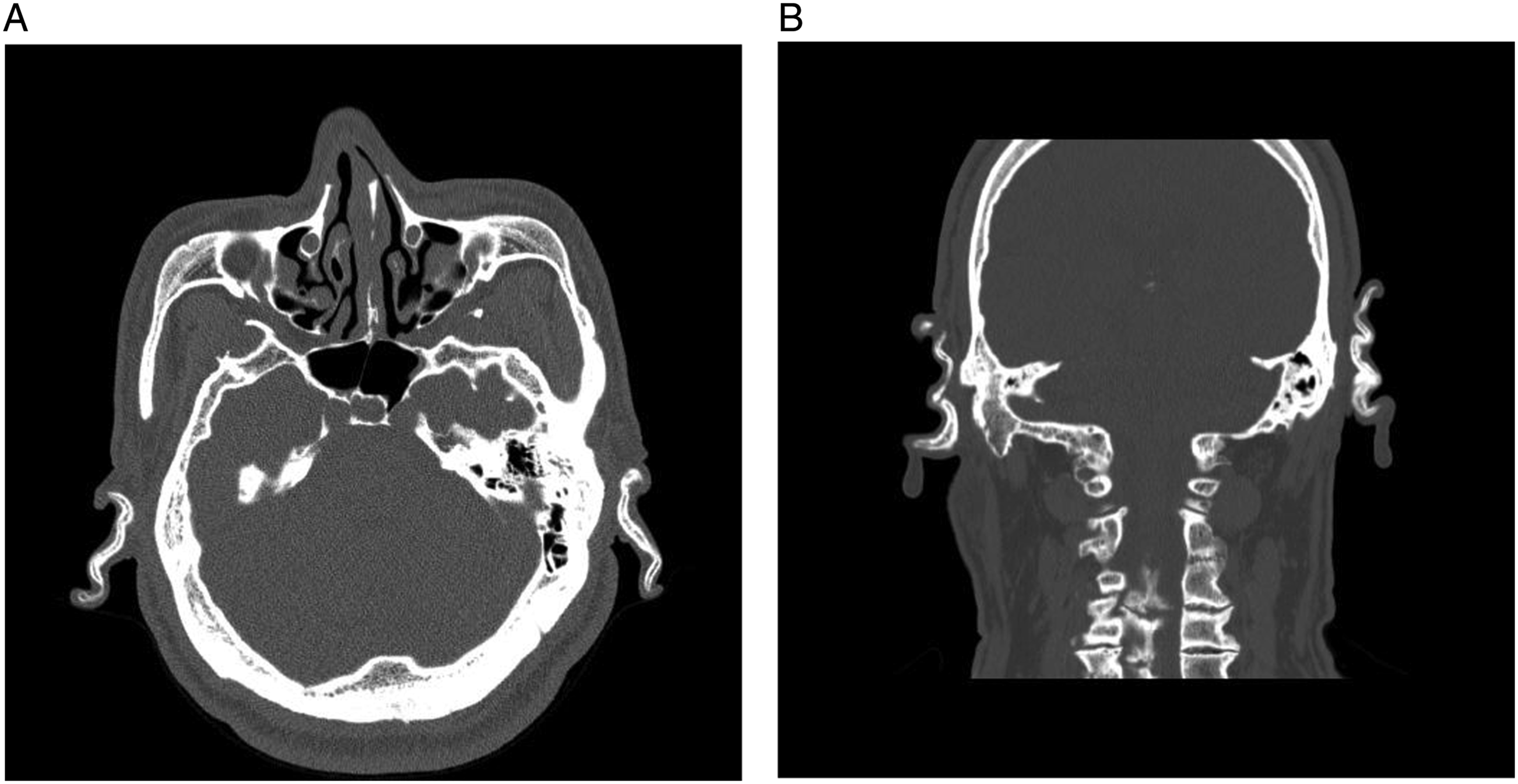
A 58-year-old man was referred for left aural fullness over several years. The patient denied any physical trauma, inflammation, or frostbite. The patient has been treated for acromegaly after a pituitary gland surgery 24 years ago. Diabetes mellitus was first diagnosed 14 years ago. It is being controlled with medications. On physical examination, bilateral auricles were thickened and rigid. They could not be folded (Figure 1). In addition, both external ear canals were stenotic and almost occluded, especially at the left side. Palpation of nasal, thyroid, and cricoid cartilages did not reveal any abnormalities. The epiglottis and arytenoids were also normal on laryngoscopy. Laboratory evaluations including complete blood count, biochemistry profile, electrolytes, thyroid hormones, and parathyroid hormones were within normal limits. A pure tone audiometry showed a normal hearing. Computed tomography (CT) scan revealed calcification of bilateral auricular and external ear canal cartilage (Figure 2). The patient was finally diagnosed with bilateral auricular and external ear canal cartilage calcification in addition to acromegaly. We decided to follow up the patient without additional treatment because the patient had no severe symptoms. During the 7-year follow-up period, the patient continued to complain of mild aural fullness. However, calcification of bilateral auricular and external ear cartilage did not show any sign of progression. Thick and hard bilateral auricles: (A) right ear and (B) left ear. Axial (A) and coronal (B) computed tomography scans reveal calcification of bilateral auricular and external ear canal cartilage.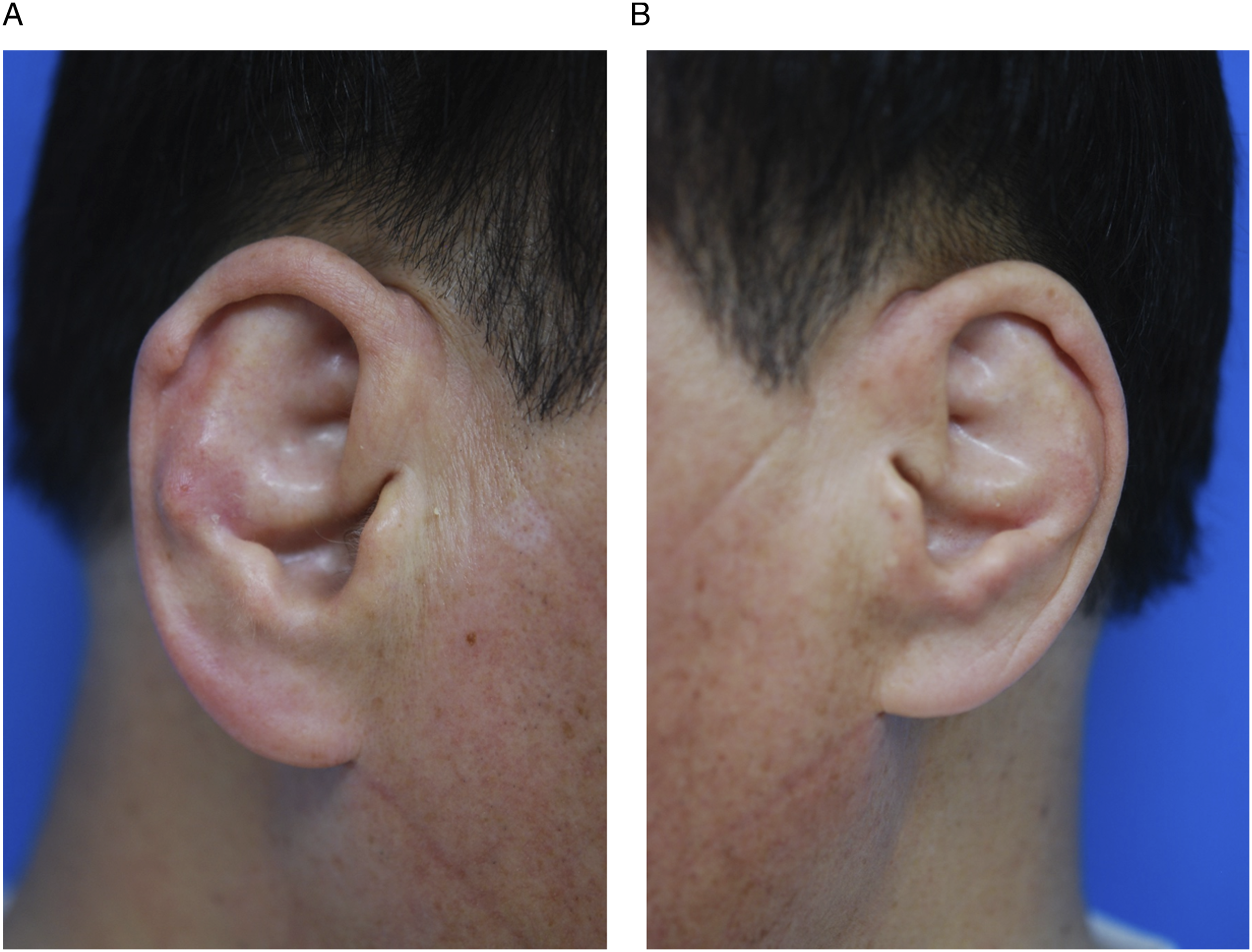
Discussion
Calcification of auricular cartilage is very rare and occurs bilaterally, mainly in men.1-7 It is the replacement of cartilage into bone tissue. 6 The most common cause of auricular cartilage calcification is frostbite.1,3,5,6 Other causes include local trauma, inflammation, neoplasms, systemic diseases such as Addison’s disease, diabetes mellitus, acromegaly, scleroderma, and other endocrine disorders.1-7 Laboratory evaluation can help differentiate metabolic or endocrine causes of this phenomenon. 4 Our patient said that he felt discomfort after the diagnosis of acromegaly. After acromegaly surgery, all pituitary hormones except for growth hormone were normal. Therefore, acromegaly was considered the cause of bilateral auricular and external ear canal cartilage calcification.
Calcification of auricular cartilage is usually asymptomatic.1-6 Some patients complain of discomfort such as pressure due to a hard auricle.1-6 When calcification progresses to the external ear canal, patients sometimes have complaints such as otalgia or aural fullness2,5 as shown in our patient.
The diagnosis of calcification of auricular cartilage can be easily made by physical examination and history taking. It should be confirmed by radiologic or histopathologic examinations.1-7 CT is a very sensitive and good method for diagnosing calcification of auricular and external ear canal cartilage. 4 In our patient, CT was performed periodically to check the degree of calcification, especially the external ear canal cartilage.
The treatment for calcification of auricular cartilage has not been clearly established due to a small number of patients.1-5 For most asymptomatic patients, observation without special treatment is recommended. If the patient complains of discomfort, surgical treatment is performed to improve symptoms. It has been reported good results can be obtained after meatoplasty in patients with calcification of the external ear canal cartilage.2-5 However, extensively calcified lesions are difficult to remove, and surgery itself may exacerbate calcification of auricular and external ear canal cartilage. 3 Our patient complained of aural fullness due to calcification progressing to the external ear canal cartilage. However, his symptoms are not severe. In addition, he has a normal hearing. Thus, he is being followed up periodically.
Calcification of auricular and external ear canal cartilage is irreversible. Thus, periodic follow-up is required to check the disease progression and the occurrence of symptoms. 1 Patients should be instructed not to irritate their ears to prevent disease progression. 2
Conclusion
We present an extremely rare case of bilateral auricular and external ear canal cartilage calcification in a patient with acromegaly. Clinicians should be aware that auricular and external ear canal cartilage calcification can occur in a patient with acromegaly.
Footnotes
Declaration of conflicting interests
The author(s) declared no potential conflicts of interest with respect to the research, authorship, and/or publication of this article.
Funding
The author(s) received no financial support for the research, authorship, and/or publication of this article.