
Abstract
We present an extreme rare case of extramedullary nasal plasmacitoma that arise from nasal septum. The mass surgically removed was analyzed by a pathologist who diagnosed an extramedullary nasal plasmacytoma. The patient did not present systemic involvement. A short cycle of radiotherapy was performed after the surgery. At 9-month follow-up, the patient is recurrence free.
Extramedullary nasal plasmacytoma (ENP) is a rare condition characterized by localized monoclonal plasma cell proliferation without apparent systemic involvement. 1 This tumor represents 5% to 10% of all plasma cell neoplasms 2 and is commonly located in the nasal cavity and/or in the nasopharynx. Extramedullary nasal plasmacytoma generally arises from soft tissues where immune cells are more diffused 3 ; ENP is generally found in the nasal cavity, 2 posterior to the turbinate 3 or in the nasopharynx, 1,2 while its presence in the nasal septum has been rarely described. 4 The aim of this article is to present a rare case of ENP arising from the nasal septum.
An 80-year-old man with a known history of hypertension presented to the outpatient service of our university hospital in May 2019 due to recurrent episodes of epistaxis resistant to medical treatment and, referring the presence of an external body in his right choana. The patient reported no history of nasal bleeding before March 2019, when he had a massive epistaxis from the right nostril. The episode was initially correlated with a sudden blood pressure spike. After this episode, the patient presented recurrent episodes of epistaxis (4 times/wk), prevalently in the morning time, and sensation of external body presence in the right nostril. He referred to his cardiologist to monitor his blood pressure and reconsider his current pharmacologic treatment for hypertension; however, blood pressure was within normal limits and no spikes or other pathological findings were present at 72 hours of Holter monitoring.
Nasal endoscopy showed in the right choana a vegetative and ulcerated mass with irregular surface, 8 mm in diameter, originating from the mucosa of the nasal septum (Figure 1). Computed tomography (CT) scan with and without contrast showed a contrast-enhancing mass located in the anteroinferior part of the nasal septum that did not invade the surrounding tissues (Figure 2). In addition, several submandibular and laterocervical microlymph nodes were identified bilaterally. The mass was surgically removed; histological analysis showed the presence of dense infiltrate of plasma cells staining positive to IRF4/MUM1 typical of plasmacytoma (Figure 3). To complete the diagnostic process, the patient underwent hematologic screening, protein electrophoresis, Bence Jones proteinuria, and bone marrow analysis. Total body magnetic resonance imaging and positron emission tomography (PET/CT) scans were performed to exclude a systemic condition. All examinations were negative; the definitive diagnosis was extraosseous (extramedullary) plasmacytoma 4 of the nasal septum. 5 On September 2019, the patient underwent a cycle of radiotherapy, 5 times/wk for 4 weeks. The last follow-up visit performed 9 months after the surgery showed no disease recurrence.
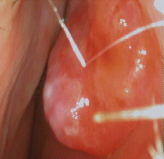
Endoscopic vision of a mass arising from the nasal septum in the right choana. The mass presents diffuse hyperemia and a central ulcerated area covert by fibrine. The lesion is clearly separated from the inferior turbinate.
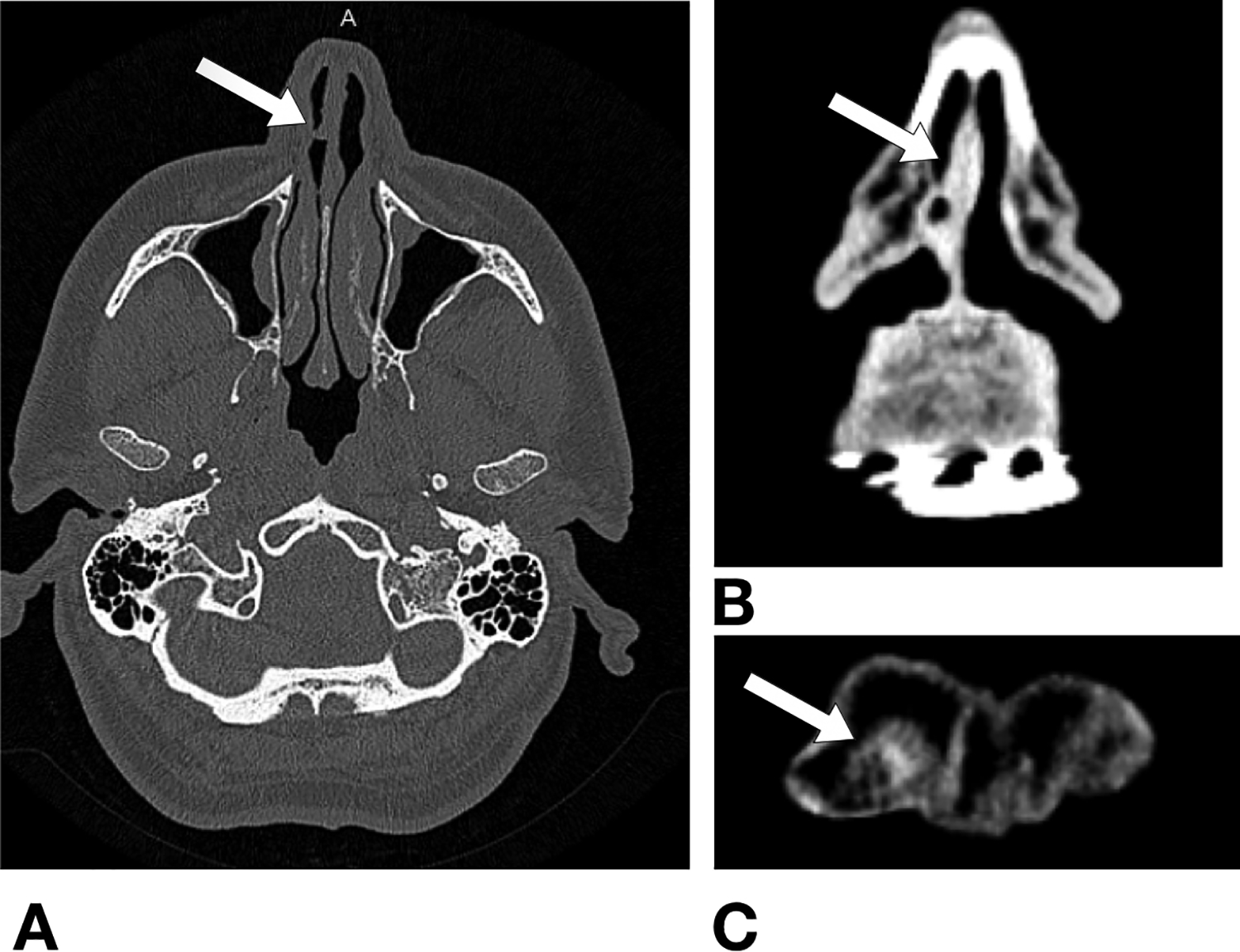
High-resolution computed tomography scan. A, Axial view without contrast: The white arrow shows the area where the ENP was identified. B, Coronal view with contrast: the white arrow indicates the area where the tumor was with an inhomogeneity of the signal. C, Detail of coronal view with contrast: A contrast-enhanced mass measuring 8 mm in diameter is visible in the anteroinferior part of the right choana (white arrow). ENP indicates extramedullary nasal plasmacytoma.
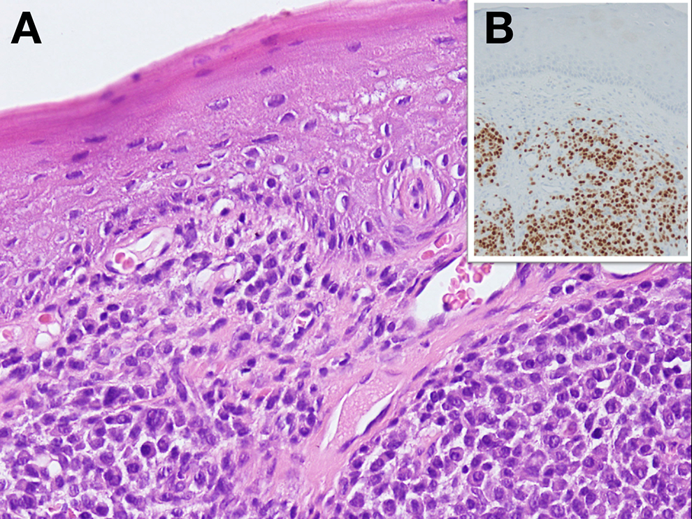
A, Tissue after surgical removal at ×30 magnification. A dense infiltrate of mature plasma cells is present. B, The cell nuclei (top right side of the image) positively react to IRF4/MUM1 staining (×400 magnification).
Extramedullary nasal plasmacytoma of nasal septum is a very rare condition that should be always considered in the differential diagnosis of the nasal cavity masses. The tumor appearance may mimic other nasal masses, especially when it is in a very early stage. 4 In case of an ulcerate mass, the differential diagnosis with squamous cell carcinoma based on accurate histological analysis is of paramount importance. 6
Footnotes
Authors’ Note
The data sets used and/or analyzed during the current study are available from the corresponding author on reasonable request.
Declaration of Conflicting Interests
The author(s) declared no potential conflicts of interest with respect to the research, authorship, and/or publication of this article.
Funding
The author(s) received no financial support for the research, authorship, and/or publication of this article.