
Abstract
Article 23(2) of EU Directive 2010/63 on the protection of animals used for scientific purposes requires staff involved in the care and use of animals to be adequately educated and trained before carrying out procedures. Therefore, the 3Rs (refinement, reduction, and replacement) and knowledge of alternative methods should be part of the education and training itself. For this purpose, the digital learning concept “Virtual Reality (VR) in Biomedical Education” evolved, which successfully combines VR components with classical learning content. Procedures, such as anesthesia induction, substance application, and blood sampling in rats, as well as aspects of the laboratory environment were recorded in 360° videos. The generated VR teaching/learning modules (VR modules) were used to better prepare participants for hands-on training (refinement) or as a complete replacement for a live demonstration; thus, reducing the number of animals used for hands-on skills training (reduction). The current study evaluated users’ experience of the VR modules. Despite little previous VR experience, participants strongly appreciated the VR modules and indicated that they believed VR has the potential to enhance delivery of procedures and demonstrations. Interestingly, participants with previous experience of laboratory animal science were more convinced about VR’s potential to support the 3Rs principle, and endorsed its use for further educational purposes. In conclusion, VR appeared to be highly accepted as a learning/teaching method, indicating its great potential to further replace and reduce the use of animals in experimental animal courses.
Introduction
With the implementation of EU Directive 2010/63 1 on the protection of animals used for scientific purposes, the 3R (i.e., refinement, reduction, and replacement) principle, proposed by Russel and Burch, 2 was incorporated into national animal welfare law in Germany and declared to be a goal. In line with the modular training framework recommended by the European Commission, the 3Rs and knowledge about alternative methods should therefore be part of the educational training of any persons who carry out procedures on animals (EU Function A), design procedures and projects (EU Function B), and/or take care of animals (EU Function C), or kill animals (EU Function D). 3 Therefore, compulsory educational training courses are provided for personnel performing animal experiments, depending on their functions (e.g., Federation of European Laboratory Animal Science Associations (FELASA) Function A for persons who carry out animal experiments). 4 In addition to theoretical knowledge, it is mandatory for all participants of FELASA courses to acquire the necessary practical skills. Strikingly, regardless of their professional qualifications, the actual environment of an animal laboratory and the practical application of the theoretical knowledge acquired can often seem unrelated to the students, particularly in relation to operating rooms, where certain procedures must be followed and knowledge about the use of surgical instruments plays an important role. Such aspects can only be conveyed to a limited extent in theoretical lectures. The acquisition of competencies is often only possible through demonstrations and practical skills training. However, this type of training is very time-consuming, requires spatial and personnel resources, which are often limited, and eventually entails the use of live animals. Improving the usage of time slots for hands-on skills training and the application of the 3Rs is therefore essential for optimal trainee preparation. This might be achieved using digital teaching methods.
Virtual reality (VR) training can help improve students’ preparation for real-life situations. VR is a digital simulation of events/environments that replaces perceptions of the real world by creating an immersive and interactive experience for users.5–8 In the field of health education (e.g., nursing, dentistry), VR has been used for more than 20 years, with simulations of colonoscopies and upper gastrointestinal tract endoscopies being some of the first applications. 9 VR simulations have consistently been proven to increase students’ performance.6,10 To date, VR has been primarily used in biomedical education to generate surgical simulators, three-dimensional (3D) anatomical models, virtual dissection tables, and virtual worlds.6,11–13 However, a few VR projects relating to the 3Rs have already been carried out. One notable example is the courseware ViSi (i.e., a virtual animal-holding gamified simulator), which simulates the handling of laboratory animals in VR with gamification elements. 13 Furthermore, 360° videos have been successfully in VR applications used in veterinary medicine in courses on pathology and surgery. 14 In this respect, 360° VR videos resemble VR simulations, the difference being that this technology uses real-world footage rather than virtual, computer-generated scenarios, and the opportunity for interaction is thus considerably reduced. However, the developmental cost of VR simulations is higher, which is why the distribution of these technologies in educational organizations is currently not particularly high. Although the cost of the hardware has continued to decrease in recent years, development of hyper-realistic and interactive environments requires either a high level of programming knowledge or considerable financial resources to have them developed externally. In comparison, the use of 360° VR for developing realistic and immersive learning experiences is much more affordable. 15
RWTH Aachen University offers courses in laboratory animal science. For scientists and technicians performing procedures on animals, FELASA-accredited courses (addressing Functions A including D) are offered. For medical students, the FELASA Function A incl. D course is offered as a “qualification profile course” (QP) as part of their curriculum in the ninth semester, the completion of which is a requirement for their MD thesis if procedures on animals are to be performed. Additionally, RWTH Aachen University offers a master’s degree program (Master of Laboratory Animal Science; MLAS) for experienced veterinarians and biomedical scientists to acquire the FELASA’s certificate for laboratory animal science specialists. In all courses, practical skills are trained step-by-step, from theory to video, to using models such as the Koken rat® or mouse toy, to cadavers, to anesthetized animals, and finally to cover awakening animals to reduce stress on both the participants and animals. Although the procedures are discussed and visualized in the theoretical sessions, instructors still need to demonstrate the procedures on live animals during practical skills training, with the attendant increase in the number of animals required. To circumvent this, we resolved to generate VR modules for procedures that are only demonstrated to participants and not required to be performed by them personally. In the case of MLAS students, the 360° VR videos should be used to better prepare them for procedures they will be performing by themselves. The study presented here investigates the use of these VR modules in laboratory animal science courses in terms of their general acceptability and potential to contribute to the 3Rs.
Material and methods
Ethical approval
The questionnaires were answered anonymously and voluntarily. Informed consent was obtained from all participants before completion of the questionnaires and all research was performed in accordance with the relevant guidelines and regulations. The study was approved by the Ethics Committee of the Medical Faculty of RWTH Aachen University (EK 087/21).
VR headset and 360° VR video recording
To record the 360° VR videos, each room had to be set up for a spherical recording, as every angle is visible to the camera, and the staff producing the videos had to leave the room to control the camera remotely. The Insta360 Pro 2 spherical camera was used for the 360° VR video recordings, which utilizes six lenses to record up to 8K 3D high dynamic range videos. 16 For some of the videos, an extra head-mounted camera (Insta360 ONE R or GoPro Fusion) was used to record close-ups of the procedures. The recorded 360° footage was stitched using the camera’s stitching software and edited with Adobe® Premiere Pro® CC. 17
After completion of the post-production processes, the videos were uploaded to the VR learning and multi-user collaboration platform, Wonda, 18 to be used in presentations and discussions during the courses. For this purpose, the Oculus Quest 2 VR Head-Mounted Display (HMD) (developed by Oculus, a subsidiary of Meta) was used. The main advantage of such headsets is that they are standalone devices that can run videos and software on an Android-based operating system without being connected to a computer. Further, the device uses a fast-switch LCD display for each eye, each with a resolution of 1.832 × 1.920 pixels and a refresh rate of 60, 72, or 90 Hz. 19 Using the corresponding Oculus Touch controllers, users interacted with the graphic user interface of the Oculus, the Wonda platform, and the videos.
Study sample
The study was conducted among participants of the regular FELASA Function A incl. D course, participants of the MLAS, and of the QP courses offered to medical students. Participants of the regular FELASA course included scientists and technicians, with different levels of experience in the field, pursuing basic training in laboratory animal science in order to participate in animal experiments. Participants from the MLAS course were experienced in laboratory animal science, while participants from the QP courses consisted solely of medical students with almost no experience in laboratory animal science. Between January 2021 and March 2022, a total of 63 participants used the VR course materials in groups of approximately four to eight students. Each student had an Oculus Quest at their disposal for the duration of the course.
Pre-/post-VR experience questionnaires and study design
All participants were informed, and asked about study participation prior to commencement of practical training in the courses. Pre-/post-questionnaires were sent via email or provided as a QR code in the lecture hall. Participants completed the online questionnaires anonymously using an anonymized identifier (i.e., a random number provided to each participant that related pre- and post-questionnaire answers), through the LimeSurvey platform. 20 Both questionnaires were presented in the English language, and gathered data on demographics, demonstration needs, media affinity, usability, as well as including questions on acceptance and benefits of the VR learning unit and components. The pre-questionnaire (i.e., completed before VR learning unit experience) contained 18 items and the post-questionnaire (administered after successful participation) contained 16 items. Items relating to demographic information and previous VR experience were only included in the pre-VR training questionnaire. Detailed demographic data are shown in Supplemental Material S1.
Several items on media affinity and experience were presented in the form of multiple-choice questions. All items concerning agreement and ratings of VR experience were assessed utilizing five-point Likert scales. To assess VR usability, the System Usability Scale (SUS)21–23 was included in the post-VR learning unit questionnaire. Additionally, several open-ended questions allowed feedback on demonstration needs, media support, and VR use. For the complete questionnaires see Supplemental Materials S2 and S3.
Procedure
After the theoretical part of the course, but before procedures were demonstrated live on live animals during the practical course element, students used the VR module to watch 360° VR videos on anesthesia, application through the penile vein, and blood sampling through the sublingual veins in rats (for details see Supplemental Material S4). The anesthesia VR module was implemented either in a live VR session, where the students and lecturer met in the VR room (Figure 1(a) and (b)), or course participants could watch the VR module independently at home or in the practical course room before the subsequent practical training (Figure 1 (c) and (d)).
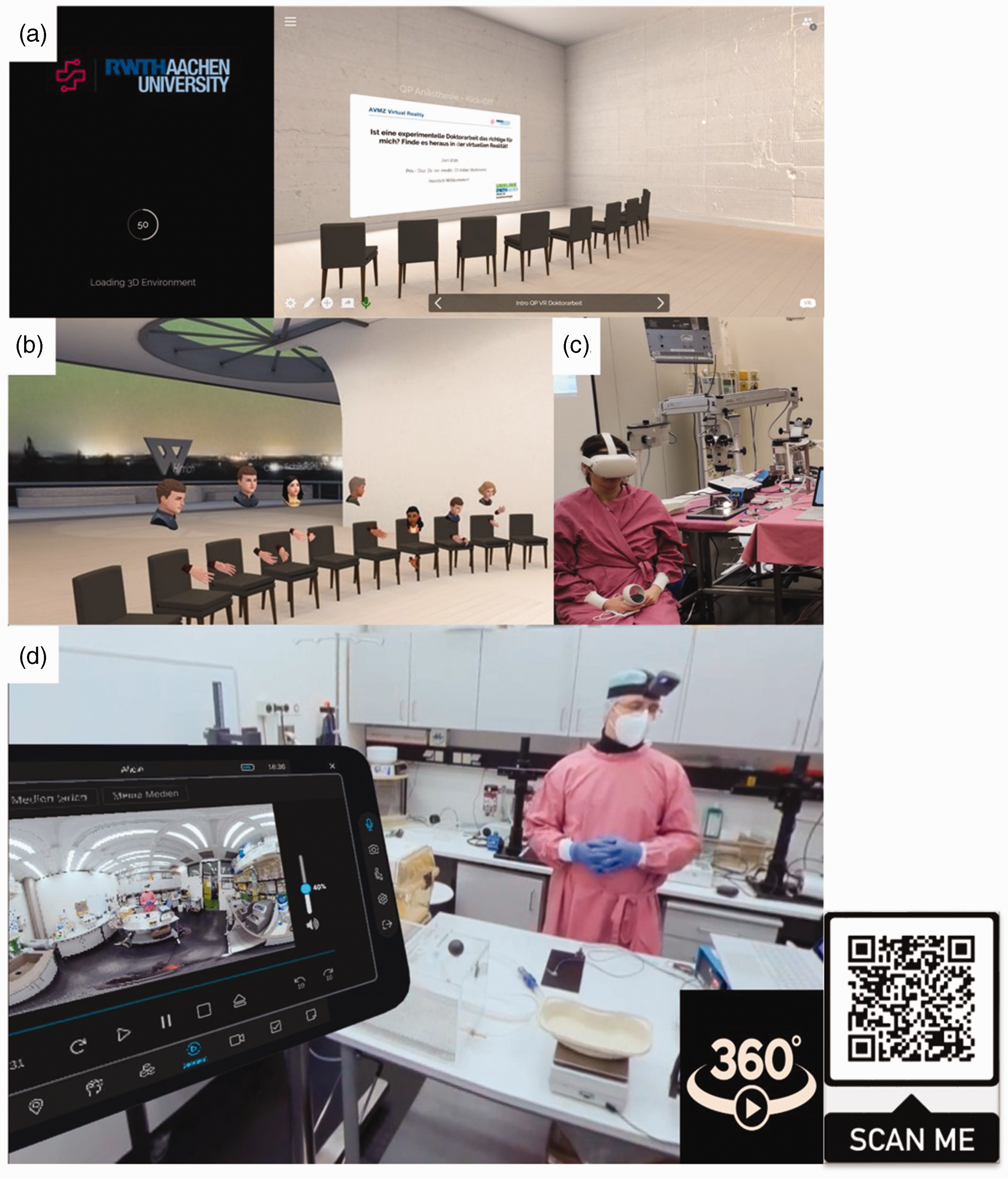
(a) and (b) a live virtual reality (VR) session where the students and lecturer gathered virtually in the VR meeting room; (c) participants were able to rewatch the immersive VR videos in the practical course room before the practical training; and (d) watch an expert explaining the procedure.
Statistical analysis
Statistical analyses were performed in R 24 with the level of significance set at α = 0.05. To explore whether participants viewed VR as being complementary to demonstrations and as contributing to the 3R principle, linear mixed models were computed using the R package, lme4. The full model included the courses (FELASA, MLAS, QP) as a three-level, within-subjects factor, and time (pre-/post-questionnaire), or as a two-level, repeated within-subjects factor. The interaction terms “course” and “time” were included to test for possible dependencies. The anova function of the lmerTest package was used to obtain p-values for the fixed effects. Models were fitted using Restricted Maximum Likelihood (REML) method and the effect size, R2 beta, was calculated using the r2glmm package. Post hoc comparisons of significant effects, using the emmeans package, were FDR (False Dicovery Rate)-corrected and Hedge’s g calculated using the esvis package. To investigate the relationship between experience of animal experimentation and VR, Spearman’s rank correlation was calculated. The ggplot2 and ggstatsplot25,26 packages were utilized for data visualization.
Results
Of the 63 participants, 59 (30 FELASA, 16 MLAS, and 13 QP) and 43 took part in the pre- and post-evaluation, respectively, of the presented digital learning concept.
As shown in Figure 2(a), previous VR experience was quite low among all participants. Similarly, participants’ experience of animal experiments was low, with slightly more experience in the MLAS group and the least experience being found in the QP group (Figure 2(b)).
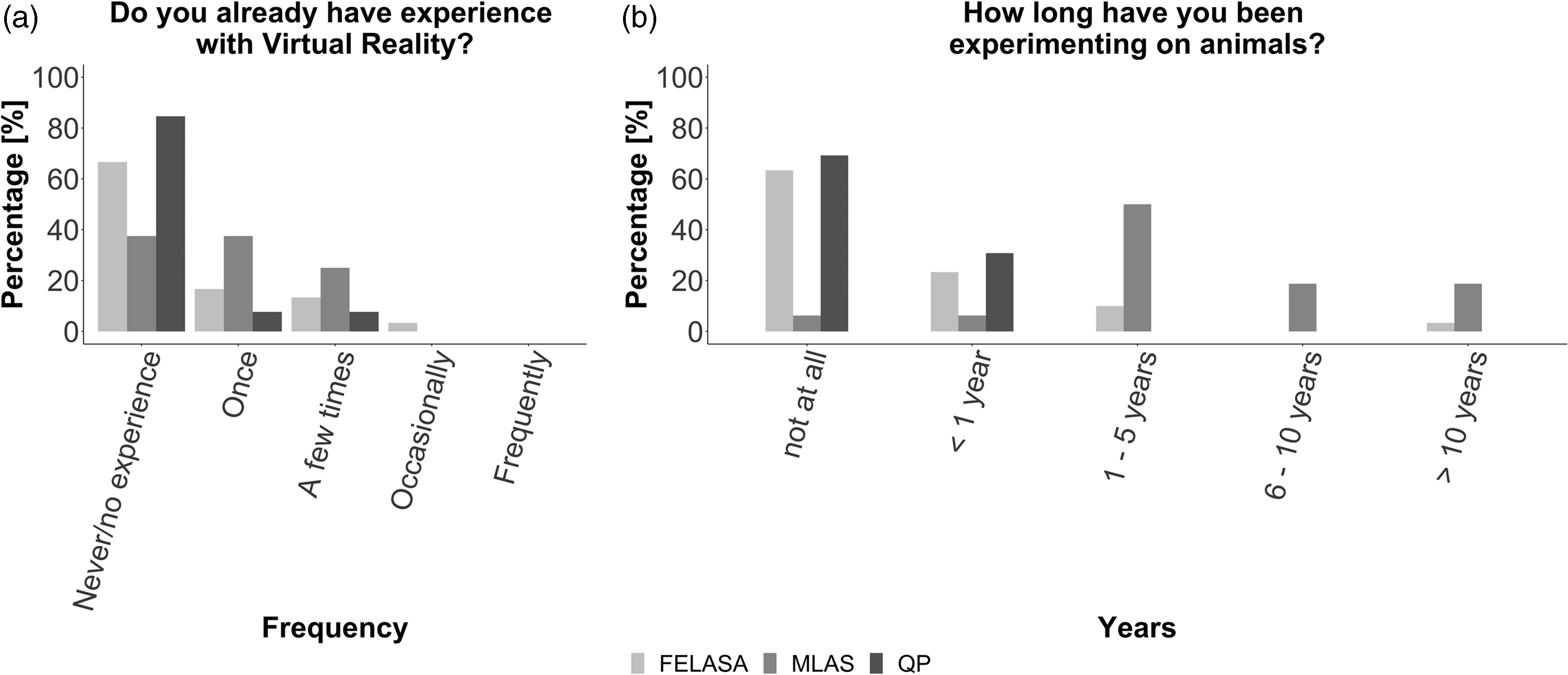
Experience of the FELASA, MLAS, and QP laboratory animal science course participants in relation to (a) virtual reality and (b) experimentation on animals.
In considering the general aspects of VR, 62.70% (pre-VR) and 79.10% (post-VR) of participants stated that they could imagine VR as a medium to support demonstrations of procedures. Other media that could be imagined to support demonstrations are presented in Supplemental Material S5.
Despite slight difficulties encountered during participation, participants generally rated application of VR to be in the marginally high acceptability range of the SUS score (M = 66.67; SD = 10.53). Some inconveniences and adverse effects reported included initial technical difficulties due to this being the participant’s first use of VR (18.60%), disorientation (39.50%), dizziness (27.90%), headaches (27.90%), loss of time/space (25.60%), and eyestrain (25.60%). According to the participants, an improvement in the display quality (44.20%) and the addition of haptic features (34.90%) could enhance the VR experience in the future (Supplemental Material S6).
Further to an overall high acceptance of VR as a teaching/training method, both before and after VR module participation, participants largely agreed that VR could be used to support/complement demonstrations and contribute to the 3Rs principle (Figure 3).
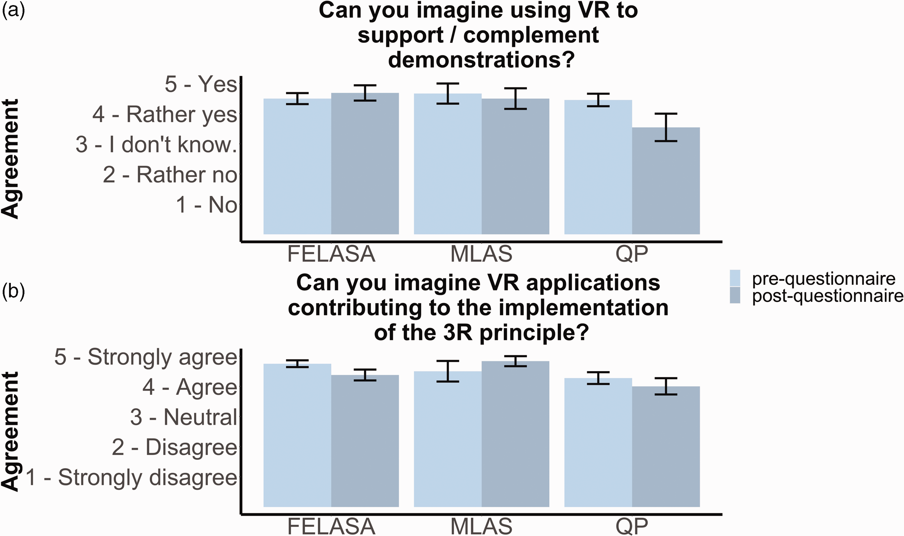
Potential of virtual reality (VR) to (a) support/complement demonstrations and (b) contribute to the 3Rs principle. Error bars represent the standard error of the mean.
In the evaluation of VR as a supporting method for demonstrations, a course (MLAS, FELASA, QP) and time (pre-/post-VR) interaction was identified, F(2, 30.81) = 3.71, p = 0.04, R2 = 0.15 (Figure 3(a)). No post hoc comparisons were significant. However, while participants’ ratings pre- and post-evaluation did not differ, the QP group following VR module participation appeared slightly less likely to agree that VR could be used as a supportive method (p = 0.07, padj = 0.22, Hedge’s g = 0.75; Figure 3).
In evaluating VR as a potential contributor to the 3R principle, a trendwise main effect of course was found, F(2, 34.55) = 2.80, p = 0.07, R2 = 0.17 (Figure 3(b)). While post hoc comparisons were not significant, the QP group rated the contribution of VR to the 3R principle as slightly lower than the FELASA and MLAS groups did (FELASA–QP: t(40.2) = 0.44, padj = 0.09, Hedge’s g = 0.61; MLAS–QP: t(31.3) = 0.55, padj = 0.09, Hedge’s g = 0.71; Figure 3). For detailed ratings per group, see Supplementary Material S7.
Interestingly, while overall agreement that VR could contribute to the 3R principle was high, prior to VR module participation participants with more experience in animal experimentation indicated higher levels of agreement than those with less experience (rs(33) = 2860.44, p = 0.002, rho = 0.52; Figure 4(a)). This relationship tended to persist after participation, but without reaching significance (rs(33) = 4013.90; p = 0.06, rho = 0.33), given that the general high agreement about VR’s potential to contribute to the implementation of the 3R principle remained unchanged (pre: M = 4.33, SD = 0.78; post: M = 4.55, SD = 0.62; V = 36, p = 0.15). In addition, agreement was high that VR should be used more extensively for educational purposes in the future, and again, participants with more experience in animal experimentation expressed this agreement even more strongly than those with less experience (rs(33) = 3663.10, p = 0.03, rho = 0.39; Figure 4(b)).
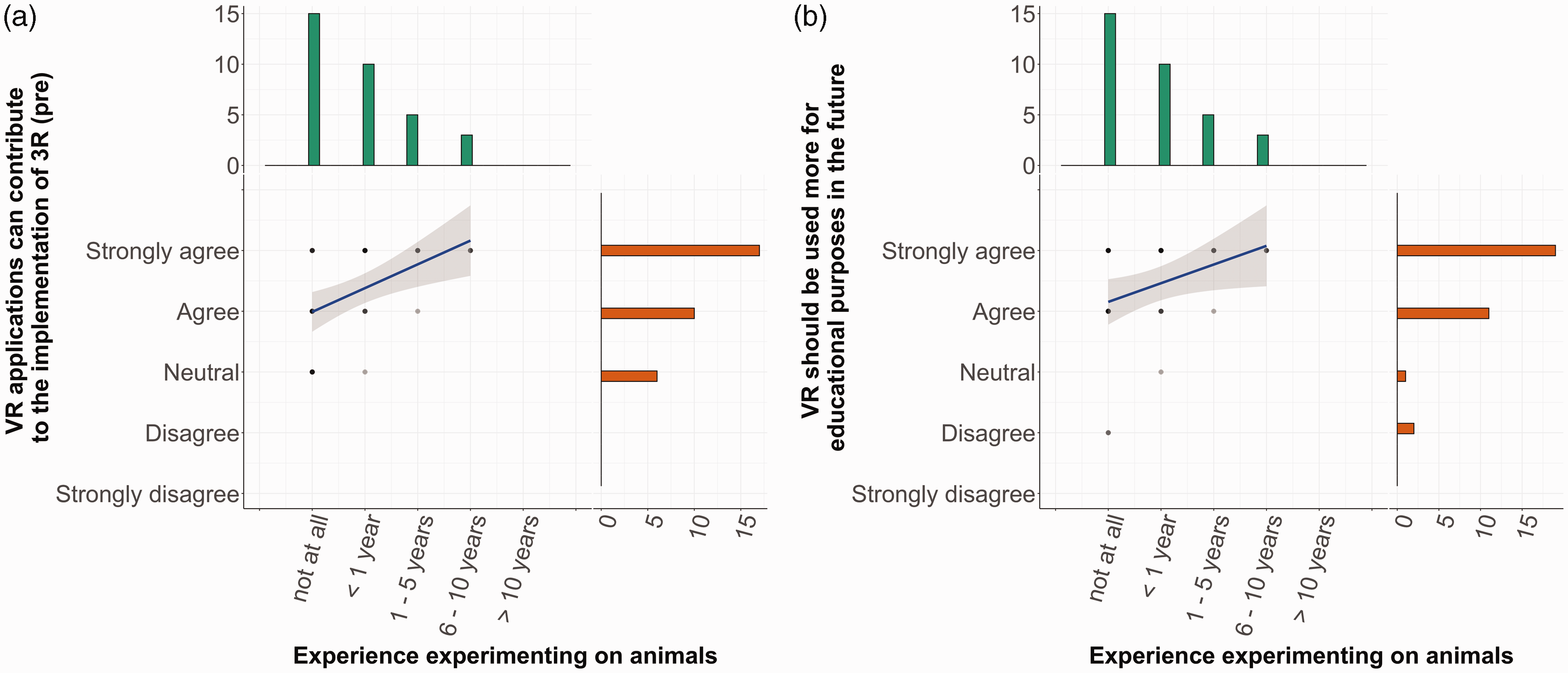
Relationship between experience with animal-based experiments and VR acceptance: (a) VR as a contribution to the implementation of the 3R principle and (b) VR being used more for educational purposes in the future.
Discussion
In the study presented here of VR training modules covering various animal experimentation procedures, we evaluated the experiences of participants pursuing different laboratory animal science courses, specifically, in relation to VR acceptance and its potential to contribute to the 3R principle. The study showed that acceptance of VR and 360° VR videos in laboratory animal science education for learning experimental skills was very high, even before participation in the VR modules. Given the overall low level of VR exposure, participants might have had high expectations of the technology, hence, it cannot be ruled out that the high acceptance rates observed in the current study were partially due to a novelty effect.27,28 Aside from its novelty appeal, the high VR module acceptance might be attributed to participants valuing the superior visualization possibilities of VR and its potential for the realistic teaching/training of procedures. Furthermore, the novelty effect and expectations for this technology appear justified, as indicated by the post-evaluation of the method, especially among the more experienced participants.
Participants with a high level of knowledge and experience in the field of animal experimentation were more likely to indicate that VR contributed to the implementation of the 3Rs principle and that it should be used more extensively for educational purposes in the future. Their judgment carries particular weight, as more experienced participants might be expected to be better able to assess whether VR training would be beneficial or not. Interestingly, although the overall high acceptance rates remained unchanged, participants with less experience (QP) tended to rate VR’s potential to complement demonstrations as lower after participation. Further investigation of this aspect in larger samples is warranted.
Even though VR has been used for over a decade in various fields (e.g., medical and surgical education), 11 only a few studies have reported on the use of VR in veterinary education.29–31 For this purpose, Espitia et al. developed a virtual multi-user learning environment for training students, to measure clinical competence and evaluate students’ active engagement in critical thinking and complex problem solving. 31 Others have developed open-access VR anatomical programs for students to use for home education, especially during the COVID-19 pandemic, when professional medical and veterinary colleges and universities were unable to offer conventional lessons.29,30,32 In the field of laboratory animal science, only Tang et al. have reported the development and implementation of ViSi as an animal-handling training method for biomedical science undergraduates.13,33 Publications in this field describe newly generated VR teaching formats and their use in evaluating acquired competencies, but consistently fail to include any systematic evaluation of VR use to replace, reduce, or refine the use of animals in education. The current study has convincingly filled this gap by representing newly generated VR modules that, for the first time, are focusing on investigating the potential to replace demonstrations on animals used in laboratory animal science courses.
It should be noted that Tang et al. previously recognized the need to awaken the learners’ critical thinking and the potential of incorporating VR into pedagogical concepts at the undergraduate student level.13,33 Furthermore, Kanzler et al. showed that medical-/biology students’ knowledge of animal experimentation is limited, regardless of educational stage. 34 Certainly in the early phase of education, VR modules could replace working with live animals and generate sufficient initial experience in the field of laboratory animal science.
Crucially, demonstrations still play a central role in the education of laboratory animal science practitioners, as well as that of surgeons during their residency. 35 However, this teaching method often causes weaknesses in the trainees’ learning process due to discrepancies in the learner–teacher ratio. 36 This problem could therefore be similarly addressed with digital teaching methods. A recent study showed that use of 180° VR videos might enhance the learning outcomes of undergraduate students studying stereotaxic surgery, and that this technology could help prevent exacerbation of the learning gap created by the COVID-19 pandemic. 32
Another recent study, evaluating VR as a training adjunct for surgical skills among medical students, showed that VR adds value to the teaching but cannot replace high-quality, hands-on practical training. 37 Furthermore, Hunt et al. did not observe differences in veterinary students’ first canine surgical performance after undertaking a minimally interactive VR training module. 38 This finding is consistent with the current evaluation; however, opinions about VR as a full replacement, or only as an additional teaching strategy, strongly depend on the profession/medical field of the participants. In the field of anatomy, VR is highly accepted and has been shown to improve medical students’ skills in assessing and understanding computed tomography scans and interpreting surgical images. 39 In contrast, in a recent evaluation of robotic surgical skills learning on a VR-based platform, further improvements and expert knowledge were identified as needed before VR robotic training courses could be implemented in curricula.39,40
In summary, VR technology has gained acceptance in diverse areas, such as in nursing schools, 40 as a support for oral hygiene in elderly/disabled people, 41 and as a replacement for demonstrations, especially if these methods do not have to be performed hands-on by participants later in their studies. To replace conventional training courses for more complex skills, where interaction and hands-on practice are mandatory, a further improvement in the technology is needed, and must be evaluated in terms of the competency to be acquired, before integration.
Limitations
In the current study, the sample size was quite small due to course enrollment limitations. Furthermore, not all students had the opportunity to watch all three videos because of the curricula of the different courses. Gender was not equally distributed in the QP group.
Notably, the cost of the latest generation of HMDs may be an important factor in the wider use of this technology. The effect of VR novelty should be considered in larger future evaluations of similar VR modules.
It should be recognized that some participants experienced motion/simulator sickness and nausea, which may prevent them accessing VR teaching/learning modules (see current data and as described by others6,13,38,42); for those individuals, alternatives options need to be provided.
In addition to organizational and VR-specific issues, future studies are required to measure the effect of the practical skills acquired through 360° VR videos and to assess the skills transferability to hands-on training in a laboratory setting, as well as to traditional videos (without a laboratory VR setting). This was not done in the current evaluation since the VR videos were primarily used as a substitute for demonstrations.
Conclusion
Overall, the VR modules found application and high acceptance in the FELASA courses, in the QP for medical students, and in the MLAS program. The VR teaching/learning unit will now be applied as a substitute for live demonstrations in these courses. Thereby, 25 rats will be saved per year in the Institute for Laboratory Animals Science (ILAS) courses alone. Considering that laboratory animal science courses are offered at numerous universities in Europe and around the world, the use of VR training modules has significant potential to replace animals used for demonstrations. Furthermore, VR modules could contribute to the refinement of animal procedures by shortening the learning curve of educational courses and, thereby, further reduce the number of animals used.
Supplemental Material
sj-pdf-1-lan-10.1177_00236772221128127 - Supplemental material for Virtual Reality in Biomedical Education in the sense of the 3Rs
Supplemental material, sj-pdf-1-lan-10.1177_00236772221128127 for Virtual Reality in Biomedical Education in the sense of the 3Rs by Martin Lemos, Laura Bell, Susanne Deutsch, Leonie Zieglowski, Lisa Ernst, Daniel Fink, R. Tolba, Christian Bleilevens, Julia Steitz in Laboratory Animals
Supplemental Material
sj-pdf-2-lan-10.1177_00236772221128127 - Supplemental material for Virtual Reality in Biomedical Education in the sense of the 3Rs
Supplemental material, sj-pdf-2-lan-10.1177_00236772221128127 for Virtual Reality in Biomedical Education in the sense of the 3Rs by Martin Lemos, Laura Bell, Susanne Deutsch, Leonie Zieglowski, Lisa Ernst, Daniel Fink, R. Tolba, Christian Bleilevens, Julia Steitz in Laboratory Animals
Supplemental Material
sj-pdf-3-lan-10.1177_00236772221128127 - Supplemental material for Virtual Reality in Biomedical Education in the sense of the 3Rs
Supplemental material, sj-pdf-3-lan-10.1177_00236772221128127 for Virtual Reality in Biomedical Education in the sense of the 3Rs by Martin Lemos, Laura Bell, Susanne Deutsch, Leonie Zieglowski, Lisa Ernst, Daniel Fink, R. Tolba, Christian Bleilevens, Julia Steitz in Laboratory Animals
Supplemental Material
sj-pdf-4-lan-10.1177_00236772221128127 - Supplemental material for Virtual Reality in Biomedical Education in the sense of the 3Rs
Supplemental material, sj-pdf-4-lan-10.1177_00236772221128127 for Virtual Reality in Biomedical Education in the sense of the 3Rs by Martin Lemos, Laura Bell, Susanne Deutsch, Leonie Zieglowski, Lisa Ernst, Daniel Fink, R. Tolba, Christian Bleilevens, Julia Steitz in Laboratory Animals
Supplemental Material
sj-jpg-5-lan-10.1177_00236772221128127 - Supplemental material for Virtual Reality in Biomedical Education in the sense of the 3Rs
Supplemental material, sj-jpg-5-lan-10.1177_00236772221128127 for Virtual Reality in Biomedical Education in the sense of the 3Rs by Martin Lemos, Laura Bell, Susanne Deutsch, Leonie Zieglowski, Lisa Ernst, Daniel Fink, R. Tolba, Christian Bleilevens, Julia Steitz in Laboratory Animals
Supplemental Material
sj-jpg-6-lan-10.1177_00236772221128127 - Supplemental material for Virtual Reality in Biomedical Education in the sense of the 3Rs
Supplemental material, sj-jpg-6-lan-10.1177_00236772221128127 for Virtual Reality in Biomedical Education in the sense of the 3Rs by Martin Lemos, Laura Bell, Susanne Deutsch, Leonie Zieglowski, Lisa Ernst, Daniel Fink, R. Tolba, Christian Bleilevens, Julia Steitz in Laboratory Animals
Supplemental Material
sj-jpg-7-lan-10.1177_00236772221128127 - Supplemental material for Virtual Reality in Biomedical Education in the sense of the 3Rs
Supplemental material, sj-jpg-7-lan-10.1177_00236772221128127 for Virtual Reality in Biomedical Education in the sense of the 3Rs by Martin Lemos, Laura Bell, Susanne Deutsch, Leonie Zieglowski, Lisa Ernst, Daniel Fink, R. Tolba, Christian Bleilevens, Julia Steitz in Laboratory Animals
Supplemental Material
sj-pdf-8-lan-10.1177_00236772221128127 - Supplemental material for Virtual Reality in Biomedical Education in the sense of the 3Rs
Supplemental material, sj-pdf-8-lan-10.1177_00236772221128127 for Virtual Reality in Biomedical Education in the sense of the 3Rs by Martin Lemos, Laura Bell, Susanne Deutsch, Leonie Zieglowski, Lisa Ernst, Daniel Fink, R. Tolba, Christian Bleilevens, Julia Steitz in Laboratory Animals
Footnotes
Acknowledgments
The authors would like to thank Sebastian Fedrowitz for assistance during data acquisition, and all students for their participation in the evaluation.
Data availability
Original datasets are available from
Declaration of conflicting interests
The author(s) declared no potential conflicts of interest with respect to the research, authorship, and/or publication of this article.
Funding
Supplemental material
Supplemental material for this article is available online.
References
Supplementary Material
Please find the following supplemental material available below.
For Open Access articles published under a Creative Commons License, all supplemental material carries the same license as the article it is associated with.
For non-Open Access articles published, all supplemental material carries a non-exclusive license, and permission requests for re-use of supplemental material or any part of supplemental material shall be sent directly to the copyright owner as specified in the copyright notice associated with the article.