
Abstract
The objective of the present study was to evaluate the occurrence of pathogens and diseases in laboratory fish over a 10-year period at the Centre for Experimental Fish Pathology of Sicily, University of Messina. This report also emphasizes the adverse effects of subclinical infections on research endpoints, as well as the importance of animal health with respect to welfare. Infections in fish used for research can alter experimental outcomes, increase the variability of data, and impede experimental reproducibility. For this purpose, 411 diseased fish of different species (out of a total of 2820 fish) that belonged to four marine species (Dicentrarchus labrax, Sparus aurata, Argyrosomus regius and Mugil cephalus) and to four fresh water species (Danio rerio, Carassius auratus, Xiphophorus variatus and Poecilia reticulata) were examined in this study. Our results showed that mycobacteriosis and myxosporidiosis were the most important diseases found in our research fish, and the results represent a useful tool to obtain wider knowledge on the incidence of various diseases in different fish species. Further studies in this field are necessary to improve knowledge on the state of the health of fish used for research.
Introduction
Several pathogenic microorganisms are responsible for clinical diseases in laboratory animal species and can influence research by confounding experimental results. For this reason, the health surveillance of animals used for research is necessary to maintain an optimal state of health in laboratory animals. 1
It is well known that many infectious agents harm the welfare status or reduce the breeding performance of animals such as rabbit and rodents, which are commonly used in laboratory experiments. 2
There is growing interest in the use of fish as experimental animals for biomedical research, both for human research and for aquaculture purposes. However, little is known about the pathogens of fish compared to mice, rats and rabbits; information regarding pathological surveys or the health status of fish used in experimental trials is scarce. Despite the increasing use of fish in research, the relative dearth of information about fish health reflects inadequate attention to health management of laboratory fishes, including a lack of suitable pathogen control in research institutions.3–8 This aspect, which has unfortunately been neglected, could be a useful tool to control the biological and experimental variability of fish used in research procedures, providing useful information for the correct interpretation of results. 9 However, given that pathogens can affect fish in a very similar manner to rodents, current regulations are intended to improve health management in fish research facilities.
To date, only a few studies regarding the prevalence and impact of some microorganisms on animal health have been conducted.10–14
Spagnoli et al.15,16 studied the effects of Pseudoloma neurophilia, one of the most serious pathogens in zebrafish facilities, on the nervous system and its implications for neuro-behavioural research.
Previous studies were also conducted in 2009 by Broussard et al., 11 Hegedus et al. 17 and van der Sar et al. 18 on Mycobacterium marinum. It is known that mycobacterial infections, frequently isolated from laboratory fish, significantly affect zebrafish health and transcriptome responses.19,20 In contrast, only sporadic data are available on pathogens in other species of laboratory fish.
Today, both the use of specific pathogen-free (SPF) animals for experimental protocols and the application of appropriate pathogen control strategies should be essential in the field of fish research. 21 Currently, other fish species used for research come mainly from the aquaculture sector, and the absence of pathogens in their traded fish lines cannot be guaranteed.22–24 However, some SPF zebrafish lines are available. A more detailed study of the prevalence and distribution of microorganisms within fish research facilities is necessary to enhance pathogen control strategies and prevent the entry and spread of such noxious agents. Furthermore, the effects on the anatomy and physiology of research fish caused by the presence of an infectious agent must be carefully evaluated, improving experimental result predictivity and reducing the number of animals used.
The aim of this study is to provide an overview of the prevalence, clinical presentation and pathological findings of the main diseases encountered at the Centre for Experimental Fish Pathology of Sicily (CISS) laboratory. Our research offers a valuable contribution to knowledge about the prevalence of fish disease and the effects of different fish disease processes on research. 13
Materials and methods
The facility
This study was carried out at the CISS at the University of Messina. The laboratory has been recognized by the Italian Ministry of Health since 2006 and was authorized as an establishment for the production of fish for experimental purposes in 2010. The CISS facility has three different laboratories: a quarantine laboratory, a zebrafish breeding facility and a main laboratory in which all experiments are performed. The main laboratory is divided into different dedicated areas (one for saltwater species, one for freshwater species and one for experimental procedures) and is equipped with 50 fibreglass and plastic tanks with a capacity of 120 to 800 l; each tank is provided with independent biological-sand and activated carbon filters, aerator pumps and automated monitoring systems for continuous water parameter controls (Oxywifi2, Tecnos, Chioggia, Italy). Zebrafish maintained only in the zebrafish breeding room are reared in a ZebTEC Active Blue Stand Alone system (Tecniplast). In this recirculating housing system, the water is disinfected by ultraviolet treatment (minimum dose 135,000 µWsec/cm2); it derives from reverse osmosis-treated city water and salt is added to a conductivity of 450 µS. Environmental conditions at the primary enclosure (water tanks) are maintained at 27 ± 1℃, pH 7.2 ± 0.3, and a dissolved oxygen content (DO) of 6.00 ppm for freshwater species. Marine species are held in seawater, artificially reconstituted by the addition of salt (Blue Treasure Sea Salt, Qingdao Sea-Salt Aquarium Technology Co. Ltd, China), and in which the following parameters are guaranteed: temperature of 20–22℃, pH 8, DO 7 ppm and a salinity of 1035 g/l. Moreover, freshwater and marine fish are exposed to a light/dark cycle (14 light/10 dark) and fed twice daily with Artemia Nauplii (JBL Artemio Pur, BL GmbH & Co. KG, Germany) in the morning and a commercial diet (GEMMA) in the afternoon.
The health status of all fish housed in CISS is checked daily through clinical observations of animals and by histopathological evaluation of retired stocks (older than 18 months for zebrafish) or the occasional dead fish. Fish sentinel monitoring is carried out every 6 months through bacteriological, parasitological, molecular and immunohistochemical screenings of pre-filtration and post-filtration sentinel specimens from the zebrafish facility. Fish species acquired from the aquaculture industry or ornamental fish trade or animals used for experimental purposes without proper health certification are examined after the determination of a realistic sample size from the fish stock. Fish are examined by the aforementioned lethal diagnostic tests according to the population size and researchers' needs during the quarantine period or at the end of the trials. 25
Animals
Spontaneous diseases registered for each fish species during 10 years of activity of the facility and their impact level on experimental research.
None: diseases that do not have particular effects on research procedures; low: diseases with a low impact on research procedures and do not change results of the study; severe: diseases with a high impact on research, this pathology can invalidate the analysis and the study; mortality: diseases causing mortality and consequently invalidate the study; exclusion: diseases that need the exclusion of fish from research because they could invalidate the study.
Experimental procedures
A complete necropsy was performed on all specimens included in the surveys (moribund fish, animals that died within the previous 12 hours and live fish euthanized during or at the end of the studies).
After decontamination of the external surface with 70% ethanol, carcasses were analysed to highlight gross changes. Microscopic examination of smears from the skin, gills, blood and internal organs (intestine, liver, gall bladder, gonads, kidney and heart) was carried out; specimens were also processed for histological evaluation and microbiological and molecular assays. All biological samples were collected aseptically.
For blood sampling, imaging and/or experimental procedures, live fish were anaesthetized with MS-222 (Sigma Aldrich, Milano, Italy) at a concentration of 200 mg/L (buffered with 400 mg/L of sodium bicarbonate). 26 In some cases, after surgery antibiotic therapy was administered using enrofloxacin (0.16 mg/L) dissolved in water. Fish euthanasia was performed using an overdose of buffered MS-222 (500 mg/L) for all adult fish or hypothermic shock in an ice bath (five parts ice to one part system water at a constant temperature of 2–4℃) for zebrafish (fish were not in direct contact with ice). No adjuvant method was used. Adults were exposed for at least 10 min after cessation of opercula movements, and fry 4–7 dpf were exposed for at least 20 min after cessation. 27 Small samples from all organs and tissues analysed were subjected to histopathological and immunohistochemical examinations.
For histopathology, samples from all organs and tissues of larger fish or the entire body of zebrafish and similar-sized fish were fixed in 10% neutral buffered formalin for 72 h following routine methods. Only in the presence of calcified tissues were samples decalcified using Electrolytic Decalcifier Bio-Optica for several hours (3 h to 6 h) based on size, followed by dehydration in a series of graded alcohol then embedding in paraffin. Wax tissue samples were rinsed in tap water, dehydrated by rinsing in an alcohol solution, clarified in xylene and finally embedded in paraffin wax. Tissue sections 5 µm thick were stained with haematoxylin-eosin (HE), Masson's Trichrome, Alcian Blue periodich acid-Schiff (PAS), Ziehl Neelsen and Grocott stains and photographed under a light microscope (Zeiss Axiophot). For immunohistochemistry, samples of organs and tissue were formalin fixed and processed for paraffin embedding. The blocks were cut into 10 µm thick sections, mounted on gelatine-coated microscope slides, deparaffinised, dehydrated and processed for indirect peroxidase immunohistochemistry, as described in Marino et al. 28
The antibodies used were rabbit polyclonal antibody against S-100 (Dako, Glostrup, Denmark, diluted 1:1000), vimentin (clone Vim 3B4, Boehringer-Mannheim, Germany, diluted 5 µg ml-1) and calretinin (Chemicon, Temecula, CA, USA, diluted 1:500).
A polyclonal antibody against Mycobacterium avium (NeoMed, Casorezzo, Italy, diluted 1:100) was used to recognize the genus.
In some cases, a bacteriological examination was performed. On isolated microorganisms, biochemical tests were performed to identify the microbial species.
For the bacteriological examination, liver and kidney samples were collected. Tissue samples were cultivated on the following media: brain heart infusion (BHI) and BHI agar + 1.5% NaCl, Marine Agar 2216 E, blood agar + 1.5% NaCl, thiosulfate citrate bile salts sucrose (TCBS) + 1.5% NaCl, and the cultures were incubated at 24° C for 24–48 h. The following tests were performed on all the isolates: growth at different salinity and temperature, Gram stain and strain identification by the miniaturized system, and API (Analytical Profile Index) 20E. The Bionor Elisa kit specific against Photobacterium damselae subspecies piscicida was used to confirm the results.
Regarding biomolecular analysis, DNA was investigated on wax embedded tissue samples prepared for histological examination. DNA was extracted using the Gene Elute kit (Sigma Chemical). The PCR was targeted to the internal transcribed spacer using universal primers. To detect specific DNA in the genus Mycobacterium, the primers Int1 and Ext2 (5'-CCCCATCGACCTACTACG-3'; 5'-CCCGGACAGGCCGAGTTT-3') were used. 29 PCR products were analysed by gel electrophoresis and sequenced by the Applied Biosystems 3.1 version kit and 3130 genetic analyser Applied Biosystems 3130. The sequence data collected were compared with known sequences in GenBank using WU BLAST 2 software.
Lymphocystivirus infection was confirmed by a molecular assay. Viral DNA was detected, extracted from cutaneous tissue and quantified by real-time PCR.
On cadaver specimens, radiographic examinations were performed for zebrafish and goldfish. Standard radiograph equipment used for small animal patients is suitable for fish radiography. Radiographs were obtained using exposure settings of 40 kV and 4 mAs for fish ranging from 9 to 14 cm in length, and exposure settings of 40 kV and 6.5 mAs were used for fish between 17.7 and 25.5 cm in length. Fish were radiographed in three projections in the following order: ventral-dorsal, 30° right lateral-dorsal-left lateral-ventral oblique and 30° left lateral-dorsal-right lateral-ventral oblique.
Results
Both clinical and subclinical spontaneous diseases found in each species over a 10-year period at the CISS facility are summarized in Table 1.
Danio rerio
A total of 24 adult zebrafish from transgenic lines raised at CISS showed different congenital abnormalities (cranioschisis attributable to incomplete bone welding of the cranial vault, albinism and microcephaly associated with microphthalmia).
Several zebrafish embryos showed numerous skeletal abnormalities, such as the presence of vertebral axis abnormalities and pectoral and caudal fin anomalies.
Eight adult male, wild-type zebrafish were affected by Mycobacteriosis and showed serious emaciation and moderate ascites. All specimens were euthanized and processed for histological, immunohistochemical, bacteriological and molecular examinations.
Organs and tissues showed no macroscopic changes. Histology confirmed a classic systemic granulomatosis; several granulomas were located on the parenchyma of the spleen.
Microbiological and molecular examinations confirmed the presence of Mycobacterium spp.; further speciation revealed the presence of M. marinum, M. chelonae and M. fortuitum in the examined tissue.
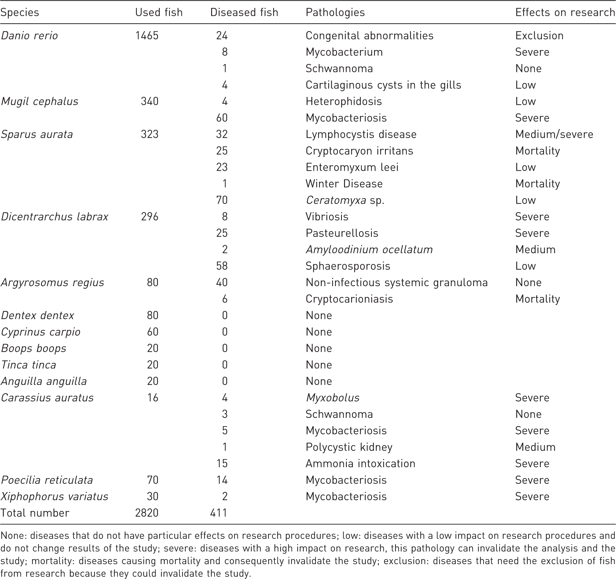
Four adult female, wild-type pond zebrafish, imported from Singapore and acquired for reproduction purposes from the ornamental fish trade before the new EU Directive 63/10, were positive for cartilaginous cysts in the gills during histological examination. On the basis of morphological criteria, the parasites were identified as trematodes belonging to the genus Centrocestus (Figure 1).
30
Centrocestus sp. larvae within cartilaginous cyst in zebrafish gills (haematoxylin-eosin (HE) 20x).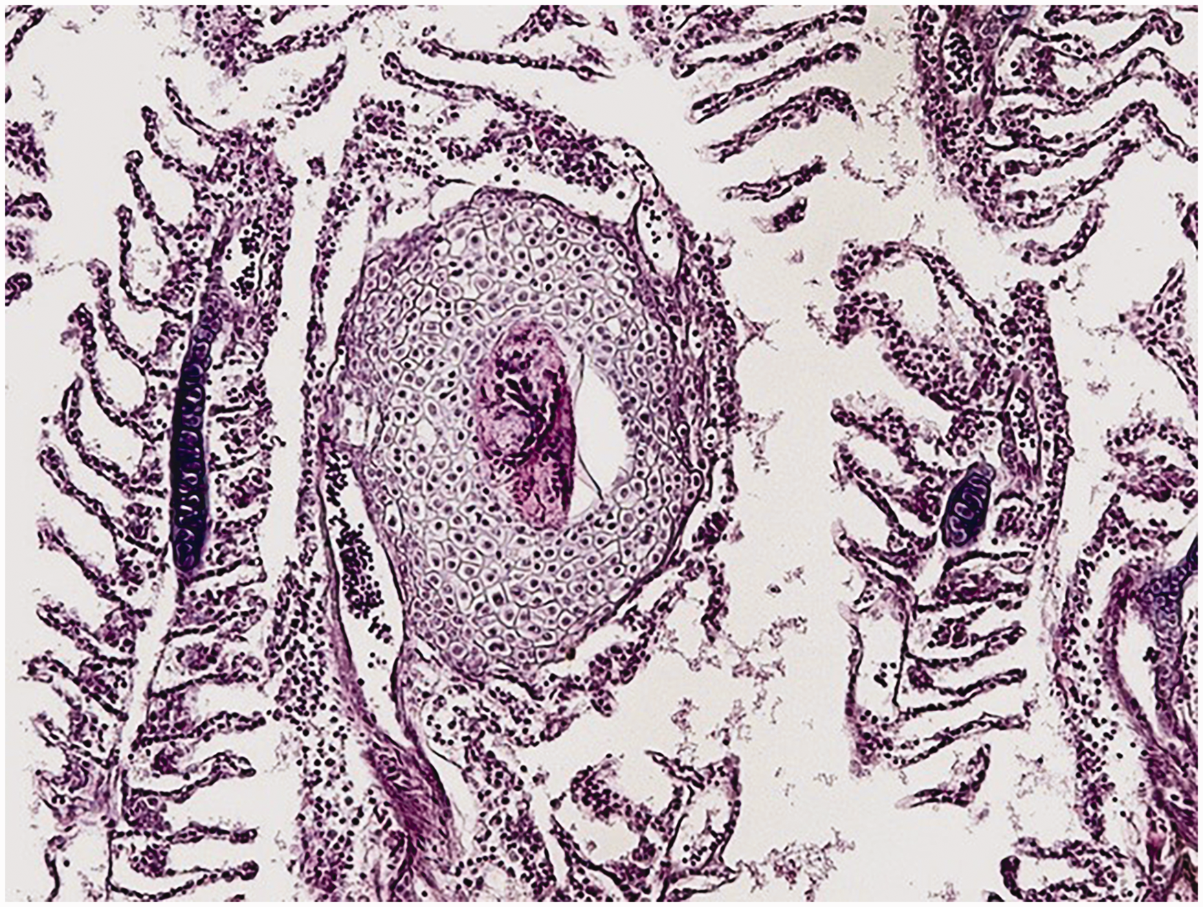
One adult wild-type D. rerio showed a black mass on the under-lip margin. Histological evaluation and immunohistochemistry were performed and a schwannoma, with a description reported by Marino et al., 31 was diagnosed.
Carassius auratus
A chemical test revealed a concentration of ammonia in the water above the normal level (0.5 mg/L) for 15 goldfish (nine males and six females) that showed symptoms of intoxication (erosions and epidermal bleeding). The fish-tank water was partially substituted and ammonia levels were back to normal in 24 h.
A goldfish showing abdominal distension was subjected to radiographic examination, which confirmed the presence of renal cysts.
In four goldfish (one male and three females) that showed white dorsal masses, a cytological examination was carried out after anaesthesia. Myxosporidiosis was diagnosed, and the genus Myxobolus was identified (Figure 2). The fish were euthanized after the identification of the parasitic spores.
Cytological imprinting smear obtained from nodules showing Myxobolus sp. in goldfish (May Grunwald Giemsa 40x).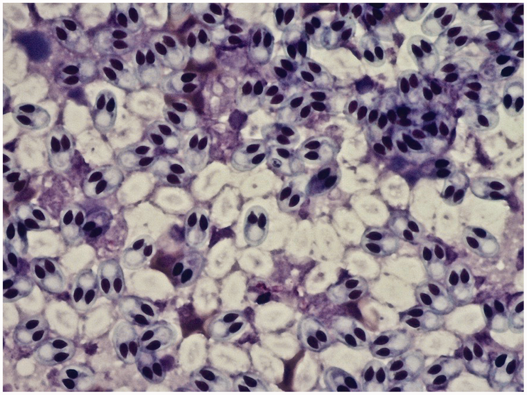
Schwannomas were diagnosed in three adult, 7-year-old goldfish (one male and two females) (Figure 3); these tumours appeared as subcutaneous soft nodular bulges which, in HE-stained sections, showed defined borders and were composed of elongated cells.
Macroscopical feature of schwannoma in goldfish fin.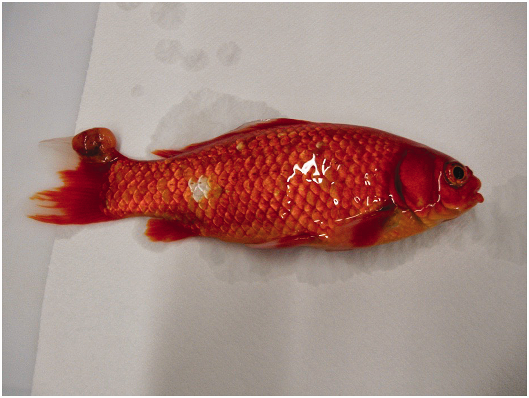
Five adult C. auratus (three males and two females) reared as broodstock in the same tank, two Xiphophorus variatus acquired from a private aquarium shop, and 14 P. reticulata (10 males and four females) reared in the same tanks in the facility and used for fish production within CISS developed a chronic infectious disease with ulcerated skin lesions and tumour-like nodules. The specimens of C. auratus and P. reticulata were euthanized, whereas those of X. variatus died spontaneously. All these fish were found positive for mycobacteriosis after histological examination showed granulomatosis; Ziehl-Neelsen staining demonstrated the presence of acid-fast bacteria, likely Mycobacterium spp.
Dicentrarchus labrax
The dinoflagellate Amyloodinium sp. was identified on fresh skin and gill samples analysed from two fish during a post-mortem parasitological examination.
Overall, 25 fish died of chronic photobacteriosis with necrotic foci and granulomas in the spleen (Figure 4), liver, kidney and heart due to the presence of Photobacterium damselae subsp. piscicida.
Photobacteriosis in seabass. Note enlarged spleen with necrotic/granulomatous foci.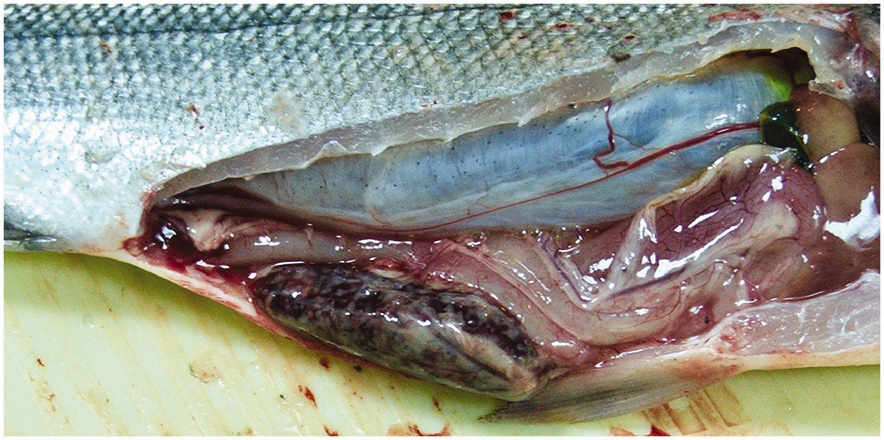
A total of 58 D. labrax were found positive for sphaerosporosis (Sphaerospora dicentrarchi) in the intestine during a post-mortem histopathological examination. Eight seabass that showed signs of acute infectious disease with skin and multi-organ haemorrhages were euthanized. These fish were positive for vibriosis (Vibrio anguillarum) during microbiological examinations.
Sparus aurata
After some months of farming, 32 apparently healthy 4-month-old S. aurata juveniles, both males and females, showed the typical clinical signs of lymphocystis virus disease with the appearance of several skin proliferations on the fins, mouth, dorsum and on the lateral side of the body. Three fish per tank were euthanized and autopsied; tissue samples were collected for histopathological and molecular analyses and confirmed the diagnosis of lymphocystis disease (Figure 5) by the presence of viral DNA in all analysed fish.
Detail showing hypertrophic dermal fibroblast with viral cytoplasmic inclusions (haematoxylin-eosin (HE) 40x). Arrows: cytoplasmic viral inclusion.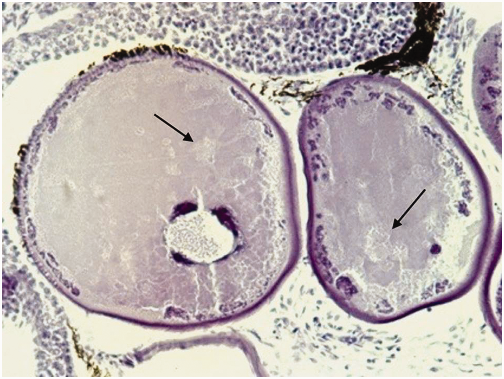
One single case of ‘winter disease’ was registered in a gilthead sea bream acquired a few days after transfer from a fish farm in early February. The fish showed pale skin, clouding eyes, abdominal swelling, pale liver, ascites and a haemorrhagic gut. The sea water on the fish farm was 14℃. A total of 23 S. aurata (10 males and 13 females) were histologically positive for enteromyxidiosis at post-mortem examination and Enteromyxum leei was identified.
During the parasitological exam, 70 adult fish (32 males and 38 females) were positive for gall bladder myxosporidian infection; Ceratomyxa sparusaurati was detected in fresh samples of the gallbladder of these fish, which produced no clinical signs. Myxosporidian parasites had colonized the mucosa of the intestine with varying severity.
Cryptocaryon irritans was seen in 25 fish after a partial water substitution with marine water. Mortalities appeared 3 days after the change, and the only obvious macroscopic signs were white spots on the skin of moribund fish.
Argyrosomus regius
At histological examination, 40 adult A. regius, both males and females, showed a chronic evolution of granulomas localized in the spleen, liver, kidney, intestine and heart. Six of these subjects died later of severe cryptocarioniasis.
Mugil cephalus
A total 64 wild adult M. cephalus (38 males and 26 females) were positive for zoonotic pathogens. Although no clinical signs were observed, four fish were histologically positive for trematode metacercaria of the genus Heterophyes and 60 were positive for mycobacteriosis.
The genus Mycobacterium was confirmed by immunohistochemistry and microbiological culture, whereas PCR confirmed that the main species was M. fortuitum. Overall, 32 mullet showed lymphocystis disease.
The other fish species (Dentex dentex, Cyprinus carpio, Boops boops, Tinca tinca and Anguilla anguilla) did not show any disease.
Discussion
In this study, the authors describe diseases found in zebrafish and other fresh and salt water teleosts, providing useful data for research.
Zebrafish (D. rerio) is the principal teleost species used at CISS. This fish can suffer from a wide range of pathologies.
In the last few years, the use of zebrafish has increased considerably in many fields of research. Low breeding costs, high fecundity of the broodstock, rapid organogenesis, transparency of embryos and the extensively studied development process have all made zebrafish a valuable animal model to study organogenesis disorders, test novel therapies and screen toxicity of chemicals and potential teratogens. 32 In particular, zebrafish embryos provide a reliable model for high-throughput screenings of chemicals, as they present the advantages of an in vivo model and the convenience of an in vitro approach.33,34 Recently, the use of zebrafish for the development of toxicology assays has been encouraged and produced the highest demonstrated concordance with rodent studies or similar data.35–38 Zebrafish developmental toxicity assays have a good predictive value for assessing chemicals or candidate drugs for human safety. However, the use of various zebrafish lines can result in a data discrepancy among different laboratories and with mammalian in vivo results because genetic background differences may affect zebrafish-strain susceptibility to a particular test substance. Therefore, in some cases, this variable could produce false positive or false negative results. 39 For the correct interpretation of results, not only the quality of the clutches used but also the rates and variations in the morphology encountered in the control groups of the zebrafish line should be considered. However, this information is rarely provided. Therefore, in light of the growing use of zebrafish for teratologic or toxicity screenings, we need to expand our knowledge of the spontaneous malformation rates in many of the generally used wild-type, transgenic and mutant lines. Some congenital abnormalities that are relatively common in zebrafish may be more frequent in transgenic lines.
In our study, the main disease was mycobacteriosis, which represents a potential risk for personnel. Numerous studies have previously been conducted on mycobacteriosis in zebrafish.40–45 However, cartilaginous cysts due to Centrocestus sp. metacercariae, despite being a potentially dangerous zoonosis, are not a health risk for laboratory personnel as the transmission of parasites occurs by ingestion of raw fish. The knowledge of ontogenetic anomaly prevalence in the different zebrafish lines is extremely important in trying to achieve high health standards and more reliable research results. Although P. neurophilia is one of the most common zebrafish pathogens, in our facility the histological and molecular examinations did not show any evidence of this pathogen in either brain or spinal cord tissues. 46 European seabass coming from fish farms can develop vibriosis and photobacteriosis. These two infectious diseases may interfere with the feasibility of experiments using this species by causing high mortality (90–100% in acute forms). Sphaerosporosis is a parasitic disease caused by the myxozoa S. dicentrarchi. This protozoan can be endemic in some fish farms; it is an opportunistic pathogen, generally with low pathogenicity, which can sometimes cause severe tissue changes and death. In our study, 58 D. labrax out of a total of 296 had several myxosporeans found in the muscularis mucosae of the intestine without any signs of inflammation.
Regarding the dinoflagellate A. ocellatum that we found in two specimens of D. labrax, the low parasitic charge, low incidence of disease and low tissue damage observed in the gills did not interfere with the results of the trial.
The protozoans C. irritans, E. leei and Ceratomyxa sp. that were found in some S. aurata fish during our study are frequently detectable in this species. C. irritans under experimental conditions can be lethal, with a very high mortality rate; thus these infections must be immediately identified and the infected fish excluded from experimental trials. E. leei and Ceratomyxa sp usually show low pathogenicity for gilthead sea bream.
‘Winter disease’ is a metabolic syndrome common in captive sea bream during the cold season; it causes gut dilation with undigested food and necrotic-haemorrhagic debris, which can sometimes be lethal.47,48 Another disease commonly detected in S. aurata is lymphocystis, a self-limiting viral disorder that usually occurs in sites less exposed to the immune system. 49
Non-infectious systemic granulomatosis, found in 40 A. regius in our experiment, is a nutritional disorder that is often detectable in coelomic organs of meagre less than 2 years in age. It is not transmissible and never causes death. No pathological agents have been related to this disease. It has been suggested that these granulomatous changes are caused by the deposition of tyrosine crystals related to a nutritional deficiency, possibly poly-hypovitaminosis. 50
The results showed that mycobacteriosis and myxosporidiosis are the most important risks found in this study and for fish research at CISS. These two diseases must be prevented and mitigated, as they can cause mortality and cannot effectively be treated. For this purpose, particular care should be used when new fish are introduced into a facility, including a quarantine period for mycobacteriosis (at least 30 days), 51 checking the fish for pathogens by antemortem diagnosis (often hard to apply in adult zebrafish), 52 or sacrificing a statistically significant number of fish for histopathology and molecular diagnostics. Production of fry and alevins within the facility allow avoidance of the abovementioned risk of transmission of infectious disease via egg bleaching. Most of the diseases encountered in our study could cause failure of experimental trials or the necessity to exclude specimens from statistical evaluation; this is damaging in terms of costs and time for researchers.
To provide a total picture of research results, it is important to report the health and welfare status of the fish, including programmes and methods applied to detect pathogens, agents to be investigated, reports of findings and interpretation of the results. Obviously, the availability of historical data on the health status of the spawners and alevins has definitive, strong value.
The diagnosis of disease in fish is sometimes difficult due to the presence of disorders with an unknown cause and diagnosis is often limited to morphological evaluation. In some cases, an ultra-structural exam must be carried out to detect specific pathogens; however, this technique has low reliability and is time consuming, which is often not compatible with the needs of the facility. 53 A sound knowledge of the pathogenesis of diseases could provide an improvement in detection techniques for pathogens that are sometimes responsible for unapparent and/or latent disease, thus replacing or reducing the use of the invasive diagnostic methods widely used to make diagnoses and to carry out health monitoring in fish populations. However, the detection of disease-causing pathogens during chronic infections has different limitations and is more difficult compared to detection and identification during the acute infection phase.
A good level of health surveillance has been achieved in our zebrafish facility due to the application of a reliable health monitoring programme, which is characterized by the following: daily checks of the fish health status; routine and non-routine total body histopathological evaluation of sentinels, dead and diseased fish that are supported by ancillary assays; close observance of hygienic procedures; and egg bleaching of incoming fish. Respect for standard operative procedures and, in particular, attention to biosecurity, i.e. animal transfers between quarantine and main rooms and bleaching eggs, will avoid infection with pathogens that cause clinical disease and could directly change results, increase variability, or result in a lack of reproducibility of experiments.
Based on our experience, outbreaks of infectious diseases and the latent presence of infectious microorganisms may be frequently recorded in fish from aquaculture industries, the ornamental fish trade and wild-caught animals (obviously the latter cannot be provided with reports on health status). Infectious diseases can affect both experimental trial feasibility and the analysis of experimental results by increasing variability in measured parameters among and within experimental units. To mitigate these challenges, farmed fish that have received periodic health monitoring are preferred over wild-caught animals when no purpose-bred fish are available. For ornamental species, eggs, fry and alevins produced under quarantine conditions are preferred over direct introductions from the ornamental fish trade. Recently, regulation in the EU has promoted a new perception of animal care and welfare. Almost all the diseases here described were registered before the application of the new Italian law D. Leg. 26/2014, in application of the EU Directive 63/2010. Certainly, the new regulations will require more attention during all phases, from production to rearing and experiments with laboratory fish, safeguarding the health of animals and guaranteeing the quality of experiments and results.
Footnotes
Declaration of Conflicting Interests
The author(s) declare no potential conflicts of interest with respect to the research, authorship, and/or publication of this article.
Funding
The author(s) received no financial support for the research, authorship, and/or publication of this article.
Ethical considerations
This article does not contain any studies involving human participants or animals performed by any of the authors.
Acknowledgements
The authors are particularly grateful to Prof Gabriella Gaglio and Francesco Macrì, and Dr Fabrizio Vitale, Stefano Reale, Daniele Macrì, Monique Mancuso, Giovanni De Benedetto, Fabiano Capparucci and Enrico Volpe for their support.