
Abstract
To understand the anatomical characteristics of microminipigs, one of the smallest miniature pigs, as a large animal model, we measured the body and organ sizes of four-, five-, six-, and seven-month-old microminipigs (n = 4, females) using computed tomography. In addition, the results were compared with those of young mature beagles (10 months old, two males and three females), which have been widely used as a large animal model. The microminipigs at 4–6 months of age were much smaller than the beagles. However, when the microminipigs reached seven months of age, their overall size was similar to that of the beagles. The thoracic cavity volume of the seven-month-old microminipigs was less than half that of the beagles, and the cavity was largely filled by the heart. The liver size of the seven-month-old microminipigs was approximately half of that of the beagles. Moreover, the spleen of the seven-month-old microminipigs was different in morphology, but not different in size from that of the beagles. In addition, although their volumes were the same, the kidneys of the seven-month-old microminipigs, unlike those of the beagles, were flattened in shape. Collectively, the major abdominal organs of the seven-month-old microminipigs were either the same size or smaller than those of the beagles, but the abdominal cavity volume of the seven-month-old microminipigs was larger than that of the beagles. Thus, the abdominal cavity of microminipigs is assumed to be filled with the gastrointestinal tract. The anatomical characteristics of the young mature microminipigs revealed in our study suggest that microminipigs could have great potential as a large animal model for biomedical research.
Keywords
Rodent models have established a special position among laboratory animals because rearing costs are low and handling is easy; and because various research tools and methodologies, such as transgenic techniques that enable the animals to mimic human diseases, are available. However, anatomical and physiological differences between rodents and humans complicate direct extrapolation of study results to humans. Therefore, experiments using animals other than rodents are necessary in pharmacological and toxicological studies. Thus, studies using large animal models are essential and their development is inevitable.
Because of similarities with humans, dogs and pigs have been selected as large animal models, and are most commonly used in biomedical research.1–4 However the phrase ‘a dog is man’s best friend’ dominates discussions in the field of animal experiment; thus, using dogs for experiments is becoming more difficult each year. 2
By contrast, the use of pigs for experiments in biomedical research is increasing, and 60,000 pigs are used annually in the European Union.5,6 This is because in addition to their similarities with humans, pigs are recognized as ‘food’ animals, and the emotional hurdles for their experimental use are not very great. 2 Therefore, pigs are highly suitable as an experimental animal which bridges the physiological gap between humans and rodents which then enables findings to be extrapolated to humans.5,6
Compared with domestic pigs, miniature pigs require low maintenance costs and small quantities of test reagents, and can be handled with relatively little effort, making them very attractive as laboratory animals. Thus, miniature pigs, including NIH(MGH), Göttingen, Yucatan, Sinclair, Hanford, and Clawn, have been developed at various research institutions. However, just as domestic pigs weigh over 200 kg, miniature pigs are also sometimes just too big for experimental use.
However the microminipig developed by Fuji Micra Inc (Fujinomiya, Shizuoka, Japan) is one of the smallest miniature pigs, with an adult weight of 20 kg.7,8 Their size is about the same as a medium-sized dog and they are of a suitable size for laboratory use as a large animal model. Thus, the use of microminipigs in experiments is expected to increase.
To obtain reliable experimental outcomes and perform sophisticated animal experiments, the biological and genetic backgrounds of the subject animals need to be defined. Microminipigs certainly have high utility for their smallness. However, to realize the full potential of the newly developed pig breed as a laboratory animal, more needs to be known about microminipigs as compared with other domestic pigs. At the moment no difference is known other than their smallness, and sufficient information is yet to be collected.8,9 Therefore, in this study, we measured the body and major organ sizes of microminipigs using computed tomography (CT) to document some of their anatomical characteristics. In addition, results were compared with those of young mature beagles, which have been widely used as large animal models, in order to provide images of the size of microminipigs.
Animals
Microminipigs
Young mature animals are generally considered appropriate for experimental use, so microminipigs are commonly shipped to laboratories at the age of 4–6 months. 6 In this study, sizes of the bodies, limb bones, and major organs of 4–7-month-old female microminipigs were measured on a monthly basis.
Rearing of microminipigs
Four newly weaned female microminipigs, their initial weight and height at three months of age was 3.3 kg (2.8–4.4 kg) and 23 cm (19–24 cm) (median [minimum–maximum]), were purchased from Fuji Micra Inc and kept in a controlled room with a temperature of 24 ± 3℃ and a humidity of 50–70%. Five microminipigs, including four used in this study, were kept in a 4.0 m × 2.8 m sized pen, and a 1.8 m × 1.2 m sized rubber mat was prepared for their bedding. Lighting in the room was set on an 11 h light/13 h dark cycle starting at 08:00 h. The microminipigs were fed MMP pellets (3% BW, Marubeni Nisshin Feed Co, Ltd, Tokyo, Japan), a feed specially developed for microminipigs, which comprise total digestible nutrients (TDN, >74%), crude protein (>13%), crude fat (>2.0%), crude fiber (>8.0%), crude ash (<1.0%), calcium (>1.1%), and phosphorus (>0.9%), and had free access to water.
Beagles
Five 10-month-old TOYO beagles were purchased from Kitayama Labes, Nagano, Japan (two males and three females). 10 These young mature beagles, which have been widely used as large animal models, were used as a reference to understand the sizes of the microminipigs.
The beagles were reared for at least one month, and were then used in this study after a veterinarian confirmed the absence of any clinical abnormality. Throughout the study, the dogs were given food (Select Skin Care; Royal Canin Japon, Inc, Tokyo, Japan) daily, according to the amount instructed on the product, and had free access to water.
Materials and methods
This study was conducted with the approval of the Animal Care and Use Committee of Gifu University. Also, the care and use of laboratory animals were conducted in compliance with the guidelines of Good Laboratory Practice.
Determination of body sizes
From 4–7 months of age, the body weight, body height, chest width, and chest circumference of the microminipigs were measured on a monthly basis. The body weight, height from the fore-hoof to the back at the mid-scapular, and the chest width and circumference at the mid-scapular were measured using a scale, a ruler, and a measuring tape. 11
Determination of bone lengths and organ sizes using CT scanning
CT scanning was performed monthly after intramuscular administration of 0.02 mg/kg medetomidine (Domitor; Nippon Zenyaku Kogyo Co, Koriyama, Japan), 0.2 mg/kg midazolam (Dormicum injection 10 mg; Astellas, Tokyo, Japan), and 0.15 mg/kg butorphanol (Vetorphale; Meiji Seika Pharma Co, Tokyo, Japan) to sedate the microminipigs during the procedure. The entire body of each microminipig was analyzed using a four-detector helical CT (Asteion TSX-021B; Toshiba Medical Systems, Tochigi, Japan). Parameters for image acquisition were a 512 × 512 matrix, medium-scan field of view, 2 mm slice, 120 kVp, 100 mA, and helical pitch of 5.5.
We defined the length of the limb bones as the maximal length between the epiphysial extremities, and the diameter as the mid-portion width between the epiphysial extremities. The length and diameter of the humerus, radius, femur, and tibia were measured with a volume rendering image in a DICOM viewer (OsiriX MD; Pixmeo Sàrl, Bernex, Switzerland). 12 In order to determine the volume of organs, i.e. the heart, liver, spleen, and kidneys, regions of interest (ROI) on CT images were individually segmented by manual drawing and reconstructed into a 3D ROI model.
Statistical analysis
The differences in physical size, length of limb bones, and volume of major organs among each age group of microminipigs and the beagles were evaluated using Mann–Whitney U-tests. Differences yielding P values <0.05 were considered significant.
Results
During this study, all the microminipigs used showed normal weight gain for microminipigs, 6 and their daily gain was 0.09 kg (0.08–0.11 kg). Also, no clinical abnormality was found in any animal.
Body weight, height, chest width, and chest circumference of four-, five-, six-, and seven-month-old microminipigs and 10-month-old beagles.
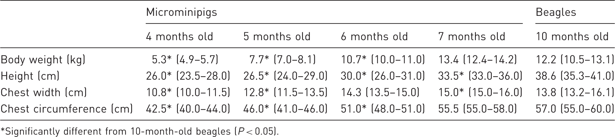
*Significantly different from 10-month-old beagles (P < 0.05).
Left limb bone length and diameter of four-, five-, six-, and seven-month-old microminipigs and 10-month-old beagles.
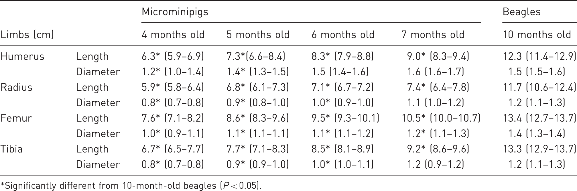
*Significantly different from 10-month-old beagles (P < 0.05).
The thoracic cavity volume of the seven-month-old microminipigs was only half that of the beagles (Table 3). The heart volume of the four-month-old microminipigs was approximately 30% that of the beagles, but it increased to 60–70% at seven months of age. The heart of the microminipigs was situated more cranially than that of the beagles, and the thoracic cavity of the microminipigs appeared to be largely filled by the heart (Figure 1).
Reconstructed 3D ventral–dorsal (upper) and L–R lateral (lower) images of a 10-month-old beagle and a seven-month-old microminipig. Reconstructed heart, liver, spleen, and kidneys are shown in volume rendering images. Volume of thoracic and abdominal cavities, heart, liver, spleen, and kidneys in four-, five-, six-, and seven-month-old microminipigs and 10-month-old beagles. *Significantly different from 10-month-old beagles (P < 0.05).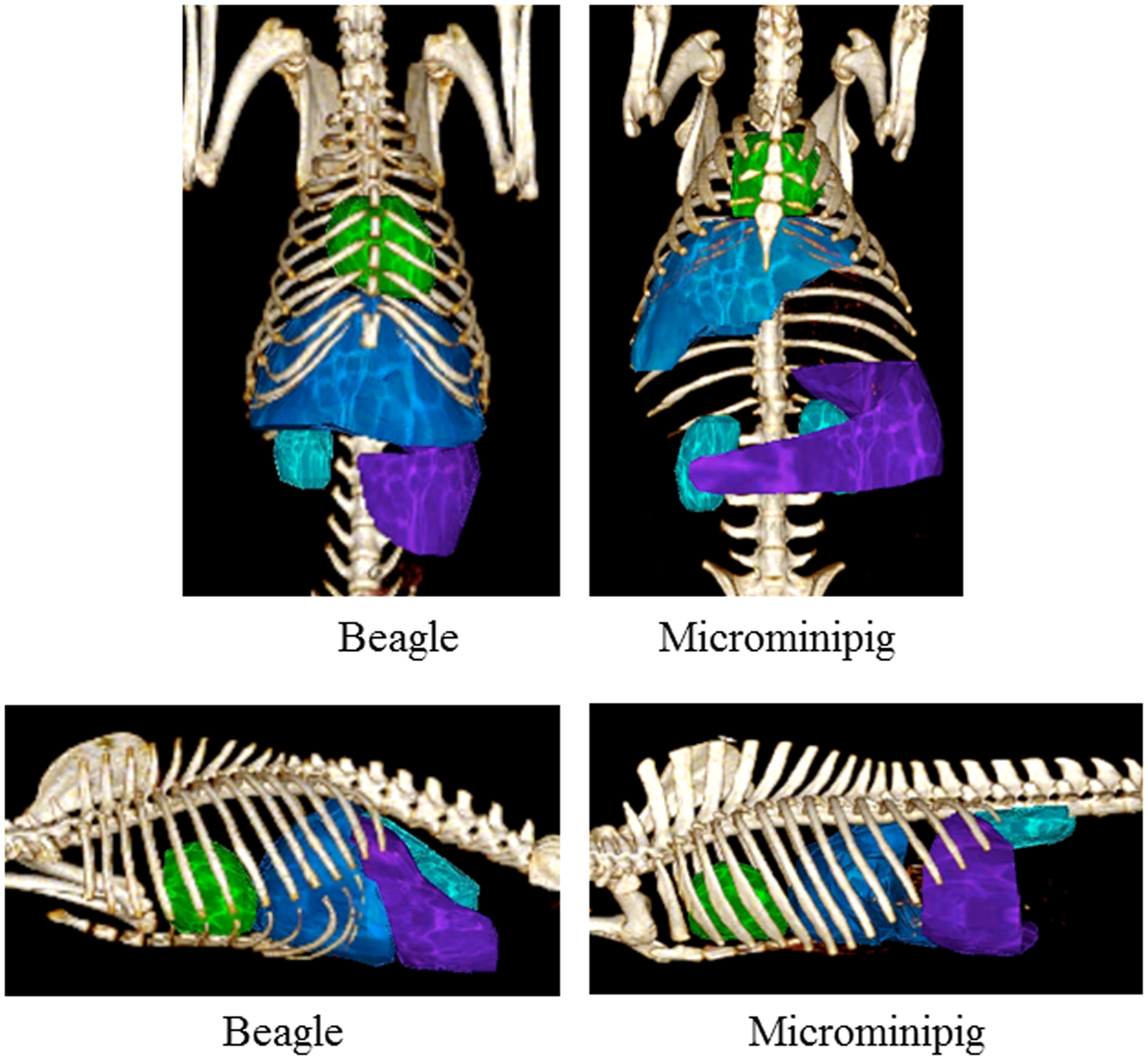
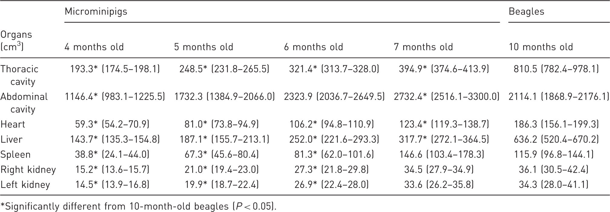
Even at seven months of age, the liver of the microminipigs was half that of the beagles. While each spleen of the microminipigs exhibited different shapes (Figure 2), their size at seven months of age was no different from that of beagles. The kidney volumes of the microminipigs and the beagles were the same, but the kidneys of the microminipigs, unlike those of the beagles, were flattened in shape (Figure 3). Although the physical size of the seven-month-old microminipigs was similar to that of the beagles, their abdominal cavity volume was larger. The major abdominal organs of the seven-month-old microminipigs were comparable in size with those of the beagles, except for the livers of the seven-month-old microminipigs which were half the size of those of the beagles. These results suggest that the gastrointestinal tract filled out more space in the abdominal cavity of microminipigs compared with that of the beagles.
Reconstructed images of the spleens of seven-month-old microminipigs (No. 1 to No. 4). Reconstructed ventral–dorsal (left) and L–R lateral (right) images of kidneys of a 10-month-old beagle and a seven-month-old microminipig.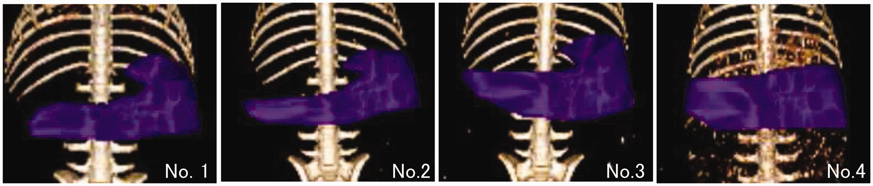

Discussion
In this study, the body size, CT images of organs, organ positions, and their volumes of four-, five-, six-, and seven-month-old microminipigs were obtained, and the results were compared with those of 10-month-old beagles. This study is unique in two ways. First, anatomical traits of microminipigs were measured from live individuals using diagnostic imaging techniques in order to understand their sizes, which differ from other studies where body weight gain and weight of excised organs have been used as indicators.13,14 Second, the results obtained from microminipigs were compared with those of beagles which are widely used as laboratory animals. The results clearly showed the smallness and anatomical traits of microminipigs, and suggested their great potential as a large animal model for biomedical research and for medical training.
The 10-month-old TOYO beagles used in this study, which were slightly larger than the beagles produced by Marshall Farm, NY, USA (average 8.7 ± 1.1 kg at 9–10 months of age), but similar in size to the beagles produced by Harlan France (average 11.3 ± 1.0 kg at 9–10 months of age) or by the Centre of Laboratory Animals Production, Havana, Cuba (average male 11.8 ± 1.9 kg and female 10.3 ± 1.7 kg at 8–10 months of age),15,16 were standard size beagles and were appropriate as a reference for understanding the sizes of microminipigs. Therefore, researchers and technicians who have used beagles in experimental studies or medical training should be able to easily image the body and organ sizes of microminipigs. The rearing facilities and laboratories used for beagles could also be easily adapted for use with microminipigs.
Ethical concerns make it difficult to use dogs in experiments, so they have been replaced by other animals in many studies. Under these circumstances, miniature pigs have become a leading substitute for dogs. 2 However, it is difficult to shift to miniature pigs from dogs because they are still relatively large, weighing 30–40 kg under controlled feeding. Of course, pigs are not always suitable substitutes for beagles in all experiments because dogs and pigs are very different species and differ both anatomically and physiologically.17,18 Nevertheless, because of their similarity in physical size to young mature beagles, which are widely used for biomedical research, young mature microminipigs are considered good candidates as a substitution for dogs as a large animal model.
Needless to say, the selection of animals with defined biological backgrounds and phenotypic and genetic homogeneity is a key aspect of a reliable and sophisticated experiment. As shown in this study, microminipigs are small-sized and have a high potential as a non-rodent model. Choices of animals will increase by establishing microminipigs as a laboratory animal, enabling more refined experiments by choosing the optimal type of animal. Consequently, to enhance the value of microminipigs as a laboratory animal and facilitate their wider use in the biomedical field, the characteristics of microminipigs must be further researched.
Footnotes
Declaration of conflicting interests
The authors declare that there is no conflict of interest.