
Abstract
We performed immunogold labeling with an ST-1 monoclonal antibody (IgM), specific for intact heparin, to define the subcellular localization of heparin in mast cells. Rat peritoneal mast cells were fixed by a modified Karnovsky method and embedded in Araldite. Ultrathin sections were first treated with sodium periodate and then sequentially incubated with MAb ST-1, rabbit anti-mouse IgM, and protein A-gold. By transmission electron microscopy, gold particles were localized inside cytoplasmic granules of peritoneal mast cells. In contrast, with the same procedure, no labeling was observed in mast cells from rat intestinal mucosa. Control sections of rat peritoneal or intestinal mucosa mast cells treated with an irrelevant MAb (IgM) did not show any labeling. Treatment with nitrous acid abolished the reactivity of MAb ST-1 with peritoneal mast cells. These results show that different mast cells can be identified regarding their heparin content by immunochemical procedures using MAb ST-1.
M
Immunocytochemistry is an accurate method for subcellular localization and characterization of different molecules, and this approach has been used successfully to define the presence of several glycoconjugates (Straus et al., 1993, 1996; Irani et al., 1989; Craig et al., 1993). In a similar fashion, using a postembedding immunogold labeling with guinea pig antihistamine antiserum, Login et al. (1992) were able to determine the subcellular localization of histamine in granules of rat peritoneal mast cells.
In this study, using the monoclonal antibody (MAb) ST-1 specific to nonmodified heparin molecules (Straus et al., 1992), we demonstrate by immunocytochemical methods the localization of heparin in granules of rat peritoneal mast cells. Furthermore, the results presented here corroborate data indicating absence of heparin in MMCs.
Materials and Methods
Animals. Four male Wistar rats of approximately 300 g were obtained from Central Animal Care of Universidade Federal de São Paulo/EPM.
Mast Cells. Rat peritoneal cells (n = 4) were harvested by washing the peritoneal cavity with 15 ml of 0.9% NaCl. The peritoneal exudate containing the mast cells was centrifuged at 800 X g for 10 min at 4°C.
The small intestine was removed (n = 4), flushed of fecal material with 0.9% NaCl, and cut into small fragments for ultrastructural studies.
Fixation, Processing, and Embedding for Electron Microscopy. Cell suspension and small intestine fragments were fixed by immersion in Karnovsky's fixative (2% formaldehyde freshly prepared from paraformaldehyde, 2% glutaraldehyde, 0.025% CaCl2, 0.1 M sodium cacodylate buffer, pH 7.4) for 1 hr at 25°C, washed in sodium cacodylate buffer twice for 15 min each, dehydrated through a graded series of ethanol, and embedded in Araldite 502 Resin (EMS; Fort Washington, PA). Approximately 90-nm sections were cut on diamond knives on an ultramicrotome (Reichert Ultracut; Leica, Austria) and placed on nickel grids (EMS) for immunogold labeling.
Postembedding Immunogold Labeling. For murine heparin detection, immunogold staining method using Protein A conjugated to gold (Bendayan, 1984) was performed as follows. Sections (90 nm) of peritoneal mast cells and intestine on nickel grids were floated section side down on 30-μl drops of the following reagents at 20°C: (a) 0.1% sodium metaperiodate for 1 hr; (b) washed in distilled water; (c) 1% bovine serum albumin (BSA) in PBS (137 mM NaCl, 10 mM Na2HPO4, pH 7.4) for 1 hr; (d) primary antibody: MAb ST-1 (culture supernatant) or an irrelevant mouse IgM for 24 hr at 4°C; (e) washed five times for 3 min each in 1% BSA/PBS, 0.1% Tween 20; (f) secondary antibody: rabbit anti-mouse IgM (Dako; Carpinteria, CA) diluted 1:50 in 1% BSA/PBS for 1 hr (the secondary antibody, rabbit anti-mouse IgM, is essential for the protein A-gold immunolabeling because protein A does not bind to mouse IgM); (g) washed three times in 1% BSA/PBS, 0.1 Tween 20; (h) protein A conjugated to 15-nm colloidal gold (E-Y Labs; San Mateo, CA) diluted 1:10 in 1% BSA/PBS, for 1 hr; (i) washed twice in 1% BSA/PBS, 0.1% Tween 20, and washed twice in distilled water. Controls were carried out using either an irrelevant IgM (culture supernatant) or omitting the primary antibody. Unstained sections were examined in a JEOL 1200 EXII electron microscope at 60 kV.
Nitrous Acid Treatment. Nickel grids containing mast cell sections were incubated with 0.24 M NaNO2 in 1.8 M acetic acid for 80 min at room temperature as described by Höök et al. (1975). Under these conditions, glucosamine residues containing both free amino groups or sulfoamino groups are deaminated. In control experiments, NaNO2 was omitted and the sections were incubated with 1.8 M acetic acid instead.
Morphometric Analysis. Randomly photographed sections of rat mast cells (enlarged to X 34,000) were used. The area and number of gold particles over 146–188 granules in 5–10 representative cells (2–3 cells from each sample) were determined for each labeling protocol. Granule areas were determined with a point-counting method using a square test grid with 87-mm spacing. The number of gold particles was calculated and expressed per μm2 of granule area. Statistical analysis was done with a modified t-test (Williams, 1977).
Results
Heparin Immunocytochemistry
MAb ST-1, specific for heparin molecules, was used in an immunogold label to define the subcellular localization of heparin in rat peritoneal mast cells. By electron microscopy, ST-1 labeling was restricted to granules of rat peritoneal cells (Figure 1A). A higher magnification of peritoneal mast cell granules immunolabeled with ST-1 is shown in Figure 1B. No gold particles were detected over the nuclei or other organelles, and only low labeling at background level was observed over the cytoplasm. No labeling was observed in cells other than mast cells in the peritoneal exudate.
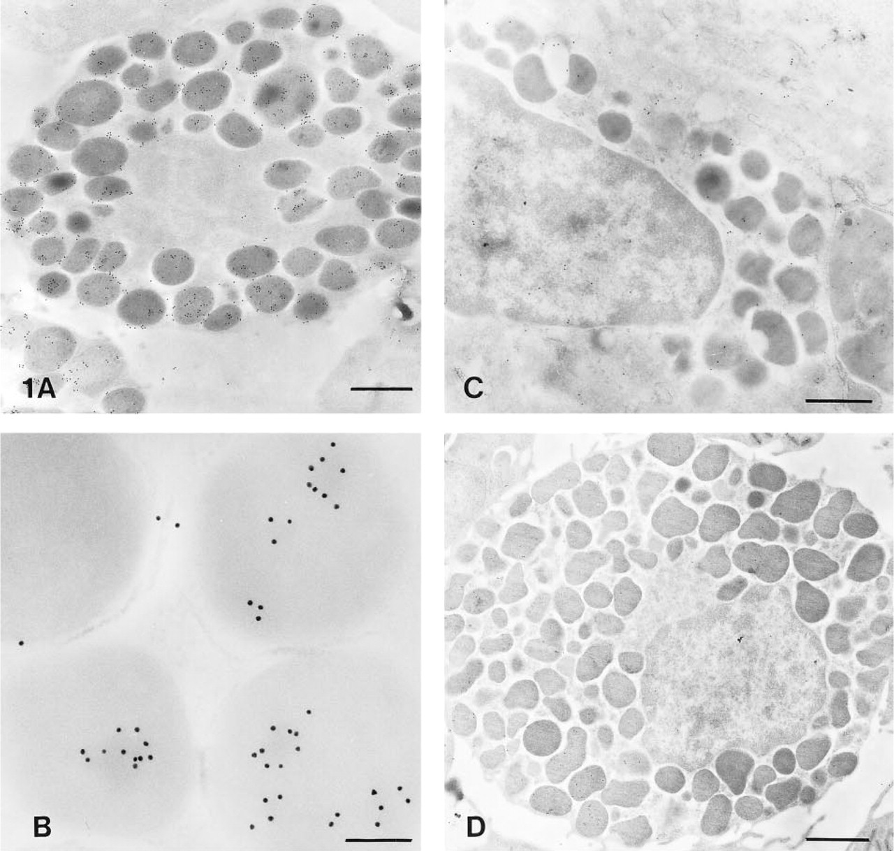
Electron micrography of rat mast cells.
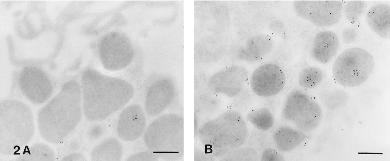
Electron micrography of peritoneal mast cells labeled with ST-1 after incubation with nitrous acid
In intestinal mucosal mast cells no significant labeling was observed with MAb ST-1 (Figure 1C), indicating the absence of heparin in these cells. In control experiments in which MAb ST-1 was replaced by an irrelevant antibody (IgM), no significant labeling was observed in peritoneal mast cell sections (Figure 1D).
The number of gold particles after ST-1 labeling on granules was determined by morphometric analysis of randomly photographed sections of rat peritoneal and intestinal MMCs. Peritoneal mast cells labeled with MAb ST-1 exhibited an average of 51 gold particles/μm2, whereas intestinal MMCs exhibited 5.1 gold particle/μm2, as shown in Table 1.
Effect of Heparin Deamination on ST-1 Binding to Peritoneal Mast Cell Granules
As shown by Straus et al. (1992), MAb ST-1 does not react with deaminated heparin. To confirm that immunogold labeling was specific for the heparin present in mast cell granules, the grids were treated with nitrous acid, as described in Materials and Methods. A significant decrease of labeling was observed after deamination (Figure 2A) compared with control experiments treated only with acetic acid (Figure 2B). As shown in Table 1, by morphometric analysis it was determined that rat peritoneal mast cell granules after nitrous acid treatment exhibited 6.7-fold less ST-1 labeling (7.4 particles/μm2) than untreated mast cell granules.
Discussion
This is the first report of an immunocytochemical study using an MAb against the intact heparin molecule. As reported by Straus et al. (1992), MAb ST-1 does not crossreact with other GAGs such as heparin sulfate, chondroitin sulfate, or dermatan sulfate. MAb ST-1 reacts with heparin from different sources as well as with heparins with high or low anticoagulant activities obtained by affinity chromatography in a Sepharose-anti-thrombin III (AT-III) column. In commercial heparin preparations, approximately one third of the molecules contain the AT-III binding region and exhibit high anticoagulant activity. These results clearly indicate that the putative AT-III binding site of heparin represented by the pentasaccharide (Lindahl, 1989),
Immunogold labeling of rat mast cells with MAb ST-1
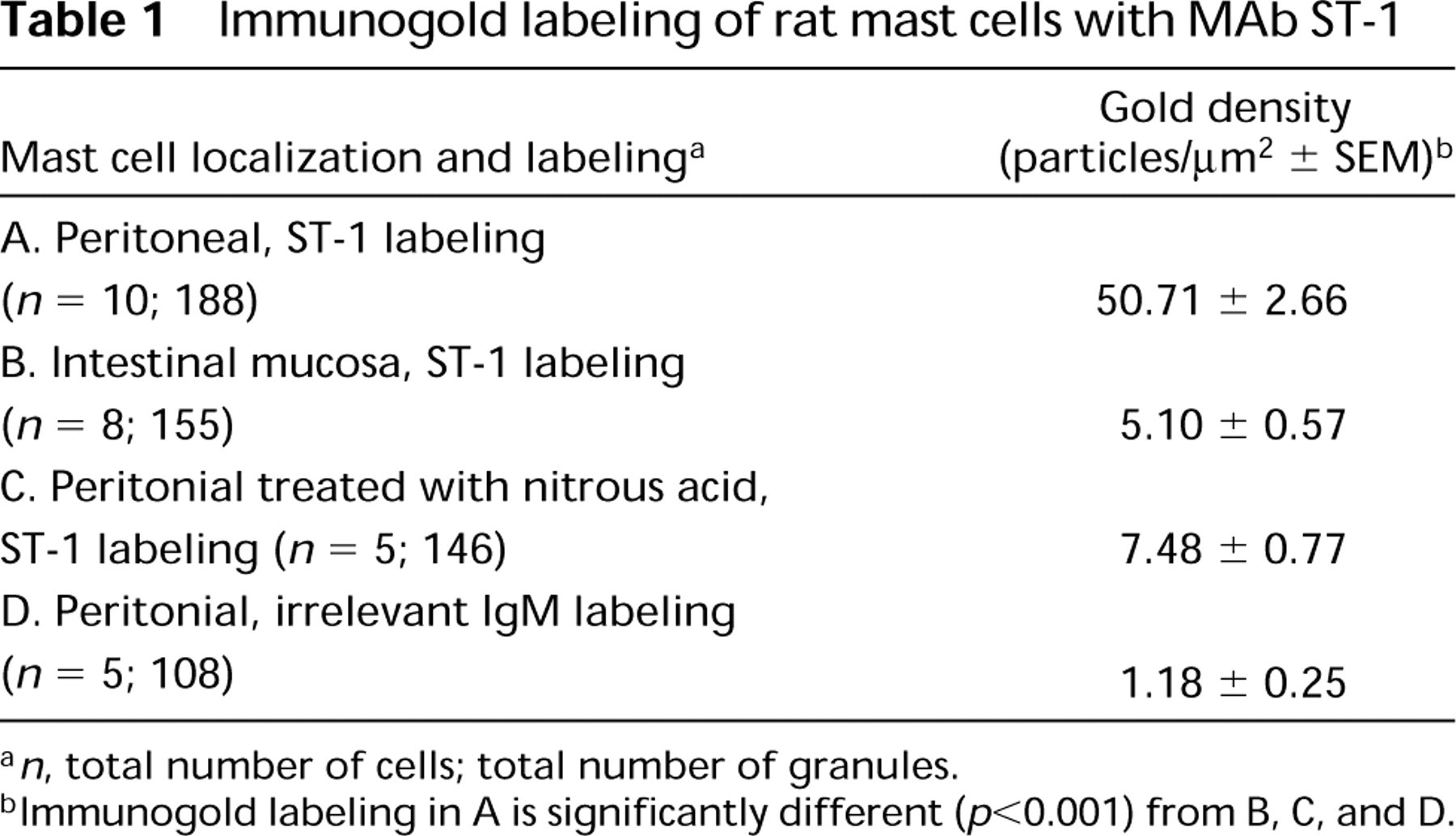
a n, total number of cells; total number of granules.
bImmunogold labeling in A is significantly different (p<0.001) from B, C, and D.
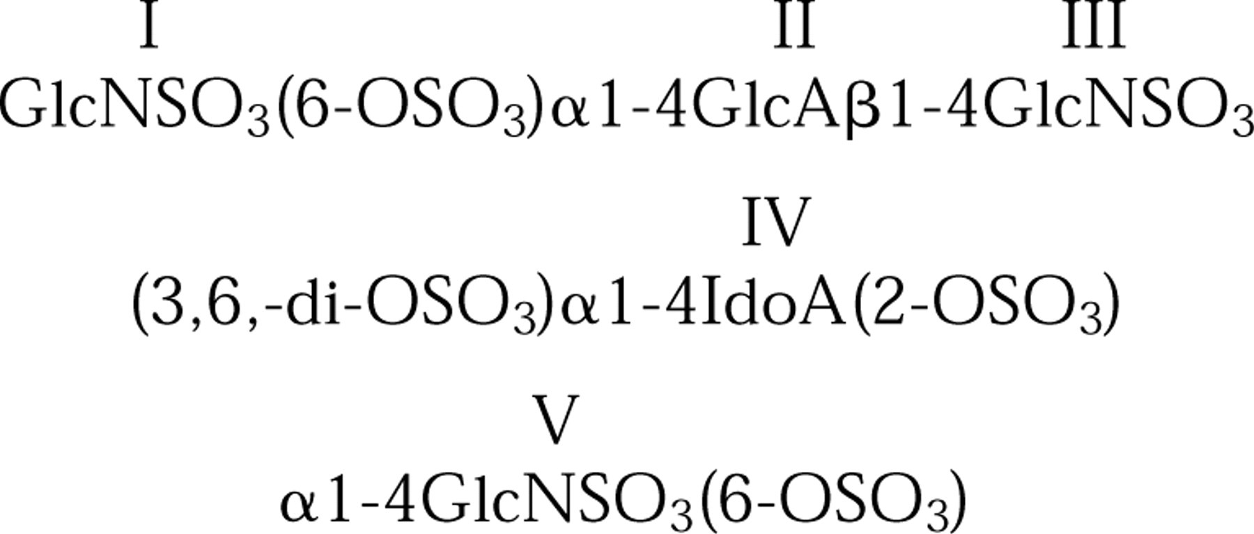
is not required for MAb ST-1 binding to heparin. The data presented here show that MAb ST-1 can be a potential tool for evaluation of heparin content in mast cells. Immunocytochemical analysis shows positive labeling of rat peritoneal mast cells but not of intestinal MMCs. The labeling was restricted to cytoplasmic granules of mast cells, and no significant labeling was observed in eosinophils or other cells of the peritoneal cavity. Control experiments using an irrelevant antibody were performed and significant labeling of granules was not observed, indicating that the binding is not due to a nonspecific binding of immunoglobulins to cytoplasmic granules. In addition, treatment of mast cells with nitrous acid, which deaminates heparin (Höök et al., 1975) significantly decreased (p<0.001) mast cell labeling with ST-1. On the other hand, as expected, control experiments using peritoneal mast cells incubated with acetic acid showed strong labeling when incubated with MAb ST-1. Recently, Craig et al. (1993), using AT-III-gold, showed labeling of rat peritoneal connective tissue type mast cells but not of mucosal mast cells. Our data are in agreement with these results, and because MAb ST-1 recognizes heparin regardless of its affinity for AT-III, it appears correct to conclude that lack of labeling with AT-III was not due to the absence of heparin molecules with high affinity for AT-III but rather was due to the absence of heparin molecules. The method presented here permits analysis of the expression of heparin in cells from different sources and can be useful to study the biosynthetic and degradative pathways of heparin during the differentiation processes of mast cells.