
Abstract
Osteoclasts are the only bone-resorbing cells. In addition to other specific properties, osteoclasts are characterized by their expression of tartrate-resistant acidic phosphatase (TRAP), which is usually detected using a histochemical method for light microscopy. Using ELF97 phosphatase substrate, this study describes a new fluorescence-based method for TRAP detection. The fluorescence-based ELF97 TRAP stain not only results in a better resolution of the TRAP-positive granules, because confocal microscopy can be applied for image acquisition and analysis, but it reveals additional and more specific information about osteoclasts because it can be combined with other fluorescence-based methods.
O
A histochemical method, commercially available as a kit by Sigma Diagnostics (St Louis, MO), is widely used and has proved to be the standard method for specific detection of osteoclasts. This histochemical TRAP staining results in a color precipitate that can be easily detected using light microscopy. Although this classical TRAP stain is easy to perform, it has the disadvantage that it is rather difficult to combine with other specific stains such as those used in immunohistological methods. Because of this problem, a fluorescence-based TRAP staining method was developed and tested that could be combined with other fluorescence-based stains, including a nuclear DNA stain, an immunofluorescence stain, and an actin stain using fluorescence-labeled phalloidin. For that purpose, ELF97 phosphatase subtrate (Molecular Probes; Eugene, OR) was tested because it has been used successfully for the detection of alkaline phosphatase activity with fluorescence microscopy (Cox and Singer 1999).
First, the ELF97 TRAP protocol was established and compared with the classical TRAP method. Human osteoclast-like cells were generated in vitro from adherent peripheral blood mononuclear cells (PBMCs). Briefly, PBMCs were isolated from buffy coats [Australian Red Cross Blood Service (ARCBS); Perth, WA, Australia] through Ficoll-gradient centrifugation. The PBMCs were cultured (37C, humidified, 5% CO2) in 25-cm2 culture flasks (Sarstedt; Nuernbrecht, Germany) in RPMI 1640 medium (GIBCO; Auckland, New Zealand), supplemented with antibiotics (GIBCO) and 10% human A serum (ARCBS). After 1 hr in culture, the non-adherent PBMCs were discarded and the adherent cells washed twice with PBS before being cultured in osteoclast differentiation medium composed of RPMI 1640, antibiotics, 5% human A serum, and cytokines (recombinant human M-CSF, 10 ng/ml; recombinant human RANK-L, 10 ng/ml; R&D, Minneapolis, MN). Freshly isolated adherent PBMCs and cells, cultured for 1–30 days were fixed with 1–3% paraformaldehyde in PBS for 30 min to 24 hr and subsequently processed for cytospins and TRAP staining. After 3–30 days, the cultured cells were stained using the classical TRAP protocol to confirm that they were TRAP-positive (Figure 1A). The kit from Sigma Diagnostics was used according to the recommended protocol and it produced positive TRAP staining in all cultures (blood samples from more than 20 individuals were tested). The results were analyzed using a Leica DM RBE microscope and recorded by a LEAF digital camera (Leica; Wetzlar, Germany). In parallel, using the same cells, the ELF97 protocol was established. The same buffer used for the classical TRAP staining was used for the ELF97 protocol (110 mM acetate buffer, pH 5.2, 1.1 mM sodium nitrite, 7.4 mM tartrate). Different dilutions and incubation times were tested, indicating that a 200-μM concentration of ELF97 and a 15-min incubation time were the most appropriate conditions. These conditions were used for all future fluorescence TRAP stains. It should be noted that, similar to the classical TRAP staining, no cell membrane permeabilization step is needed for the ELF97 TRAP staining. The fluorescence signal was analyzed and documented by conventional fluorescence microscopy or by confocal microscopy (Bio-Rad MRC 1024, Coherent Enterprise argon ion 250-mW multi-line UV, American Laser 100-mW argon ion multiline laser, Melles-Griot 0.5-mW green helium-neon laser; Bio-Rad, Hercules, CA). By omitting the ELF97 substrate (data not shown) or by staining freshly isolated TRAP-negative cells (Figure 1B), no fluorescence signal for TRAP was detected. However, when cultured over 3 days or longer in osteoclast differentiation medium, the adherent PBMCs became TRAP-positive. This indicates that the cells had differentiated towards osteoclast-like cells under these culture conditions. Comparing the resulting morphology of the classical and the ELF97 TRAP stains, ELF97 gave much better results. The fluorescent precipitate was clearly restricted to the area of the granules, whereas the classical TRAP staining resulted in a rather diffuse granular pattern in which the single granules were difficult to separate from each other. The accuracy of the ELF97 TRAP stain was enhanced and clearer when confocal microscopy was used.
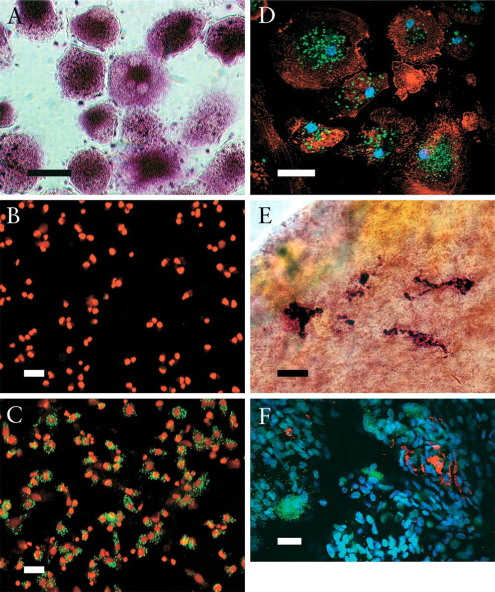
TRAP staining of human in vitro generated osteoclast-like cells (
Second, the ELF97 TRAP method was combined with other staining protocols. Not only fluorescence detection of surface molecules but also detection of intracellular molecules were of interest because for the latter cell membrane permeabilization is usually needed. First, a fluorescence nuclear counterstain with DAPI (1 μg/ml in PBS, 4′,6-diamidine-2′-phenylindole dihydrochloride; Roche Diagnostics, Mannheim, Germany) and Syto25 (5 nM in PBS; Molecular Probes) was tested after the ELF97 TRAP staining. No membrane permeabilization was required to get a brilliant fluorescence signal for the nuclear DNA (Figures 1B-1E). After the ELF97 TRAP staining, immunofluorescence stains of surface markers were tested, including CD45 and MHC surface molecules, which worked well and gave excellent results (data not shown). Finally, fluorescence stains for cytoskeleton elements were tested, for which membrane permeabilization (60 sec, 0.1% Triton X-100 in PBS) is needed after the ELF97 stain. The basal actin ring was stained with AlexaFluor546-labeled phalloidin (0.2 U/ml in PBS; Molecular Probes) (Figure 1D). The cellular tubulin structure was stained using an immunofluorescence protocol and a mouse anti-tubulin monoclonal antibody (data not shown). In both cases of fluorescence-based cytoskeleton staining, the results were excellent, without alteration of the ELF97 TRAP fluorescence signal.
It was of interest whether the ELF97 TRAP stain works in tissue samples and in other species. Therefore, the ELF97 TRAP protocol was tested in freshly excised rat tibial periosteum. Ten rats (Lewis; Guidelines of the Research Ethics and Animal Care Office were followed) were anesthetized before sacrifice. The periosteum from the anterior surface of both tibias was stripped from the bone and fixed in 3% paraformaldehyde/PBS. The tissue was stained as a whole tissue sample either with the classical TRAP stain (Figure 1E) or using the ELF97 TRAP protocol (Figure 1F). Omitting the phosphatase substrate resulted in negative TRAP staining (data not shown). Using either protocol resulted in clear staining of osteoclasts in the cell-rich internal layer of the rat tibial periosteum. The ELF97 TRAP stain was also combined with diverse fluorescence staining protocols, including a nuclear stain with DAPI and an immunofluorescence protocol for staining of MHC class II using OX6, a monoclonal mouse anti-rat MHC class II antibody (Serotec; Oxford, UK), followed by a polyclonal biotin-labeled sheep anti-mouse antibody (Amersham Biosciences; Piscataway, NJ) and AlexaFluor546-labeled streptavidin (Molecular Probes). This combined staining resulted in clear and specific staining of polynucleated cells for ELF97 TRAP and of mononucleated cells for MHC class II in the cell-rich internal layer of the rat periosteum (Figure 1F).
Summarizing, one can conclude that the fluorescence-based ELF97 TRAP stain provides a comparable if not even better option than the classical histochemical TRAP stain. Using the ELF97 TRAP protocol, one can take advantage of confocal microscopy to obtain high-resolution images. In addition, the ELF97 TRAP stain can probably be combined with most fluorescence stains, including immunofluorescence protocols. Elf97 TRAP stain not only works with single cells and cultured cells for cytological analysis but it also gives excellent results with tissues. Furthermore, it can probably be applied for different species, including humans and rats. Finally, fluorescence-based TRAP stain with ELF97 opens up new histological possibilities in the area of bone and osteoclast research.
Footnotes
Acknowledgements
Supported by the S.E. Ohman Medical Research Fund (Western Australia), the Dudgeon Smith Medical Research Fund (Western Australia), and by the AO Research Fund (Switzerland).
I thank Mary Lee and Steve Parkinson for their excellent technical support and Serge Camelo for providing the animal material. I also thank Paul Rigby from the Biomedical Confocal Microscopy Research Centre for his excellent technical support and advice, Guy Ben-Ary from the IAAF for his excellent technical support, and Len Freedman for critical review of the manuscript.