
Abstract
We assessed three different visualization systems used routinely in research and diagnosis of transmissable spongiform encephalopathies (TSEs) to demonstrate whether the methodology applied to immunohistochemical (IHC) examination may alter the results concerning detection of prion protein (PrPsc) in the lymphoreticular system (LRS): avidin-biotin–peroxidase (Vectastain ABC kit; Vector), Envision (DAKO), and catalyzed signal amplification (CSA; DAKO). The study aimed to determine which of these showed the highest sensitivity, with the hope of providing an accurate tool for pathogenesis and preclinical diagnosis research in TSEs. Histological sections from palatine tonsils, spleen, GALT (ileum and ileocecal valve), and lymph nodes from sheep belonging to a Spanish scrapie-positive flock were processed by IHC using L42 as primary antibody. As substrate chromogen, diaminobenzidine was used, and all slides were subjectively assessed by light microscopy. A further study using an image analyzer software system was carried out to confirm that the conclusion provided by microscopic examination was objective. The CSA system showed the highest sensitivity in all cases, increasing both variables assessed: the number of follicles that were PrPsc-positive was detected as well as the intensity of immunostaining in each of them.
S
The detection of PrPsc in the LRS (Ikegami et al. 1991; van Keulen et al. 1996) has caused this tissue to become an interesting target for diagnostic goals. Specifically, tonsil (Schreuder et al. 1996) and nictitating membrane (O'Rourke et al. 1998a,b) biopsies are the two most widely investigated options.
In this sense, both methods are a practical screen for early detection of PrPsc in living affected sheep (Kim et al. 2001) because of their capability to detect scrapie infection in lymphoid tissue even before symptoms occur (Schreuder et al. 1998; Miller et al. 1993; Muramatsu et al. 1994). However, a subset of sheep with scrapie in several previous studies have shown accumulation of PrPsc in CNS tissue but not in lymphoid tissue (van Keulen et al. 1996; O'Rourke et al. 2000). This result stresses the fact that a PrPsc-negative tonsil or third eyelid biopsy does not indicate that an animal is free of infection. The most widely accepted explanation for this is that the replication dynamics of the scrapie agent may be influenced by different variables such as stress, management practices, or husbandry, with the genotypic differences in the host being the most influencing variable (Jeffrey et al. 2001b). Nevertheless, to our knowledge, whether or not the enhancement of the sensitivity of conventional IHC approaches makes possible the detection of PrPsc in the lymphoid system of the total number of positive cases has not been yet investigated. Further studies in this field should therefore be carried out.
In this study, different visualization systems used routinely in research and diagnosis (Hardt et al. 2000; Lezmi et al. 2001; Monleón et al. 2003) were assessed to determine whether the methodology applied for IHC examination using lymphoid tissue may alter the results obtained. Furthermore, the study intended to determine which of the systems shows the highest sensitivity, with the aim of providing an accurate tool for investigation of the early pathogenesis of scrapie and, consequently, for preclinical diagnosis.
Materials and Methods
Sheep
All 16 animals included in this study belonged to a Rasa Aragonesa sheep flock in which an outbreak of natural scrapie occurred in June of 2002. Since the flock has been quarantined and regularly monitored, several more cases of scrapie have been diagnosed. All the sheep were raised under the normal conditions for a sheep-producing flock, with no physical separation between animals, and they were removed from the flock just before culling. Rules established in the National Research Council's guide for animal experimentation were followed for animal handling and care. The study involved a total of seven positive cases (diagnosis was confirmed by neurological examination, histopathology, and IHC in all of them), all of which occurred in adult female sheep of genotypes corresponding to ARQ/ARQ. The genotype of the nine negative animals was also of intermediate and high susceptibility (ARQ/ARQ, ARQ/ARH, or ARR/ARQ). The seven scrapie-positive sheep exhibited symptoms of the disease.
Necropsy and Tissue Collection
Necropsy was performed immediately after natural death or after sacrifice of the animal (by IV injection of sodium pentobarbital and exsanguination). The brain was removed from each sheep for scrapie diagnosis. The following tissues were taken and placed in fixative (formalin 10%): palatine tonsils, spleen, Peyer's patch of the ileum and ileocecal valve, mesenteric lymph node, retropharyngeal lymph node, and mediastinal lymph node. The tissues were trimmed, post-fixed, and embedded according to standard procedures. Tissue sections 4 μm thick were cut on a microtome and mounted on treated glass slides (Vectabond; Vector, Burlin-game, CA) and dried overnight at 37C. Sections were dewaxed and hydrated by routine methods before the antigen retrieval procedure.
Immunohistochemistry
Pretreatment and Antibodies. To ensure the specificity of the pathological PrP immunostaining, several pretreatments as previously described (Hardt et al. 2000) were performed. Immersion in 98% formic acid for 15 min, proteinase K treatment for 15 min at 37C (Roche, Mannheim, Germany; 4 μg/ml), and hydrated autoclaving (immersion in distilled water in a pressure cooker) were applied to all sections before use of any of the visualization systems tested in the present study.
PrPsc detection was performed using the monoclonal antibody L42 (R-Biopharm, Darmstadt, Germany; 1:500) in all cases.
Immunostaining Procedures
Avidin-Biotin–Peroxidase System. Endogenous peroxidase activity was inhibited with 15-min incubation in 3% H2O2 peroxidase in methanol. Nonspecific antigenic sites were blocked by a 20-min step in diluted normal serum (Vector) before the sections were incubated for 1 hr at 37C with the primary antibody. Then the sections were first incubated at RT with a biotinylated secondary antibody (Vector) for 30 min and second with the peroxidase-conjugated biotin-avidin complex (Vectastain ABC kit) for 40 min. Finally, the peroxidase was revealed by immersion in DAB (Vector).
Envision System. To block the endogenous peroxidase activity, a step with blocking reagent (DAKO; Glostrup, Denmark) for 5 min was included before incubation with the primary antibody for 30 min at RT. The enzyme-conjugated polymer (EnVision; DAKO, 30 min) and DAB (DAKO) were used as visualization system and chromogen, respectively.
Catalyzed Signal Amplification (CSA) System. Because of the high sensitivity of this system, an endogenous biotin blocking step by sequential application of avidin and biotin (CSA Ancillary System, DAKO; 10 min) was necessary before endogenous peroxidase activity blocking (DAKO; 5 min). After incubation for 15 min at RT with the primary antibody, sections were covered with the biotinylated secondary antibody (CSA; DAKO) and the streptavidin-biotin complex (CSA; DAKO) for 15 min in both cases. The following step consisted of the application of streptavidin-per-oxidase (CSA; DAKO) for 15 min after the incubation with the biotinyl tyramide as amplification reagent (15 min), being finally revealed by using DAB as substrate chromogen. Several additional steps were added in this protocol according to the manufacturer's recommendations to reach a greatly amplified signal (application of amplification reagent, CSA), and for reduction of background (addition of protein block, CSA).
Controls
Sections corresponding to lymphoid tissue samples in which PrPsc immunostaining had been previously observed by using the three systems tested were used in each IHC run, applying the same primary antibody (L42, positive control) as well as a non-inmune serum (mouse serum, DAKO; negative control). The reproducibility (necessary for the comparative study) of the immunostaining was ensured by including serial sections from the same tissue samples in the assay for every visualization system.
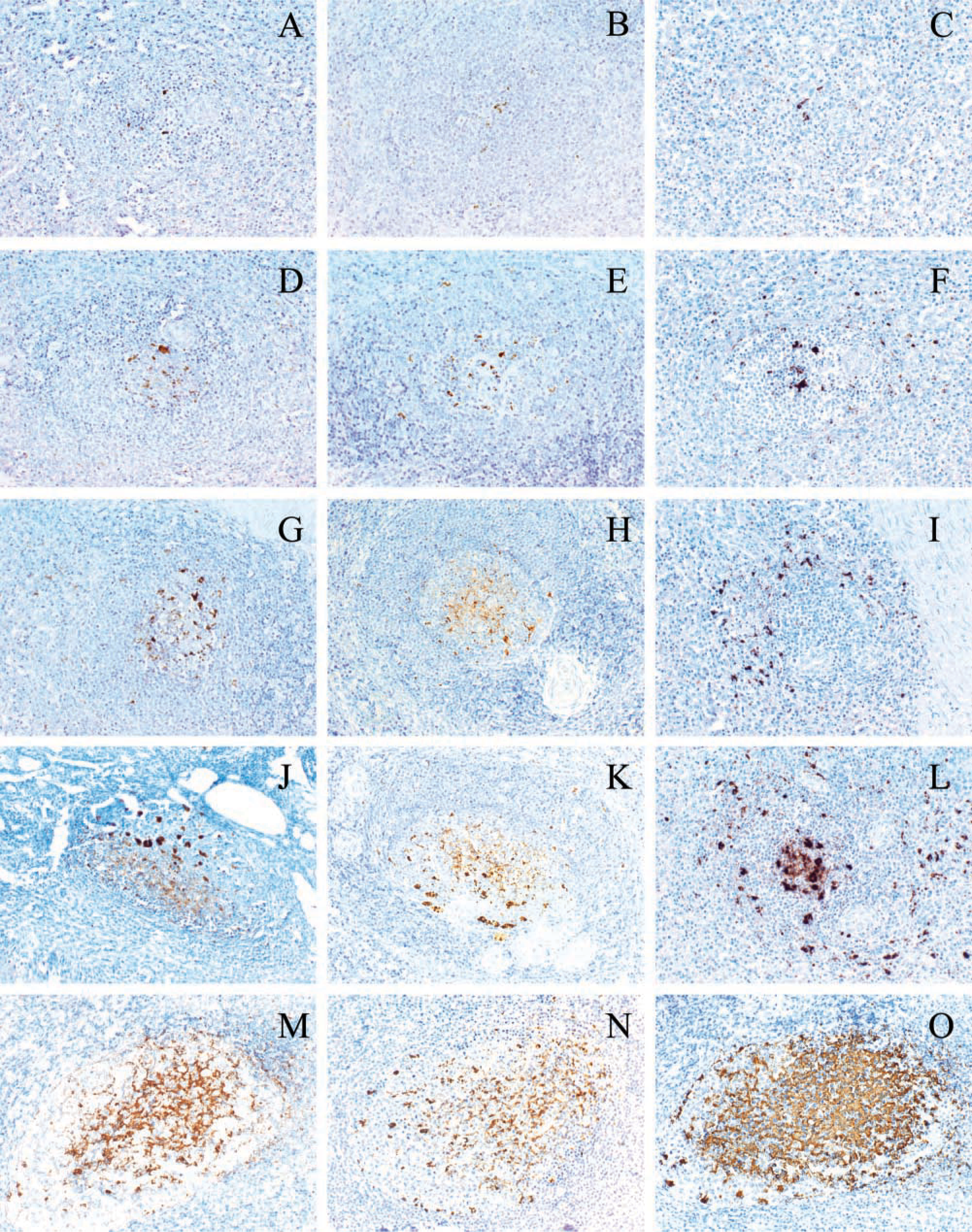
Intensity of PrPsc immunolabeling found in scrapie-positive LRS depending on visualization system used (ABC, EnVision, or CSA). (
Microscopic Assessment
All the lymphoid follicles present in the slides corresponding to all tissue samples were assessed in the present study. The follicles in which PrPsc was detected by light microscopic examination (×20 objective) were individually collected for each slide. Furthermore, each follicle was subjectively scored according to the intensity and the area to which the labeling was extended; (+, trace of light brown labeling in a reduced area of the follicle; + + + + +, intense brown labeling over most of the follicle) (Figure 1).
Image Analyzer Assessment
To confirm that the microscopic assessment provided accurate and reliable results about the sensitivity of the systems tested in the study, an objective assessment methodology was additionally applied. A further study consisting of assessing the intensity of immunostaining in the same 10 lymphoid follicles present in each sample examined using an image analyzer software system (MIP4) was carried out. A single tissue (tonsil) corresponding to three different animals and two visualization systems (EnVision and CSA systems) was chosen in this case.
Statistical Study
A Fisher's test was used to compare the intensity of immunostaining detected by light microscopy as well as the number of pixels obtained by using the image analyzer, both provided by the different visualization systems tested.
Results
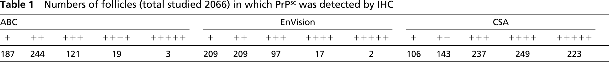
A total of 2066 lymphoid follicles were finally assessed in the present study as PrPsc accumulation was detected. No immunostaining was detected either in CNS or in lymphoid tissue of any negative sheep. No nonspecific staining was observed in the controls used in this study. Both tingible body macrophages and follicular dendritic cell labeling patterns (Jeffrey et al. 2000) were recognized in all tissues. Accumulation of PrPsc within the dark and light zones in the case of the secondary follicles was detected, although the immunolabeling was present in the dark zone to a lesser extent. The results of the IHC analyses of all samples examined from the seven positive sheep are shown in Table 1.
Numbers of follicles (total studied 2066) in which PrPsc was detected by IHC
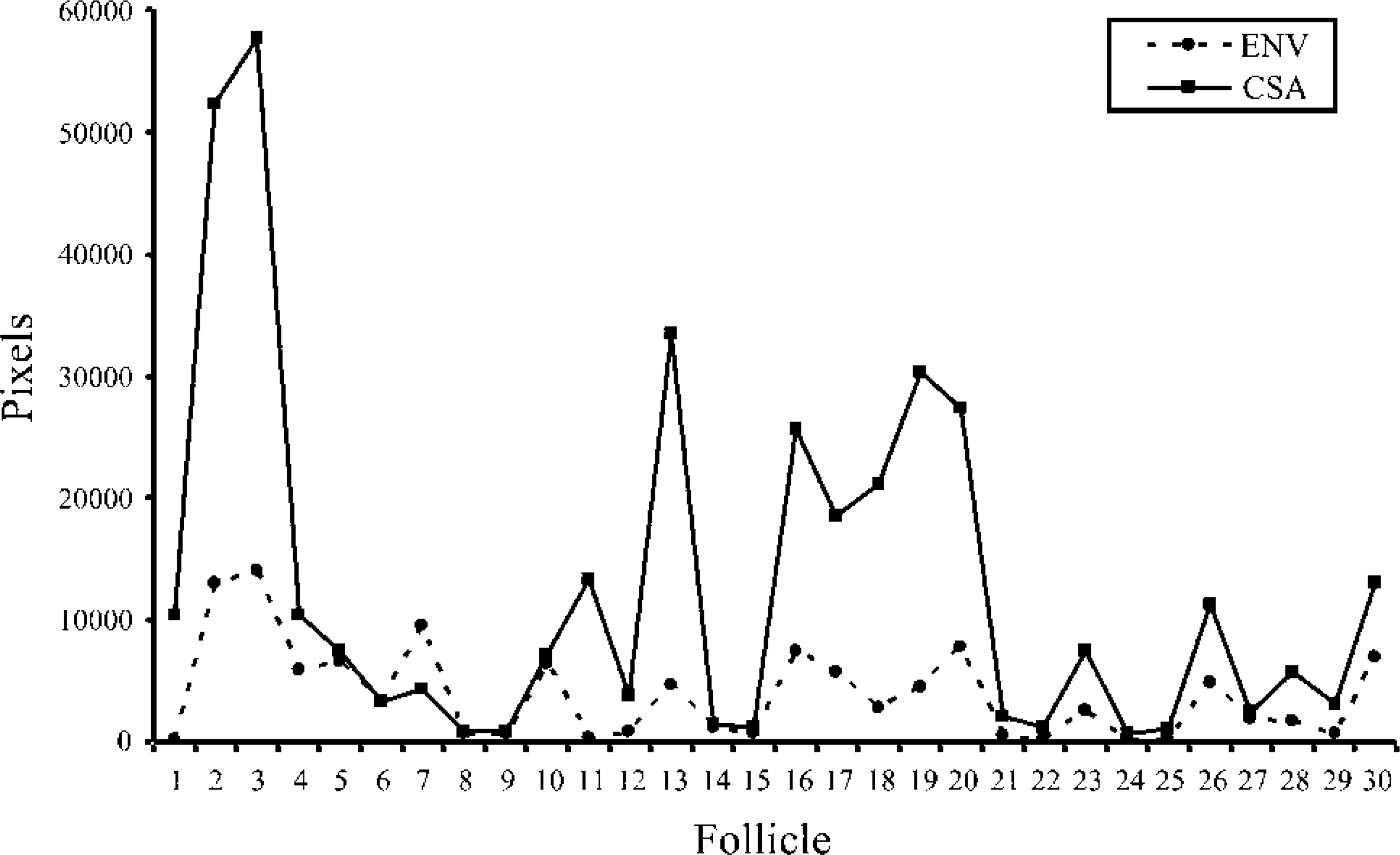
Representation of the number of pixels (using the image analyzer software system) provided by applying EnVision and CSA systems to analysis of the same 10 follicles in tonsils of three different sheep.
No statistical differences (p>0.05) between the results obtained by the ABC and the EnVision systems were observed on the basis of the results provided by this study. However, although no different results concerning presence/absence of PrPsc in any sample were found, the number of follicles in which it was detected, as well as the intensity scored in each of them, differed when the CSA system was used (p<0.0001).
Not only did CSA detect positivity in almost twice the number of follicles (958) than did other two visualization systems (574 and 534, respectively), which means an improvement in sensitivity of about 40% (40.1 and 44.3) over the ABC and EnVision systems, but the CSA system even assessed a total of 223 follicles with the highest score intensity (+ + + + +) against two or three follicles with this same score being allocated by using the other two systems. Furthermore, whereas + and + + intensity was provided by the ABC and EnVision systems in approximately 75% of the total of follicles assessed, the proportion was inverted with use of the CSA system, in which this percentage corresponded to the follicles scored with + + +, + + + +, and + + + + +, specifically, 50% of follicles with + + + + and + + + + +.
On the other hand, according to the results obtained in the present study, tonsil was the tissue in which the highest number of follicles were considered as positive (the number of follicles corresponding to lymph nodes, 1049, is the sum of those considered in three lymph nodes: mesenteric, mediastinal, and retropharyngeal), regardless of the visualization system used. This fact, in combination with the findings about significant and nonsignificant differences between the systems tested, made it possible to carry out a comparative study by image analyzer using a single source of tissue (tonsil) and two visualization systems (EnVision and CSA).
Concerning the image analyzer assessment, statistical differences (p<0.005; Figures 2 and 3) between the number of pixels provided by using both EnVision and CSA systems were found, the most sensitive system reaching a value (12,610.93 ± 2721.37) of more than three times the mean obtained with EnVision (3858.37 ± 694.58).
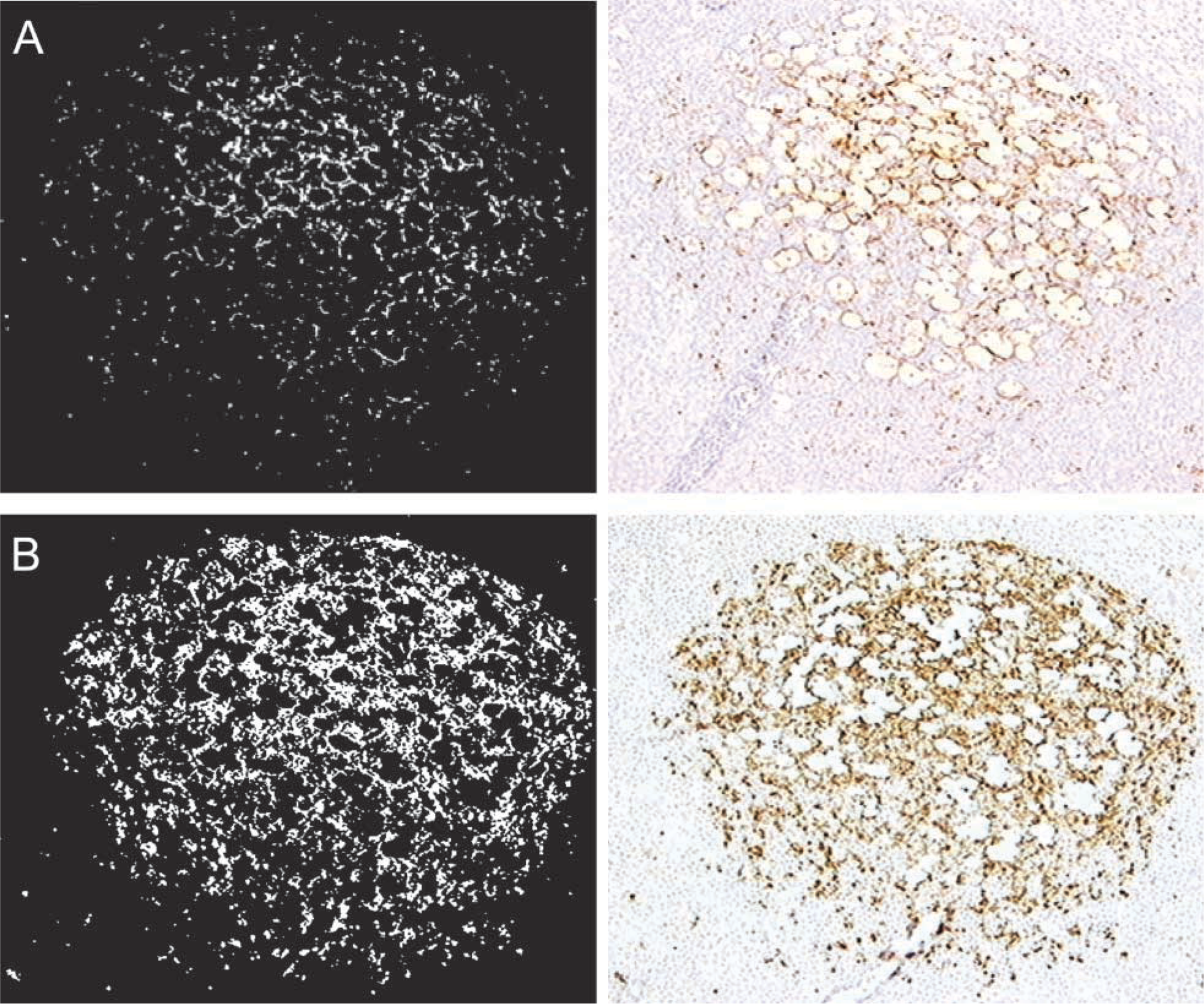
Images provided by using the image analyzer software system (MIP4) after application of EnVision (
Discussion
How TSE's infectious agent propagates from LRS tissues to the CNS in naturally occurring scrapie is not yet clear. However, it is assumed that the LRS is involved in the prion neuroinvasion (van Keulen et al. 1996,2000; O'Rourke et al. 2000). On the other hand, differences in scrapie agent strains, husbandry and management practices, weight of infection present on pasture or within the flock, all constitute possible influences on the infection rate and the disease progression (Jeffrey et al. 2001b). Nevertheless, the most widely accepted hypothesis about the most influencing factor is that the replication dynamics of the scrapie agent and rates of PrPsc accumulation differ as a result of genotypic differences in the host (Hunter et al. 1996,1997,2000; Andréoletti et al. 2000).
Although the enhancement of sensitivity of conventional methods could be considered a possible option for the detection of the whole number of positive scrapie cases (and therefore also of preclinical animals), not many works dealing with this practical alternative can be found in the literature. One study applying the tyramide signal amplification system (Heggebo et al. 2000) showed an increased sensitivity that could prove the enhancing staining to be a useful tool in research on the pathogenesis of scrapie. Concerning the main aim of the present study, to confirm whether some differences in sensitivity among routinely used protocols exist and to what extent, it has been achieved by means of an exhaustive study comparing three different visualization systems. The two systems most frequently applied in the laboratory, ABC and EnVision, have not demonstrated any evident differences in results, which is an important observation as they had never been previously compared. However, the increased sensitivity shown by the CSA system in all samples analyzed in this work, even achieving the detection of PrPsc deposits in a much higher number of follicles than the other two systems when the same sample examination is referred to (specifically, in 54 follicles as compared to 11 and two detected by ABC and EnVision, respectively, in one case; data not shown) underscores the relevance of the choice of visualization system. This would become important in the detection of preclinical scrapie cases, in which only a sparse number of follicles might be immunostained for PrPsc in a very early stage of the disease. Moreover, the relevance of this choice increases to a greater extent when a bioassay applied to tonsil or nictitating membrane biopsies is carried out because of the small size of the in vivo sample and consequently the small numbers of follicles subjected to examination.
All the clinically affected sheep showed marked and widespread PrPsc accumulation throughout all tissues sampled. Moreover, according to other authors' findings (Wadsworth et al. 2001) concentrations of PrPsc were shown to be nonuniform throughout the LRS. However, the quantitative differences among different tissues, as described in other works (Andréoletti et al. 2000; Jeffrey et al. 2001b), were not evident in the present study.
In conclusion, the lack of an absolute IHC protocol performance for this group of diseases encourages the search for new methodologies for the study of in vivo diagnostic strategies and of prion pathogenesis that would be both reliable and as sensitive as possible. According to the results provided by the present study, the CSA system may play an important role in this field. In addition, Jeffrey et al. (2001a) suggest a new method for identifying different scrapie strains by IHC PrP profiling in the CNS and LRS. The need for a very sensitive test that ensures the detection of PrPsc in the whole total of scrapie cases is therefore highlighted and, as a consequence, the relevance of the implications of the study described here is indeed demonstrated.
Footnotes
Acknowledgements
We thank the technical staff for assistance in processing the samples and the University of Zaragoza, Instituto de Salud Carlos III, and Gobierno de Aragón for financial support.
The statistical work developed by Iñaki Albizu (EXOPOL S.L.) is also gratefully acknowledged.