
Abstract
A novel antigen retrieval procedure was carried out in the post-embedding immunogold electron microscopy method to improve the stainability of the samples. This was done by weakly fixing cultured Helicobacter pylori (ATCC43504) and embedding in Lowicryl K4M. Before staining with the anti-H. pylori antibody, the ultrathin sections were mounted on a nickel grid and heated at 121C for 15 min, 99C for 40 min, and 65C for 24 hr in distilled water, 0.1 M phosphate buffer (pH 7.4), 0.01 M EDTA (pH 7.2), 0.05 M Tris buffer (pH 10.0), 0.8 M urea (pH 7.2), 0.01 M citric acid (pH 6.0), or a commercially available target unmasking fluid (S1699; pH 6.0). Antigen retrieval in the Tris buffer solution generally showed better stainability than the classical post-embedding method without any antigen retrieval. At 65C for 24 hr, better stainability of the ultrasections was observed for each of the solutions used except for the phosphate buffer compared to the control. We suggest that the antigen retrieval method should be applied for routine use even by in postembedding immunogold electron microscopy.
Keywords
E
About a decade ago, an antigen retrieval (AR) technique, which made the various antigens that had been difficult to stain in formalin-paraffin sections detectable, was introduced. It described the process of heating tissues using a microwave oven (Shi et al. 1991). Variation in both the optimal temperature and the AR solution has since resulted in establishment of several variant methods for the AR technique presently being used at the light microscopic level. The AR method had been successfully used for a slide that had been stored for more than 60 years (Cattoretti et al. 1992). Furthermore, certain staining of formalin-fixed slides after AR showed a similar degree of stainability as those of the frozen section (van den Berg et al. 1993; Iwamura et al. 1994; Taylor et al. 1996). Therefore, in view of such excellent results seen in light microscopy, the AR method was being tried at the electron microscopic level using the post-embedding method. Ultrastructural localization of intracytoplasmic proteins could be achieved if the AR of post-fixed and post-embedded specimens could be accomplished by electron microscopy. Enhancing of antigenicity of the samples at the electron microscopic level began with the silver labeling method or the heating method (Shi et al. 2001; Yi et al. 2001). In the latter method, a microwave oven is generally used to heat the specimens and a high temperature is maintained throughout the electron microscopy (Stirling and Graff 1995; Wilson et al. 1996; Rangell and Keller 2000). However, the microwave method causes the ultrathin sections to float to the surface of the solution, where they are easily destroyed by bubble formation. When grids are boiled in a microwave oven, the solution is irregularly concentrated by rapid heating and it is difficult to maintain a stable temperature (Wilson et al. 1996). Up to the present, there is no report of a comparative study on the heating conditions and the solution used for AR at the electron microscopic level using a post-embedding immunogold method.
In this study we used apparatus that is different from a microwave oven to overcome the heating problems induced by a microwave oven and statistically compared the efficiency of the AR under several heating conditions. We used Helicobacter pylori as material for AR in our experiment (Saito et al. 2003).
Materials and Methods
Bacterial Culture
The H. pylori strain (ATCC 43504), preserved at −80C, was inoculated and cultured in Brucella broth medium (DIFCO; Detroit, MI) with the addition of 10% heat-inactivated horse serum at 37C under microaerobic conditions (CO2 10%, O2 5%, N2 85%) with rolling for 24 hr.
Immunoelectron Microscopy
The bacteria were fixed in a mixture of 0.5% glutaraldehyde and 2% paraformaldehyde (PFA) in Hepes buffer (pH 6.8) for 15 min and then in 2% PFA in Hepes buffer (pH 6.8) for 14 days at 4C.
Because we attempted to elucidate the pathogenicity of H. pylori to human gastric cells, we tried to find the best fixative for both the bacteria and the adhered gastric cells. Therefore, a weaker fixative for preservation of the ultrastructure and antigenicity was needed. In a preliminary experiment, both the cultured H. pylori and the biopsied specimens from a human stomach were fixed with 2% PFA for 15 min, 4% PFA for 15 min followed by 2% PFA for 14 days, 0.2% glutaraldehyde (GA) plus 2% PFA for 15 min followed by 2% PFA for 14 days, 0.5% GA plus 2% PFA for 15 min followed by 2% PFA for 14 days, 0.5% GA plus 4% PFA for 30 min, and 1% GA plus 2% PFA for 30 min. These samples were then stained with some polyclonal antibodies without any AR. We concluded that fixation with 0.5% GA plus 2% PFA for 15 min followed by 2% PFA for 14 days was the most suitable for the post-embedding method. It preserved both the cell's antigenicity and ultrastructure in a biopsy specimen from human gut. This was demonstrated by an immunostaining experiment to examine the changes in the intracellular proteins and to observe the cell-to-cell contact area.
After dehydration with 80% ethanol, the specimens were infiltrated with increasing concentrations of Lowicryl K4M (Polysciences; Tokyo, Japan). Polymerization of Lowicryl K4M was performed by UV irradiation (wavelength peak at 360 nm) for 24 hr as previously reported (Saito 1990). Ultrathin sections were cut and then mounted on nickel grids coated with 2% Neoprene (Ohken; Tokyo, Japan). After pre-incubation in 100% ethanol for 3 min, the ultrathin sections were heated in several AR procedures.
Antigen Retrieval
After being sunk in 100% ethanol for 3 min, grids with ultrathin sections were transferred in Beem capsules and immersed in either (a) distilled water (DW), (b) 0.1 M phosphate buffer (PB, pH 7.4), (c) 0.01 M EDTA (pH 7.2), (d) 0.05 M Tris buffer (Tris, pH 10.0), (e) 0.8 M urea (urea, pH 7.2), (f) 0.01 M citric acid (CA, pH 6.0), or (g) a commercially available target unmasking fluid (pH 6.0, S1699; DAKO Japan, Tokyo, Japan) and then kept at either 121C for 15 min using an autoclave (BS-305; TOMY, Tokyo, Japan), at 99C for 40 min using a gene amplificator (Perkin-Elmer Gene Amp PCR system 2400; Norwalk, CT), or at 65C for 24 hr using a constant temperature box (SAKURA WAX-MELT; SAKURA Seiki, Tokyo, Japan).
Immunostaining was performed as follows. The grids were immersed in a mixture of 0.5% normal goat serum and 0.05% bovine serum albumin in 0.02 M phosphate buffer (pH 7.2) for 3 min and incubated in rabbit IgG fraction specific for H. pylori (DAKO Japan; diluted 1:200) for 4 hr, followed by reacting with 10-nm gold particle-labeled goat antiserum specific for rabbit IgG (GAR G10; Amersham, Arlington Heights, IL, diluted 1:10) for 1 hr. In the most effective solution, the AR effect was also compared at high, neutral, and low pH. As controls, the sections were stained without any heating soon after being sunk in 100% ethanol for 3 min (Saito et al. 1998), without the primary antibody or with the diluted rabbit IgG in place of the first antibody. The ultrathin sections were finally observed with an electron microscope after staining with uranyl acetate.
To determine the efficiency of each staining after the various conditions of AR, the gold particles per 100 granules were counted. Because the degree of antigenicity of the flagella differed among the bacteria, it was not statistically meaningful to count the gold particles there.
The above experiment was repeated five times. The difference was examined using the post-hoc test (Fisher's protected least significant difference; Fisher's PLSD). The difference among pH 10.0, pH 7.0, and pH 2.0 in the most effective solution was statistically compared by the Mann-Whitney test.
Results
In the control sections without heating, only a few gold particles showing positivity for the antibody were detected in the cytoplasm, some granules, the flagella, and the cell membrane. The antibody-positive granules contained several dense materials in which gold particles were distributed sporadically (Figures 1–3). In the sections without the primary antibody treatment, no gold particles were found. Gold particles in extracellular spaces were rarely observed.
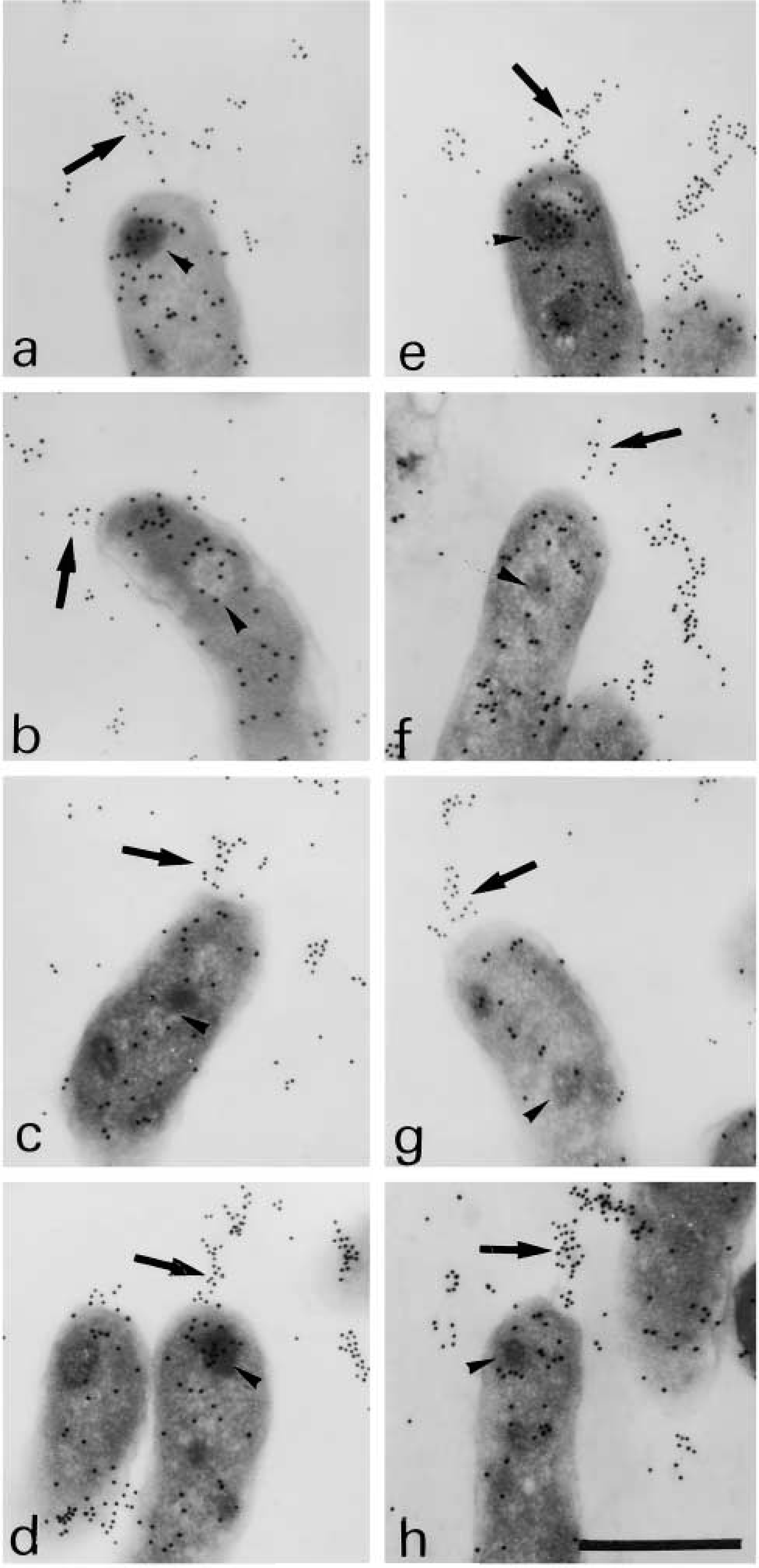
Immunoreaction after antigen retrieval (121C, 15 min). Flagella (arrow) in
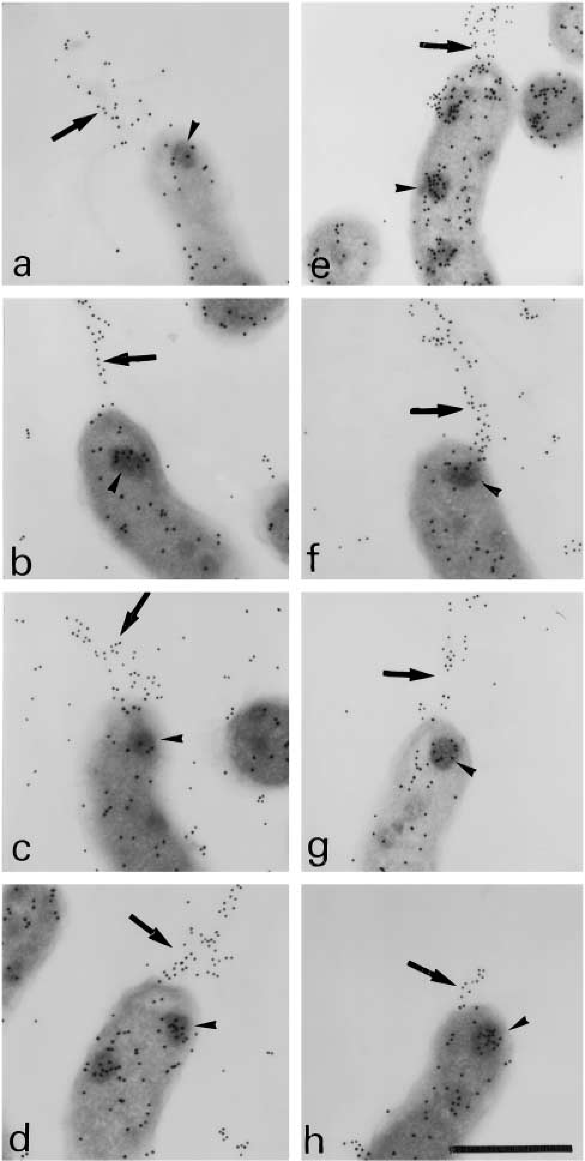
Immunoreaction after AR (99C, 40 min). Flagella (arrows) in
Under all heating conditions, better AR effects were generally observed in EDTA and Tris solution. During heating with the autoclave at 121C for 15 min, some grids floated to the surface of some fluids although some holes had been made in the lid to prevent floating by the imbalance of pressure. The sections that floated showed poorer staining than the sunken ones.
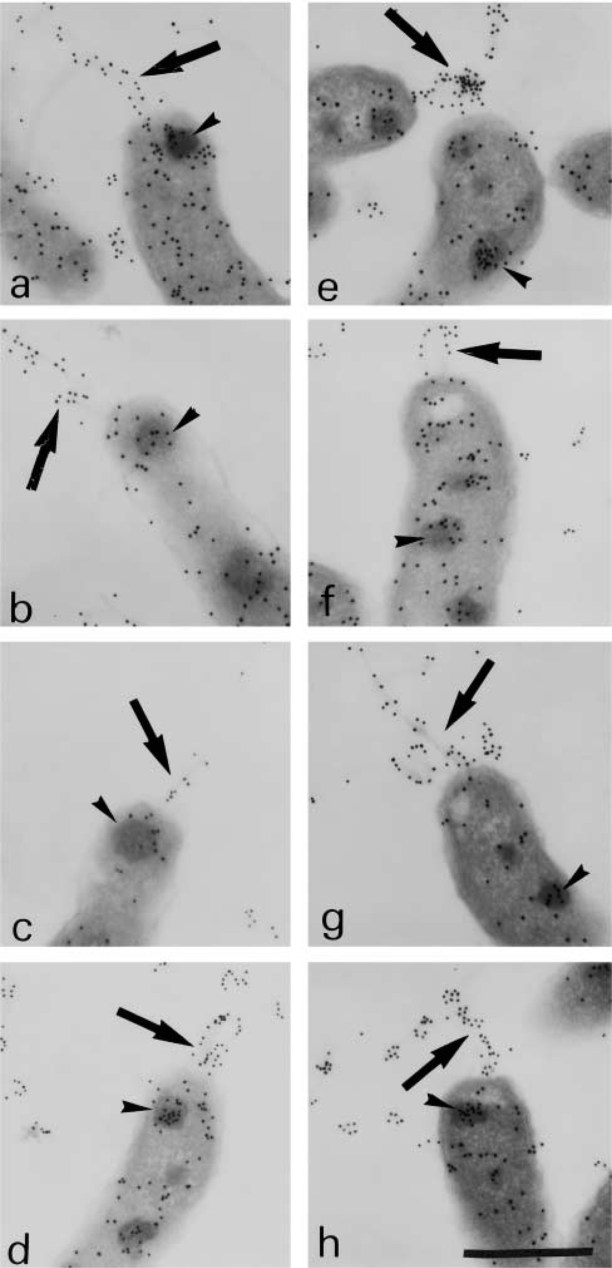
Immunoreaction after AR (65C, 24 hr). Flagella (arrows) in
The control section without any AR treatment showed 3.3 ± 2.2 particles/granule (p/g).
At 121C for 15 Min (Table 1; Figure 1)
The positivity (5.7 ± 4.5 p/g) in Tris was stronger than control (p<0.0001). The stainability on flagella seemed to be better in EDTA, Tris, and S1699 than the control. The section in PB was less positive than control.
At 99C for 40 Min (Table 1; Figure 2)
The positivity in EDTA, Tris, CA, or S1699 was stronger than the control (p<0.0001). The stainability on flagella was better in EDTA, Tris, urea, CA, and S1699 than the control. In granules, AR by Tris showed the best stainability.
At 65C for 24 Hr (Table 1; Figure 3)
In granules, except for PB, the positivity after AR was stronger than the control (p<0.0001). Except for DW and PB, the stainability on flagella was better in each solution than the control. The best staining on flagella was observed with AR by Tris. This heating condition was the most suitable for AR. Under this retrieval condition using Tris, the effect was examined at pH 10.0, 7.0, and 2.0 (Figure 4). AR at pH 10.0 showed the best effect (p<0.0001).
Under all conditions, the stainability after using PB was weaker than that of the control. At 5% of significance level, antigen retrievals at 65C in Tris, 65C in EDTA, and 99C in Tris were more effective than at 99C in EDTA and 99C in S1699, which showed better stainability than those at 65C in DW, 65C in S1699, 65C in CA, 65C in urea, 99C in CA, and 121C in Tris. The order of efficiency of the antigen retrieval method could be surmmarized as follows: Tris (65C and 99C) and EDTA (65C) > EDTA (99C) and S1699 (99C) > DW (65C and 121C), S1699 (65C and 121C), CA (65C, 99C and 121C), urea (65C and 121C), EDTA (121C) and Tris (121C) > PB (65C, 99C and 121C) at 5% significance level. The difference among antigen retrievals at 65C in Tris, 65C in EDTA, and 99C in Tris could not be statistically determined.
Discussion
In our study using the post-embedding method, the remarkable effects of AR by heating, as seen in light microscopy, were also found to be effective for immunoelectron microscopy. However, the stainability that we had expected of the immunoreaction was no better than that of the frozen sections. Staining of the samples after antigen protein had been viewed with skepticism in trials by light microscopy up to the present for fear of the induction of nonspecificity (Taylor et al. 1996). However, in our study under the conditions used in light microscopy, we showed the same localization of the antigen protein as that of the control. This demonstrated that the immunoprecipitations after AR were bona fide antibody-antigen complex. Moreover, there was less nonspecific reaction outside of the cells. This was expected of the low background staining after such treatment, which had also been reported for light microscopy (Taylor et al. 1996). The effect of AR varied among the three different temperatures tested. A microwave oven has been used as a tool for AR in both light and electron microscopy. However, we obtained poor results, such as floating by bubble formation, unbalanced fluid concentrations, and unstable temperatures during AR using a microwave oven in our preliminary experiments. Therefore, methods other than a microwave oven were selected to avoid those vulnerable points in this study. During treatment at 121C using a high-pressure steam sterilizer to avoid bubble formation, some grids with ultrathin sections also floated to the surface of the solutions by bubble formation and failed to undergo AR. Therefore, it was proposed that the grid needed to be submerged in the solutions for AR reaction by the post-embedding method despite a report that floating sections during the process were also well stained after AR by electron microscopy (Stirling and Graff 1995).
Immunoprecipitated gold particles per granule after antigen retrieval a
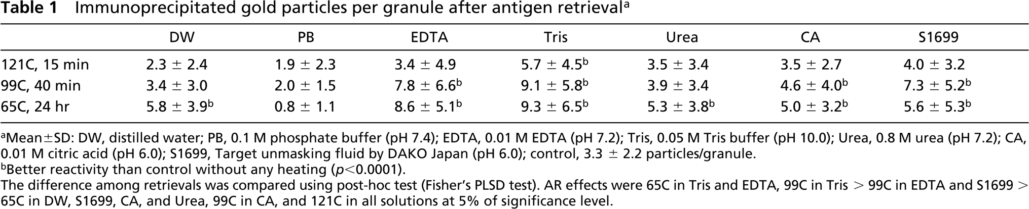
Mean±SD: DW, distilled water; PB, 0.1 M phosphate buffer (pH 7.4); EDTA, 0.01 M EDTA (pH 7.2); Tris, 0.05 M Tris buffer (pH 10.0); Urea, 0.8 M urea (pH 7.2); CA, 0.01 M citric acid (pH 6.0); S1699, Target unmasking fluid by DAKO Japan (pH 6.0); control, 3.3 ± 2.2 particles/granule.
Better reactivity than control without any heating (ρ<0.0001). The difference among retrievals was compared using post-hoc test (Fisher's PLSD test). AR effects were 65C in Tris and EDTA, 99C in Tris > 99C in EDTA and S1699 > 65C in DW, S1699, CA, and Urea, 99C in CA, and 121C in all solutions at 5% of significance level.
Various suitable temperatures for AR by light microscopy have been reported (Shi et al. 1997). In our study, treatment at 65C in Tris, 99C in Tris, and 65C in EDTA produced a better AR result by immunoelectron microscopy. Our result showed that the heating itself is important for AR, considering its effect even in DW and at a low temperature of 65C set in this study. The thermal volume of the solution used might be more important than a “high” temperature that is generally considered to be effective for antigen retrieval by light microscopy (Taylor et al. 1996). Suitable heating time should be further examined because our preliminary study showed that treatment for 24 hr produced better stainability than treatment for 2 hr at 65C.
The effect of antigen retrieval was varied for each solution. The relationship between the AR solutions and morphological preservation was not examined in this study, but morphological preservation appeared to depend on the fixative used rather than on the AR solution. In trials by light microscopy, we have used various fluids for AR. With PB, the results with all heating experiments were poorer than the control. When we tried AR with Tris using an anti-urease antibody, we failed to achieve a better result than the control (not shown).
The mechanism of action of the AR solutions is still obscure. The optimal solution might also be dependent on the antibody used. Although a good effect produced in AR was believed to be manifested under a high pH of more than 8 or a low pH of less than 3 (Shi et al. 1995), the EDTA and urea solutions at near-neutral pH also showed good effect, in addition to Tris at pH 10.0, in this study. In Tris solution, the retrieval effect was worse at pH 2.0 or neutral pH than that at pH 10.0. The most suitable pH may depend on the solution used.
Antigen retrieval by heating of the section has been generally attributed to the breakage of formaldehydeinduced crosslinks between epitopes and extraction of diffusible blocking proteins, allowing better penetration of the antibody and increasing the accessibility of epitopes for the antibody to react (Suurmeijer and Boon 1993). We fixed the materials for a few minutes in the mixture of GA and PFA to get a better ultrastructural preservation, followed by re-fixation in PFA. When gastric biopsy specimens with H. pylori were examined using the post-embedding method, a fixative containing GA is deemed suitable for preserving the ultrastructure of the materials. However, use of GA to fix biopsied viscera with H. pylori on the electron microscopic level seems to necessitate AR (Wilson et al. 1996, Rangell and Keller 2000). Because the use of GA fixative followed by AR by light (Cohen et al. 2001) and electron (Wilson et al. 1996) microscopy had been reported to produce good results, we used a low concentration of GA in our study. For only free cells, the fixatives without GA can be used for AR.
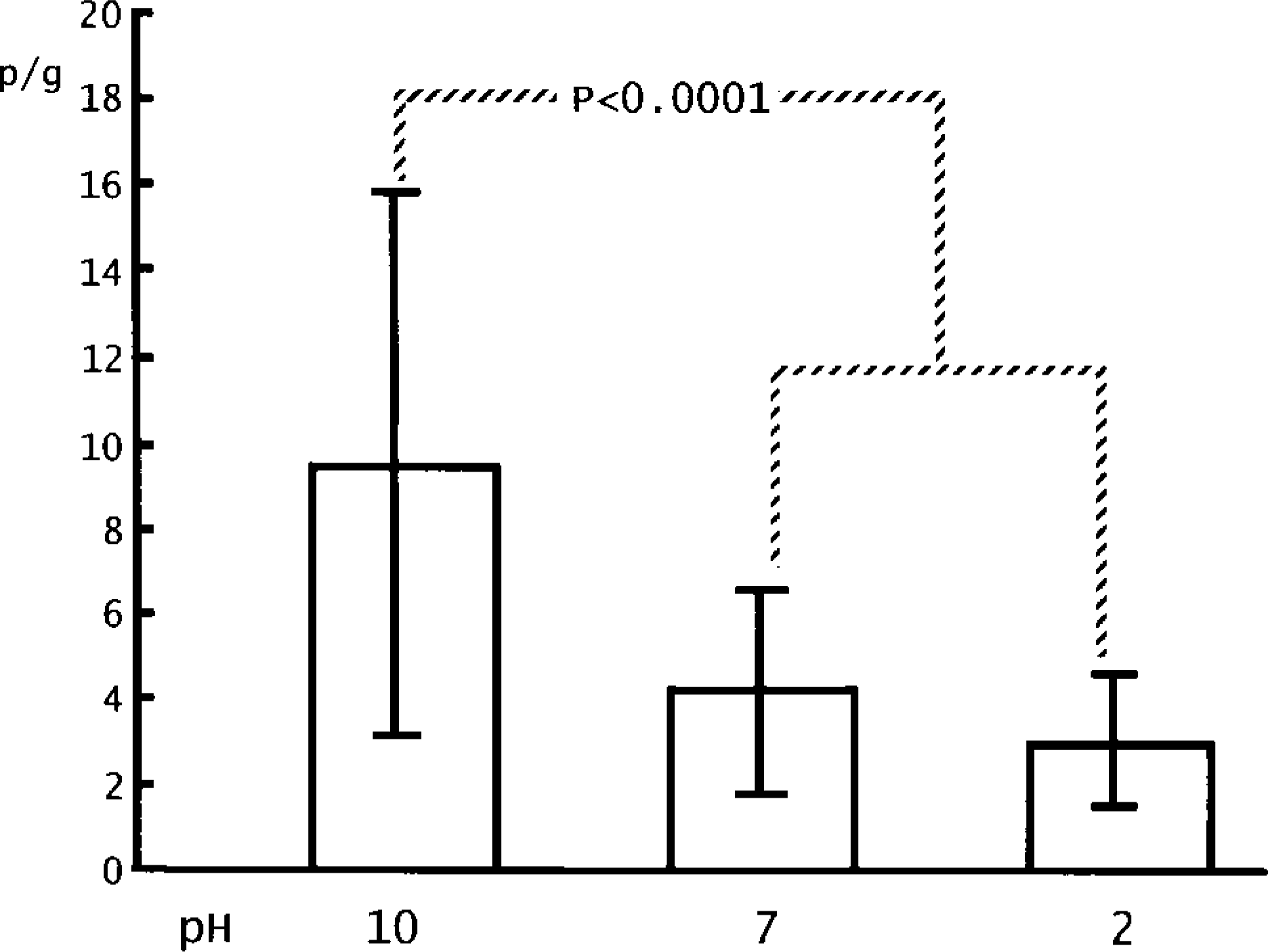
Antigen retrieval at pH 10.0, 7.0, and 2.0 in Tris under 65C for 24 hr. The difference was examined by the Mann-Whitney test (ρ<0.0001).
The major antigen fraction detected by the antibody used in this study is the heat-stable O-antigen (lipopolysaccharide; LPS) (Andersen et al. 1988). This antibody is adopted in laboratories worldwide for detecting H. pylori infection, which gives rise to various benign and malignant gastric disorders such as ulcers, lymphoma, and carcinoma by adhering to the surface of gastric cells (Marshall and Warren 1984; Parsonnet et al. 1991; Isaacson 1994). This antigen is localized in granules, in cytoplasm, on the cell surface, and on the flagella of the organism. This suggests that this protein is produced in the cytoplasm, stored in the granules, transported on the cell surface, and finally onto the flagella. It is known that there are several proteins for adherence to other cells on the flagella (Sherburne and Taylor 1995). Therefore, the protein detected by this antibody is believed to cause malignant damage to the gastric cells after adherence. This antibody could be used further in electron microscopy to shed light on the mechanism of H. pylori infection leading to gastric disorders.
For a start, ultrathin sections should be treated at 65C overnight in Tris to get better immunoprecipitation by immunoelectron microscopy using the post-embedding method. In conclusion, antigen retrieval by heating was effective at both the electron microscopic and the light microscopic level.
Footnotes
Acknowledgements
We thank Dr Subrina Jesmin (Department of Cardiology Section, Hokkaido University Graduate School of Medicine) for the worthwhile discussions, Dr Eriko Shoji and Dr Yumiko Ohkochi for helpful assistance, and Prof Dr Hong Kean Ooi (Department of Veterinary Medicine, National Chung Hsing University, Taiwan) and Dr Yoichi Tari (DAKO Japan, Kyoto) for helpful advice.