
Abstract
We recently found methacarn to be a versatile fixative for analysis of RNA and protein applicable for microdissected specimens from paraffin-embedded tissue (PET). In this study we investigated the performance of methacarn for genomic DNA analysis using microdissected rat tissues. We found that extensive portions of DNA up to 2.8 kb could be amplified by nested PCR using DNA templates extracted by a simple and rapid extraction procedure from a 1 × 1-mm area of cerebral cortex of a 10-μm-thick section. By nested PCR, a 522-bp fragment from a single cell could be amplified in 20% of cresyl violet-stained Purkinje cells, and the minimal number of cells required, as estimated using hippocampal neurons, was on the order of 10-20. Although tissue staining with hematoxylin and eosin affected the PCR, amplification of a 522-bp fragment was successful, with 150-270 cells by 35 cycles of single-step PCR. Immunostaining resulted in a substantial decrease of yield and degradation of extracted DNA. However, even after immunostaining, a 184-bp DNA fragment could be amplified with 150-270 cells by 35 cycles of PCR. The results thus demonstrate the superior performance of methacarn to that reported with formalin in genomic DNA analysis using microdissected PET specimens.
A
For histological assessment, tissue fixation and subsequent paraffin embedding are routinely employed because of the ease of handling tissues and subsequent staining, as well as the good preservation of morphology. Usually, formaldehyde-based fixatives, such as buffered formalin, are used for this purpose. However, with such crosslinking agents there is limited performance in terms of the efficiency of extraction and the quality of extracted RNA (Rupp and Locker 1988; Stanta and Schneider 1991; Finke et al. 1993), protein (Ikeda et al. 1998; Shibutani et al. 2000), and genomic DNA (Shibata 1994; Frank et al. 1996; Poncin et al. 1999; Uneyama et al. 2000), with consequent difficulty in the analysis of microdissected, histologically defined tissue areas.
Extraction efficiency and quality of molecules are critical for analysis of microdissected cells. Recently, we found that methacarn, a non-crosslinking, protein-precipitating fixative (Puchtler et al. 1970; Mitchell et al. 1985), meets critical criteria for analysis of RNAs and proteins in defined areas of paraffin-embedded tissue (PET) sections by simple extraction protocols (Shibutani et al. 2000). For DNA extraction from formalin-fixed PETs, the extraction protocol usually requires proteinase K treatment with extended incubation periods (Dietmaier et al. 1999; Murase et al. 2000; Hirose et al. 2001). On the other hand, we found that methacarn fixation allows very high yields and amplification of extensive portion of genomic DNA in PET sections by a simple extraction procedure, whereas formalin-fixed PET sections resulted in poor extraction efficiency and unsuccessful PCR of the DNA fragment that was amplifiable with methacarn-fixed specimens (Uneyama et al. 2000).
Tissue staining is essential for cell identification in practical molecular analyses using the microdissection technique (Pontén et al. 1997; Burton et al. 1998; Alcock et al. 1999; Fend et al. 1999; Murase et al. 2000; Serth et al. 2000; Gjerdrum et al. 2001), and therefore it is important to examine the effect of tissue staining on the performance of molecular analyses as reported in DNA analysis using formalin-fixed PETs (Burton et al. 1998; Murase et al. 2000; Serth et al. 2000). The present study was performed to determine the suitability of methacarn fixation for genomic DNA analysis in terms of target fragment size and number of microdissected cells required, using cresyl violet-stained sections. In addition, effects of tissue staining with hematoxylin and eosin (HE) or immunohistochemistry (IHC) for amplification of DNA fragments was assessed to clarify the performance in analysis of histologically or immunophenotypically defined cells.
Materials and Methods
Animals and Tissue Fixation
Sprague-Dawley (SD) or F344 rats, both from Charles River Japan (Kanagawa, Japan), were used in the present study. To compare methacarn-fixed PET preparations with unfixed or ethanol-fixed frozen tissue specimens for the extraction efficiency and integrity of DNA, 1-mm-thick liver slices weighing 50-60 mg were prepared from a 30-week-old SD rat. To examine the relationship between cell numbers and amplifiable fragment size, cerebral and cerebellar tissues of SD rats at 10 postnatal days of age were used. To examine the yield and quality of extracted DNA from HE-stained or immunostained tissues, the liver of a rat that had been subjected to two-stage hepatocarcinogenesis was utilized (Shirai 1997; Ito et al. 2000). For this purpose, male F344 rats were given a single IP injection of diethylnitrosamine (DEN: Nacalai Tesque, Kyoto, Japan; 200 mg/kg/0.9% NaCl solution) to induce liver glutathione-S-transferase placental form (GST-P)-positive foci (Satoh et al. 1985; Tatematsu et al. 1985). To promote formation of GST-P-positive foci, animals were treated orally with 400 ppm thioacetamide (Wako Pure Chemicals; Osaka, Japan) in a powdered basal diet 2 weeks after DEN treatment (Ogiso et al. 1990). At week 3, rats were subjected to partial hepatectomy and were sacrificed at week 8, and one animal was selected and subjected to subsequent DNA analysis. All animals used in the present study were sacrificed by exsanguination from the abdominal aorta under ether anesthesia. The animal protocols were reviewed and approved by the Animal Care and Use Committee of the National Institute of Health Sciences, Japan.
A methacarn solution consisting of 60% (v/v) absolute methanol, 30% chloroform, and 10% glacial acetic acid was freshly prepared before fixation. Liver and brain tissues were removed and fixed in methacarn for 2 hr at 4C. For embedding, 1- or 5-mm-thick liver slices and coronal slices of cerebrum and cerebellum were dehydrated three times for 1 hr in fresh 99.5% ethanol at 4C, immersed in xylene for 1 hr and then three times for 30 min at room temperature and immersed in hot paraffin (60C) three times for 1 hr, for a total of 3 hr. For preparation of frozen liver samples, 1-mm-thick slices were subjected to either quick freezing in dry ice or fixation with 99.5% ethanol for 1 hr at −30C, and were stored at −80C.
Preparation of Methacarn-fixed PET Sections and Stainings
Paraffin-embedded tissues were sectioned at 10 μm and mounted on a 1.35-μm thin polyethylene film (PALM GmbH; Wolfratshausen, Germany) overlaid on a glass slide. The sections were deparaffinized by immersing in xylene three times for 2 min, followed by 99.5% ethanol twice for 2 min. Rat brain tissue sections were subjected to staining with cresyl violet. In brief, deparaffinized sections were immersed briefly in water and stained with 0.1% cresyl violet for 20 min. Sections were then washed once with 95% ethanol that contained 0.5% acetic acid and then twice with 99.5% ethanol. Liver sections from a rat that had been subjected to two-stage hepatocarcinogenesis were stained with HE or immunostained with GST-P. For HE, sections were stained with Mayer's hematoxylin for 10 sec, washed briefly with water, stained with eosin for 10 sec, washed with 99.5% ethanol, and then air-dried. For IHC demonstration of GST-P with a standard protocol, deparaffinized sections were treated with 1% periodic acid solution for 10 min, washed briefly with water and 1 × PBS, pH 7.4, and then blocked for nonspecific binding with 0.5% casein (Merck; Darmstadt, Germany) in PBS for 30 min. After incubation with rabbit anti-GST-P antibody (Medical and Biological Laboratories, Nagoya, Japan; X2000 dilution) for 2 hr, sections were incubated with biotin-labeled goat anti-rabbit IgG and then with avidin-biotin complex (ABC) utilizing the Vectastain Rabbit Elite kit (Vector Laboratories; Burlingame, CA). The immunoreaction was visualized using the avidinbiotin system with 0.004% H2O2 as substrate and diaminobenzidine (DAB) as chromogen. After washing, the sections were air-dried. In a rapid immunostaining protocol, tissue sections were treated with periodic acid solution for 1 min and then incubated with casein-PBS for 5 min. Sections were then incubated for 10 min each with anti-GST-P antibody (X100 dilution), secondary antibody (X10 higher concentration than that of the standard protocol), and ABC. Immunostained tissue sections with a standard protocol were subjected to subsequent analysis unless stated. Autoclaved ultrapure water was used for all solutions of the above stainings.
Microdissection
Microdissection was performed with PALM Robot-Microbeam equipment (Carl Zeiss; Tokyo, Japan) as described previously (Schütze and Lahr 1998). Briefly, the film with the attached specimen was mounted in reverse (film side up) on a new coverslip. The specimens were then subjected to Robot-Microbeam dissection by laser beam and the desired cells were catapulted by laser pressure to trap into mineral oil-coated caps of a PCR tube. For large specimens (circular areas of 150-200 μm in radius or square areas larger than 60 × 60 μm), the excised cells were picked up with a thin needle tip. Transfer of microdissected specimen onto the cap of PCR tube was confirmed under a microscope.
DNA Extraction from Whole Tissue Sections or Small Tissue Blocks
When a whole tissue section was intended for DNA analysis, a 10-μm-thick section attached to polyethylene film was removed from the glass slide and digested with 500 μl of 10 mM Tris-HCl (pH 8.0), 150 mM NaCl, 10 mM ethylenediaminetetraacetic acid, 0.1% sodium dodecyl sulfate (SDS), and 1 unit of proteinase K at 55C for 2 hr. The film was then removed from the tube. Then 500 μl of Tris buffer-saturated phenol was added, mixed well, and centrifuged at 10,000 × g for 15 min. The supernatant was extracted again with 500 μl of Tris-phenol:chloroform (1:1), and the separated aqueous portion after centrifugation was transferred to a new tube and treated with 0.5 U RNase A at 37C for 1 hr. The solution was extracted with 500 μl of phenol:chloroform (1:1) and then treated with ether. Extracted DNA was precipitated by adding 1 ml cold 99.5% ethanol and, after storing at −20C overnight, was centrifuged at 5000 × g for 5 min. The pellet was washed twice with 75% ethanol, dried by a rotary evaporator (Sakuma Seisakusho; Tokyo, Japan), and resuspended in 10 μl of water. One μl of sample was used to measure DNA concentration by Hoechst 33258 and a fluorescence spectrophotometer (F4010; Hitachi, Tokyo, Japan). Extraction of DNA from methacarn-fixed, paraffin-embedded small liver tissue blocks was performed similarly as that used for whole tissue sections after deparaffinization. Frozen tissue blocks of unfixed or ethanol-fixed liver were directly subjected to DNA extraction, and deparaffinized blocks of methacarn-fixed liver PET were air-dried before extraction.
DNA Extraction from Microdissected Cells
Microdissected Purkinje cells, small areas of the hippocampal CA1 region, or liver cells were catapulted and trapped with mineral oil on PCR tube caps and subjected to extraction with 4 μl of DEXPAT (TAKARA SHUZO; Kyoto, Japan) at 95C for 10 min, and the entire extracts were used as a template for PCR by adding to the master mix of total 50 μl directly. For a 1 × 1-mm area of cerebral cortex, tissue specimens were extracted with 40 μl of DEXPAT. DEXPAT consists of detergent and absorbent beads, and we used the detergent portion alone.
PCR
For analysis of target fragment size and number of microdissected cells in brain regions, hot-start PCR of the rat genomic sequence of the α2u-globulin gene (accession no. M24108 in GenBank/EMBL Data Bank) was performed with PLATINUM Taq DNA polymerase (Invitrogen; Carlsbad, CA). First-step PCR was performed with a total reaction volume of 50 μl, and primer sequences were as follows: upstream-outside primer, 5′-ACGGATCCAGGCTTCAAGTTCCGTATTA-3′; downstream primer for the 969-bp fragment, 5′-CGTCATCTGTGGAGGAAATT-3′; downstream primer for the 2954-bp fragment, 5′-TGAAATCCTGAGACTAAGCT-3′; downstream primer for the 3995-bp fragment, 5′-CTAAATGGTGGGGAACTGTC-3′. The primer sequences for the second-step PCR were as follows: upstream-inside primer, 5′-AAAGTTAAATGGAATCAGAA-3′; downstream-inside primer for the 522-bp fragment, 5′-TAAGTCCGTCTCACATGGCT-3′; downstream-inside primer for the 1873-bp fragment, 5′-TTTTCGCACAAGGGATCCAG-3′. For the second-step PCR of the 2822-bp fragment, the downstream primer for the 2954-bp fragment in the first PCR was used. Nested PCR in a 20 μl total volume was performed using 1 μl of first PCR product as a template.
To examine the effect of tissue stainings, a 184-bp fragment of the rat GST-P gene (accession no. L29427 in GenBank/EMBL Data Bank) was amplified using upstream primer, 5′-GGAGCAGGACCCAAAAATGA-3′; downstream primer, 5′-GCAGACGAATAAAGGCCCCA-3′ by single-step PCR. Amplification of 522- and 969-bp fragments from the rat α2u-globulin gene was also examined.
To amplify target fragments smaller than 1 kb, PCR was performed with cycle parameters of 95C for 5 min, 35 cycles of 95C for 1 min, 55C for 1 min, 72C for 30 sec. The extension times for 1873, 2822, 2954, and 3995 bp were 1.5, 2.5, 2.5, and 3.5 min, respectively. The PCR reaction mixture contained 20 mM Tris-HCl (pH 8.4), 50 mM KCl, 0.2 mM dNTP, 1.5 mM MgCl2, 0.2 mM each primer and 1 U (for 20 μl total volume) or 2.5 U (for 50 μl total volume) of PLATINUM Taq DNA polymerase.
Results
Yield and Quality of Extracted DNA
Yield and quality of DNA extracted from methacarnfixed rat liver PET blocks were compared with those from unfixed or ethanol-fixed frozen tissue blocks (Table 1; Figure 1). The DNA yield from ethanol-fixed frozen tissues was similar to that from unfixed frozen tissues. On the other hand, the DNA yield from methacarn-fixed PETs was slightly reduced, the mean value being about 70% of that from unfixed frozen tissues (Table 1). Figure 1 shows the integrity of extracted DNA visualized as a smear by electrophoresis on a 1.5% agarose gel. Although slightly stronger intensity at the top of the DNA smear was observed in unfixed or ethanol-fixed frozen samples, DNA in every case distributed within the high molecular weight range, suggesting good preservation of extracted DNA. In addition, a clump of small DNA fragments appeared around 100-bp levels in frozen tissues.
Target Fragment Size and Detection Rate by Nested PCR
To estimate the relationship between the size of target fragments and the ratio of successful amplification by nested PCR in methacarn-fixed PET sections, 1 × 1 mm of cerebral cortical areas, microdissected from cresyl violet-stained rat brain tissues, was examined. Figure 2 shows the targeted 2822-bp PCR product of the α2u-globulin gene in two different samples from cerebral cortex detected by nested PCR, the 3995-bp fragment being amplified by first-step PCR. Table 2 shows the relationship between the size of template DNAs and positive detection by nested PCR. The genomic DNA fragment of 522 bp was amplified with 100% of the samples. When the template size was increased from 1873 to 2822 bp, the detection ratio decreased from 90% to 30%.
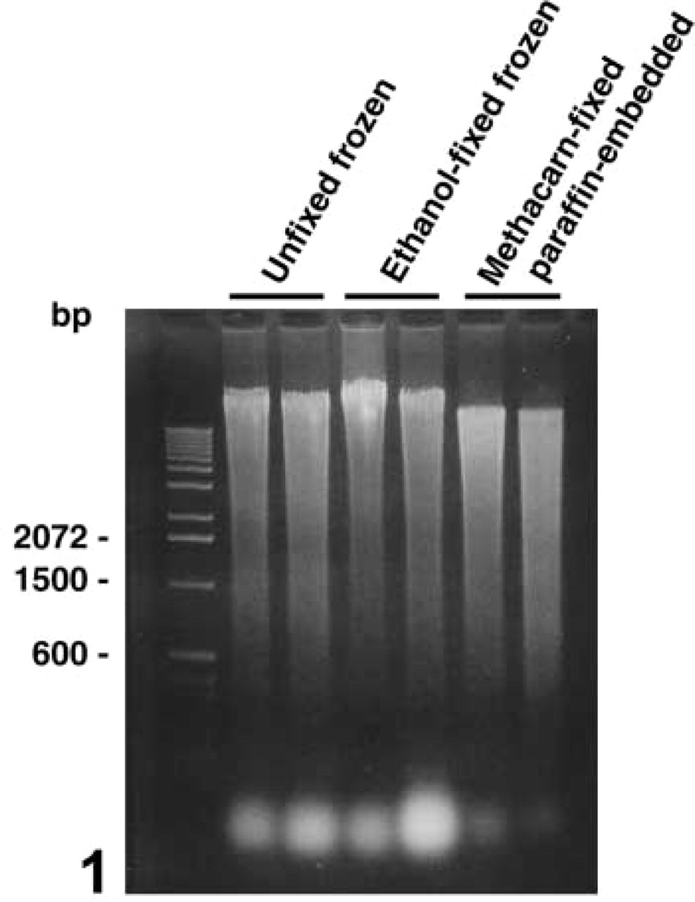
Comparison of the integrity of DNA extracted from rat liver of unfixed or ethanol-fixed frozen tissue blocks and deparaffinized methacarn-fixed tissue blocks. Tissue blocks were directly subjected to DNA extraction and 2.0 μg of extracted DNA was subjected to electrophoresis in a 1.5% agarose gel and stained with ethidium bromide.
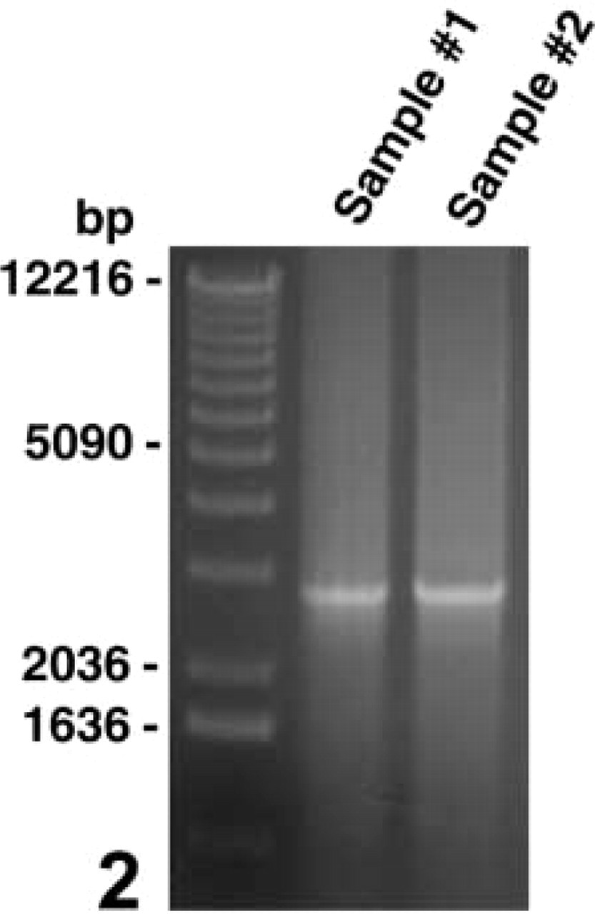
Nested PCR results for the α2u-globulin genomic sequence of 2.8 kb with DNA extracted from methacarn-fixed, paraffin-embedded rat cerebral cortex. From 10-μm-thick, cresyl violet-stained brain sections, 1 × 1-mm areas of cerebral cortex were microdissected and extracted with 40 μl of DEXPAT to extract DNA. Four-μl aliquots of cell extracts were directly applied for the first PCR reaction. With 1 μl of the first PCR product, second-step PCR was performed to amplify a 2.8-kb fraction. This figure shows results of two different samples.
Comparison of RNA yields between unfixed or ethanol-fixed frozen tissues and methacarn-fixed PETs a
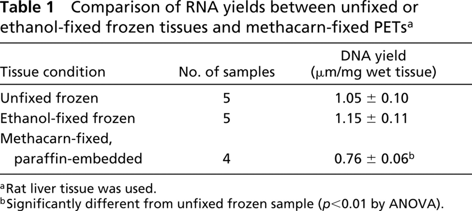
Rat liver tissue was used.
Significantly different from unfixed frozen sample (p<0.01 by ANOVA).
Cell Number and Detection Ratio by Nested PCR
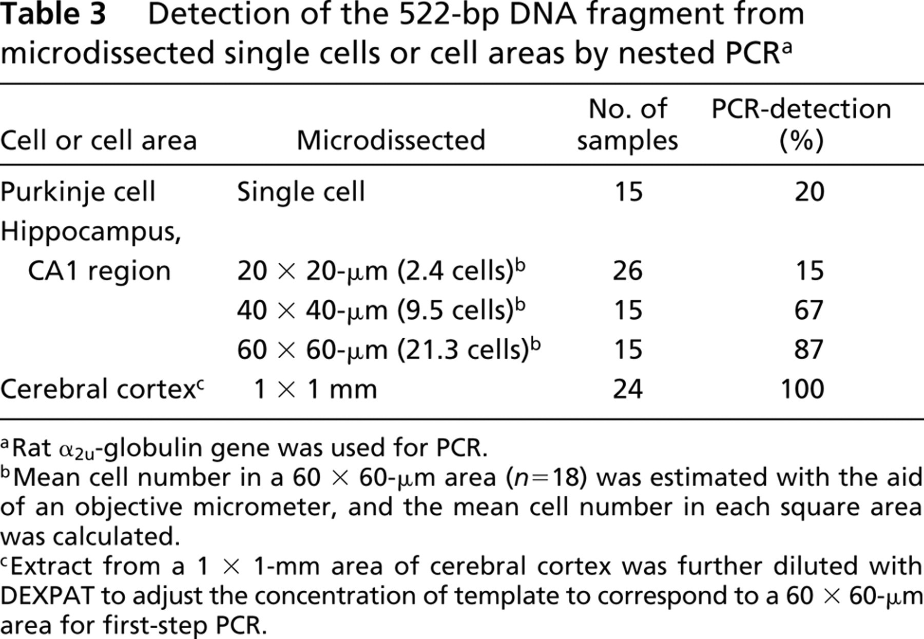
To clarify the cell number required for amplification by nested PCR in methacarn-fixed PET sections, the hippocampal CA1 region, cerebral cortex, and cerebellar Purkinje cells from cresyl violet-stained rat brain sections were microdissected. The ratio of successful detection of the target gene in single-cell DNA preparations was estimated using Purkinje cells. As shown in Figures 3A and 3B, a single Purkinje cell was microdissected and catapulted to trap in mineral oil on a PCR tube cap from a 10-μm-thick cerebellar section. The 522-bp DNA fragment of the α2u-globulin gene was successfully amplified in 20% of the PCR attempts by nested PCR (Figure 3C; Table 3). The resultant PCR product was clearer when multiple Purkinje cells were subjected to one PCR reaction (Figure 3D). Similar but less effective amplification could be obtained with microdissected areas of the hippocampal CA1 region, in which successful detection was obtained in 15% of 20 × 20-μm samples (corresponding to 2.4 cells). The ratio of PCR detection increased with the area microdissected but did not reach 100% even in a 60 × 60-μm area. However, when aliquots of cell extracts from a 1 × 1-mm area of cerebral cortex were diluted to obtain DNA templates corresponding to a 60 × 60-μm square and applied to nested PCR, the 522-bp fragment could be amplified in 100% of cases.
Detection ratio of genomic DNA fragments of different sizes from 1 × 1-mm areas of rat cerebral cortex by nested PCR a
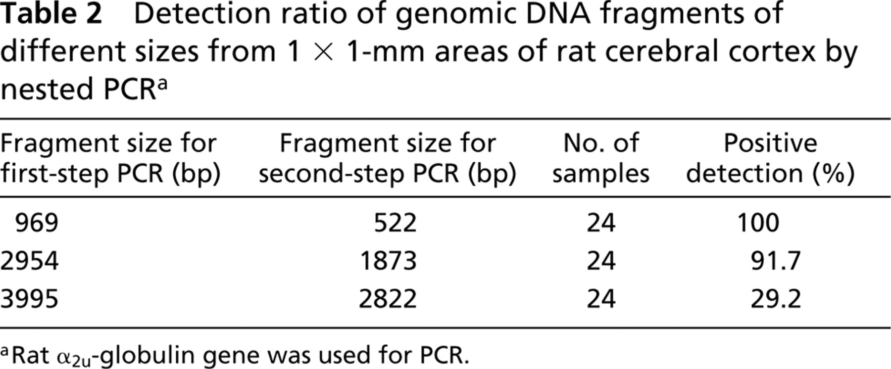
Rat α2u-globulin gene was used for PCR.
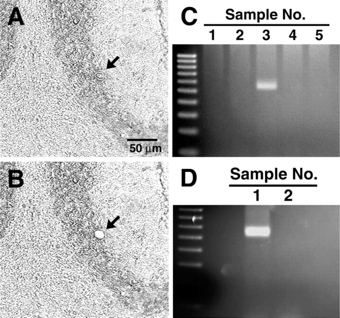
PCR amplification of a 522-bp fragment of the α2u-globulin gene from microdissected materials. (
Effect of Stainings on the Yield and Quality of Extracted DNA
To elucidate the effects of tissue stainings on the yield and quality of extracted DNA, standard HE staining and immunostaining were examined with methacarn-fixed PET sections. For this purpose, liver tissue from a rat subjected to two-stage hepatocarcinogenesis was used. Serial sections of 100 mm2 in area and 10 μm in thickness were randomized and subjected to HE staining or immunostaining with GST-P, or were left unstained. Table 4 shows the DNA yield from methacarn-fixed PET sections after tissue staining. The yield recovered from HE-stained sections did not differ from that from unstained sections. Fluorescence derived from eosin dye did not affect the DNA measurements with Hoechst 33258. Immunostained tissue sections with a standard protocol, on the other hand, resulted in a very low DNA yield, values being 12% of unstained sections. In another experiment, the DNA yield from sections immunostained with a rapid staining protocol was examined. Total time for rapid staining was 44 min, whereas it took 5 hr with a standard protocol. Rapid staining resulted in low contrast view with high background in immunoreactivity and therefore it was judged unsuitable for microdissection. DNA yield with the rapid protocol was 45% that of unstained sections.
Detection of the 522-bp DNA fragment from microdissected single cells or cell areas by nested PCR a
Rat α2u-globulin gene was used for PCR.
Mean cell number in a 60 × 60-μm area (n=18) was estimated with the aid of an objective micrometer, and the mean cell number in each square area was calculated.
Extract from a 1 × 1-mm area of cerebral cortex was further diluted with DEXPAT to adjust the concentration of template to correspond to a 60 × 60-μm area for first-step PCR.
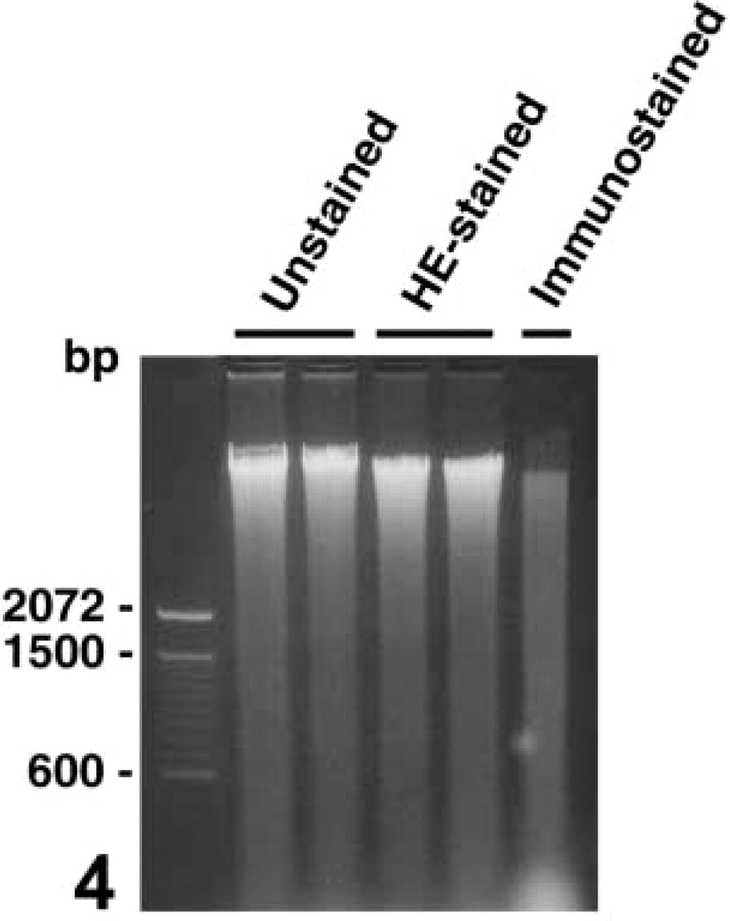
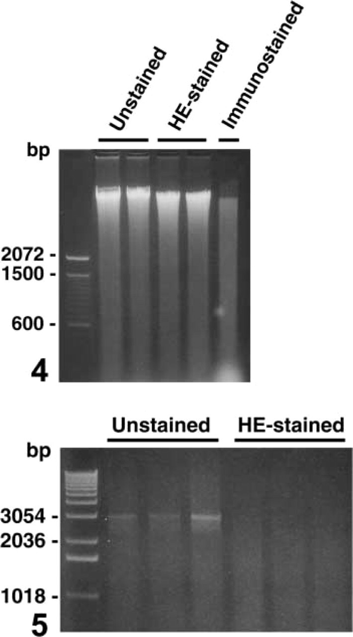
Figure 4 shows the integrity of DNA extracted from stained sections, as visualized by electrophoresis on a 1.5% agarose gel. DNA from unstained sections distributed mainly within the high molecular weight range. Similar to the unstained tissue section, HE-stained sections preserved well the integrity of extracted DNA. Compared to unstained and HE-stained cases, DNA extracted after immunostaining showed a weak homogeneous smearing within the entire molecular weight range, and a clump of small DNA fragments appeared around the 100-bp level.
Liver of a rat treated with thioacetamide at the promotion stage in the two-stage hepatocarcinogenesis model was used.
Significantly different from the unstained and HE-stained samples (p<0.0001 by ANOVA).
Tissue blocks were trimmed to obtain sections of 100 mm2 in area before sectioning, and sectioned at 10 μm in thickness.
Sections were immunostained with GST-P.
Effect of Stainings on Amplification by Single-step PCR
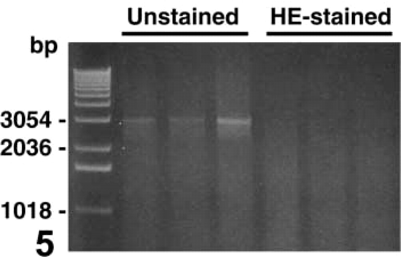
Figure 5 shows the PCR results with DNA templates from unstained and HE-stained sections. A single-step 35-cycle PCR to amplify a 2954-bp fragment of rat α2u-globulin gene was performed with 200 ng purified DNA. The 2954-bp fragment detected with unstained sections could not be amplified with HE-stained sections. Although data are not shown, 969- and 1873-bp fragments could be amplified with HE-stained sections. On the other hand, amplification of 969-2954-bp fragments was unsuccessful with immunostained section-derived DNA (data not shown).
To elucidate the performance of methacarn fixation for DNA analysis after immunostaining, GST-P-immunostained liver sections of a rat that had been subjected to two-stage hepatocarcinogenesis were examined. Figure 6A shows the GST-P-immunostained tissue section. The circled area at the center of a GST-P-positive liver cell focus was microdissected by the PALM system with a dissection radius of 150 μm by the automated mode. The dissected tissue is shown in Figure 6B. The circled area with a radius of 150 μm contained a mean of 150 liver cells when counted with HE-stained dissected areas of identical portions (Figure 6C). The number of liver cells contained in the 200-μm-radius circled area was therefore calculated as 270.
Figure 7 shows the result of single-step PCR of DNA fragments using microdissected tissues from 10-μm-thick rat liver sections stained with HE or GST-P. Circled areas with a radius of 150 or 200 μm were subjected to PCR analysis. PCRs of 35 cycles with target fragments sized 184, 522, and 969 bp were performed after DEXPAT extraction. A summary of the PCR results is shown in Table 5. In HE-stained tissue, a 522-bp fragment could be amplified with both 150-and 200-μm radius samples, although the amplification of 969-bp fragments was unsuccessful even with 200-μm radius samples. In immunostained tissue, a weak 522-bp band could be amplified only with 200-μm radius samples. For 150-μm radius samples, only a 184-bp fragment could be amplified.
Integrity of DNA extracted from stained sections of methacarn-fixed PET. Liver of a rat treated with thioacetamide at the promotion stage in the two-stage hepatocarcinogenesis model was used. Tissue blocks were trimmed to obtain sections of 100 mm2 in area before sectioning and were sectioned at 10-μm thickness. Sections were mounted on polyethylene film overlaid on a glass slide. Deparaffinized sections were either unstained, stained with HE, or immunostained with GST-P as described in Materials and Methods. One μg of extracted DNA from a whole tissue section was subjected to electrophoresis in a 1.5% agarose gel and stained with ethidium bromide.
Single-step PCR using 200 ng of DNA extracted from an unstained section and an HE-stained section from the liver of a rat that had been subjected to two-stage hepatocarcinogenesis model. DNA was extracted from a 10-μm-thick section after staining, as described in Materials and Methods, and 200 ng was used as a template for each PCR in a total reaction volume of 20 μl to amplify a 2954-bp fragment of the α2u-globulin gene. PCR results of three different samples from unstained and HE-stained sections are shown in this figure.
Discussion
In general, genomic DNA fragments sized around 200 bp can be amplified with formalin-fixed PETs (Ortiz-Pallardó et al. 2000; Poncin et al. 1999). Even with an optimal extraction protocol recently published, 959-bp fragments would be the maximal size for amplification by single-step PCR of 43 cycles using DNA templates isolated from cancer tissue sections, although the precise relationship between the area and the target fragment size is unclear (1-35-mm2 area) (Akalu and Reichardt 1999). Formaldehyde-based fixatives crosslink polypeptides to cause insolubility of nuclear proteins surrounding genomic DNA that results in poor extraction efficiency even with extensive extraction protocols (Coombs et al. 1999; Serth et al. 2000). On the other hand, in addition to the high performance in terms of yield and integrity of both RNA and protein preparations (Shibutani et al. 2000), we recently found methacarn fixation to allow very high yields of DNA and successful amplification of 4-kb fragment by single-step PCR from 10-μm-thick rat liver PET sections (Uneyama et al. 2000). Methacarn presumably gives preservation of DNA molecules by precipitation of surrounding nuclear proteins and cytoplasmic DNases. The precipitated proteins are chemically unmodified but biologically inactivated and can be extracted easily with detergents (Shibutani et al. 2000), and this feature may be linked to the high DNA yield with good integrity, as demonstrated by comparison with unfixed or ethanol-fixed frozen tissues in the present study. Furthermore, this feature may also be related to the successful amplification of genomic DNA by direct application of DNA extracts isolated with simple boiling in DEXPAT for 10 min.
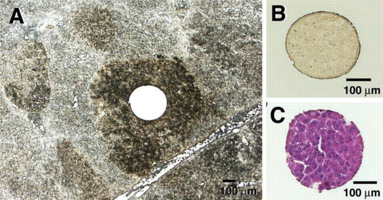
Microdissection using a GST-P-immunostained liver section from a thioacetamide-treated rat in the two-stage hepatocarcinogenesis model. (
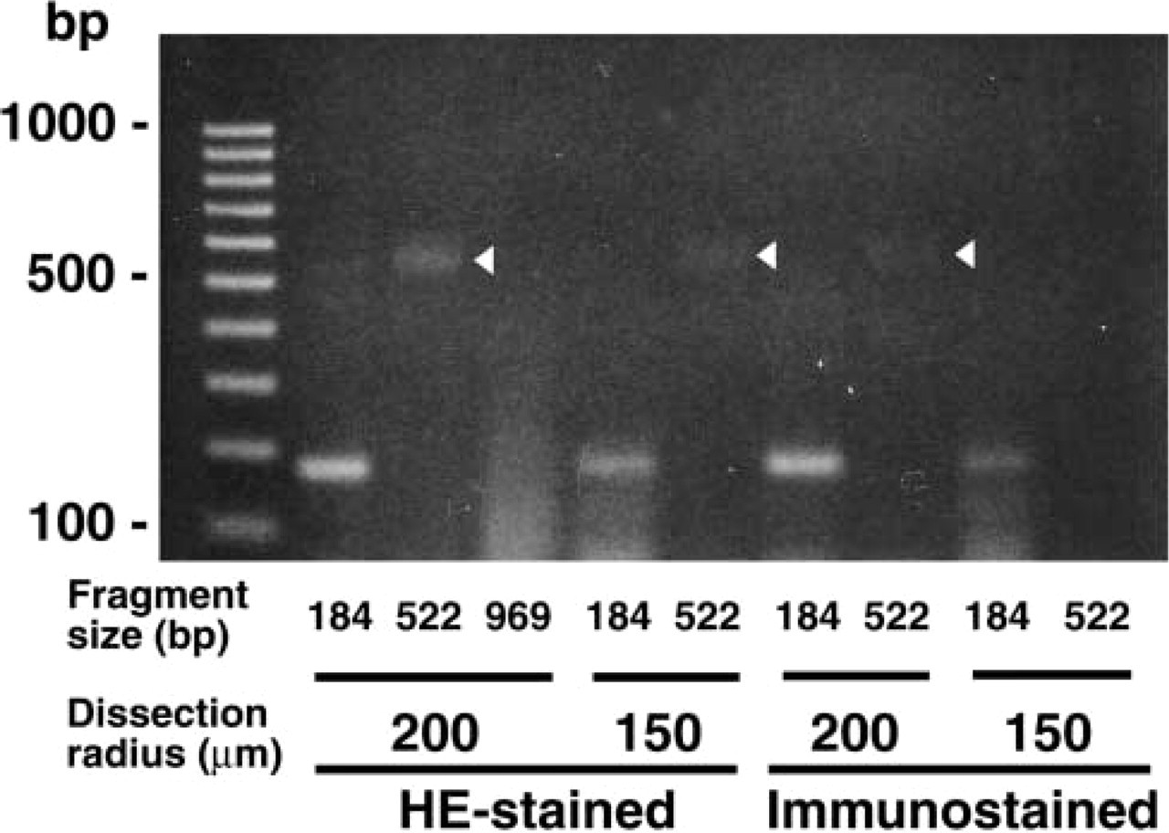
Amplification of DNA fragments using microdissected circular areas from HE-stained or GST-P-immunostained sections as described in Figure 6. Circular areas of 150 or 200 μm in radius were microdissected and solubilized with 4 μl of DEXPAT solution in PCR tubes at 95C for 10 min and whole extracts were subjected to PCR directly. PCR with 50 μl reaction volume was performed to amplify 184-, 522-, and 969-bp fragments with the same cycle parameters of 95C for 2 min, 35 cycles of 95C for 1 min, 55C for 1 min, and 72C for 30 sec, and final extension at 72C for 7 min. Eight μl of PCR product was applied to 2.0% agarose gel electrophoresis. Arrowhead indicates 522-bp PCR products.
To elucidate the performance of methacarn fixation for DNA amplification by nested PCR, we examined the relationship between the number of microdissected cells and the fragment size to be amplified. For this purpose, we selected histologically defined neuronal cells rather than tumor cells because tumor tissue is not standard owing to the considerable heterogeneity in cell size and density, with altered ploidy and chromosomal rearrangement. When the size of the tissue area was increased, amplifiable fragment size was also generally increased. Furthermore, target fragments of about 500 bp could be amplified with cresyl violet-stained single cells, but 10-20 cells were necessary for detection by nested PCR. Although the source of cells and the detection system were different from those in the present study, a similar performance was obtained when DNA from ≥25 cells of alcohol-fixed cytology specimens was used in multiplex PCR (Euhus et al. 1999). The reason why the detection rate did not reach 100% in our 60 × 60-μm area samples of hippocampus may be due to unsuccessful extraction, because the amount of lysis buffer, at 4 μl, was minimal. The 100% detection in DNA samples with the amount corresponding to a 60 × 60-μm area extracted from a 1 × 1-mm area of cerebral cortex, a site with a lower cell density than the hippocampus, may support this idea.
Detection of genomic DNA fragment by single-step PCR in HE-stained or immunostained tissue areas from methacarn-fixed rat liver PET sections a
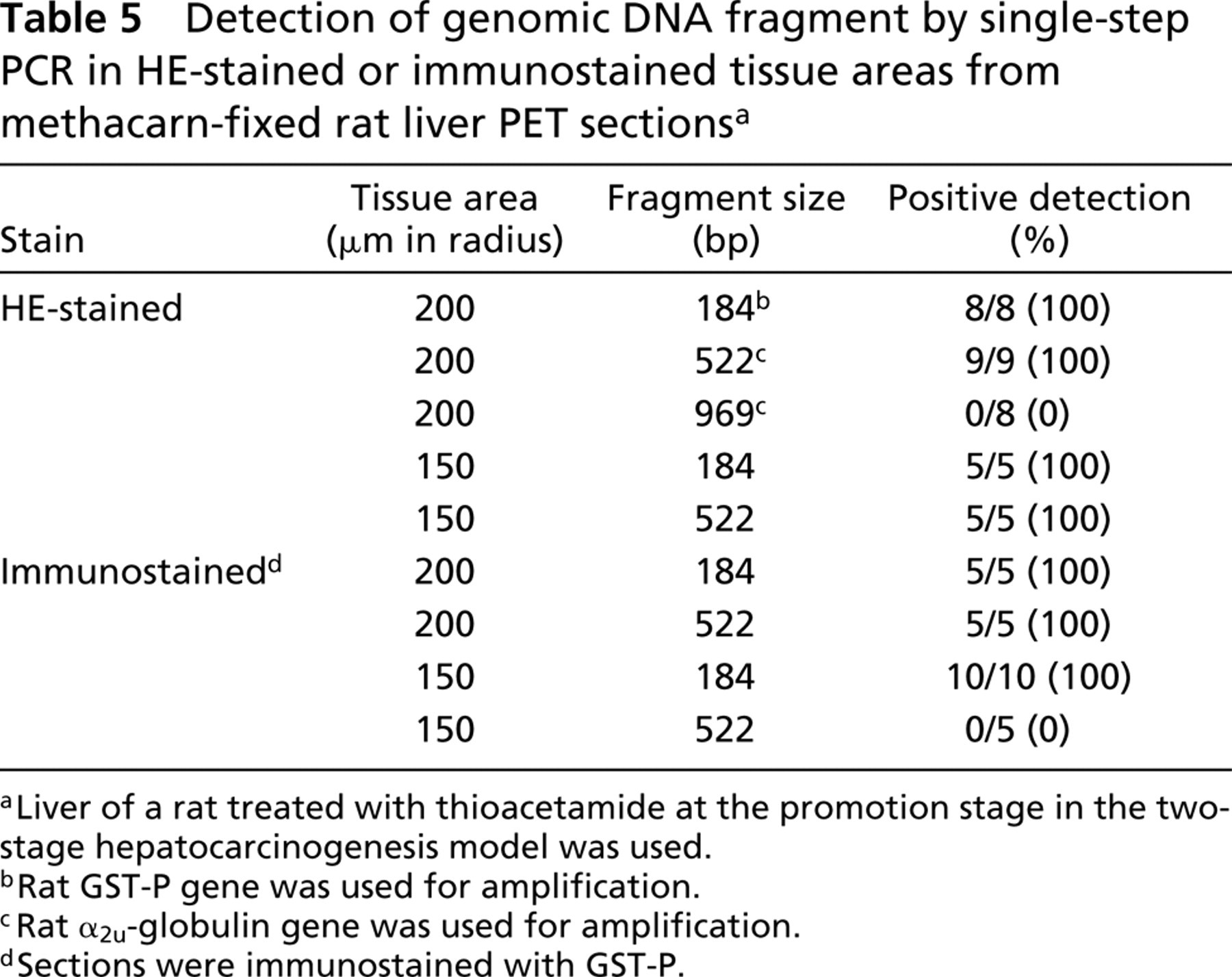
Liver of a rat treated with thioacetamide at the promotion stage in the two-stage hepatocarcinogenesis model was used.
Rat GST-P gene was used for amplification.
Rat α2u-globulin gene was used for amplification.
Sections were immunostained with GST-P.
Although experimental conditions, including cell type, generally differ from those used in our present study, Pontén et al. (1997) reported that approximately 50% of microdissected single cells from frozen, immunostained human basal cell tumor tissue sections (12-16 μm in thickness) allowed amplification of the target gene by multiplex/nested configuration, suggesting a high performance of unfixed frozen tissue sections for detection of genomic sequences. However, preparation of cryosections from unfixed frozen tissue microdissection may not be applicable for routine samples because of the inconvenience in terms of tissue storage and the skill required for cryosection preparation and subsequent microdissection.
Identification of cells/cellular architecture by nuclear staining is essential for targeted cell microdissection (Burton et al. 1998; Murase et al. 2000; Serth et al. 2000; Ehrig et al. 2001). On the other hand, PCR is the major tool for analysis of genomic DNA, and cycle numbers should be minimized to avoid amplification-derived DNA polymerization errors. Therefore, we examined the effect of standard HE staining on the performance in single-step PCR, as well as the effect on the yield and quality of extracted DNA from methacarn-fixed PET specimens. In terms of DNA yield, there was no difference between unstained and HE-stained tissues. The integrity of extracted DNA from HE-stained tissue was also well preserved. These results contrast with previous study results using formalin-fixed PET sections, showing that hematoxylin staining significantly diminishes the amount of extractable DNA (Serth et al. 2000). On the other hand, we could not amplify 3-kb fragments with HE-stained sections by single-step PCR, although it was amplifiable with unstained tissue specimens. Similar observation was reported in hematoxylin-stained manually dissected large tissue (Burton et al. 1998; Murase et al. 2000). Eosin staining, conversely, showed no deteriorating effects on PCR (Murase et al. 2000). These results suggest that hematoxylin would not be a suitable dye for DNA analysis. However, the inhibitory effect of hematoxylin staining on PCR, as observed in large tissue specimens, could be negligible when microdissected small tissue specimens are analyzed (Ehrig et al. 2001). We could amplify a 522-bp fragment from 150-270 microdissected cells of HE-stained tissue by 35 cycles of single-step PCR, indicating a superior performance of methacarn fixation for DNA analysis using HE-stained PET specimens to that reported with formalin fixation, by which a 110-bp fragment could be amplified with 38 ± 2.5 PCR cycles using DNA extracted from an unstained tissue section approximately 1 cm in diameter (Murase et al. 2000).
Analysis of expression or mutation in the immunophenotypically defined cells would be a versatile tool, especially for investigation of the interaction among molecules (Fend et al. 1999). Murase et al. (2000) reported that immunostaining by visualizing the immunoreactivity with peroxidase-DAB, as in the present study, did not affect PCR efficiency in formalin-fixed PET sections. In the present study, we found substantial loss and degradation of DNA from immunostained sections. Furthermore, immunostained sections were somewhat resistant to DNA extraction, which resulted in insoluble material. These results suggest that the increase of tissue insolubility during the immunostaining process is responsible for the substantial loss of extractable DNA, especially of high molecular weight. The progressive decrease of DNA yields, depending on the total staining period, also suggests the release of extractable DNA from tissue into water or solution during the staining process. However, successful amplification of a 184-bp fragment with about a 150-cell area by single-step PCR suggests a superior performance of methacarn fixation for DNA analysis using immunostained PET specimens to that reported with formalin fixation, by which fragments of 153 and 246 bp could be amplified with a total of 40 PCR cycles from about 1000 cells (Gjerdrum et al. 2001).
In conclusion, our present study has demonstrated that methacarn-fixed PETs retain the ability sufficient for practical genomic DNA analysis in terms of tissue handling, extraction efficiency by a simple and rapid extraction procedure, and performance in PCR analysis of stained tissues. Considering the availability of both RNAs and proteins in PET sections (Shibutani et al. 2000), methacarn should prove to be a versatile tool for multi-purpose analysis of target genes. In addition to the routine formalin fixation, it is recommended that methacarn-fixed PET specimens be prepared from surgical and biopsy materials for diagnostic and prognostic purposes, even if only for retrospective studies.
Footnotes
Acknowledgment
Supported by a grant-in-aid from the Ministry of Health, Labor, and Welfare of Japan (grant H11-Seikatsu-20).