
Abstract
Antigen retrieval (AR) methods can unmask tissue antigens that have been altered by fixation, processing, storage, or resin interactions. This is particularly important in the study of archival tissues, because primary fixatives and storage times may vary among specimens. We performed an electron microscopic study of basement membrane components of the aqueous humor drainage pathways from archival eye tissue. AR (heated citrate buffer, pH 6.0, LR White resin) increased the amount of label of collagen IV and fibronectin in tissue fixed in four different fixatives, including those containing glutaraldehyde. Labeling density was approximately doubled after AR for most fixatives, with the largest increase for tissues fixed in 4% paraformaldehyde/2% glutaraldehyde. Duration of storage time for archival tissues did not affect AR results. AR did not change the components of the extracellular matrix labeled; no “new” components were labeled after AR. We conclude that AR in citrate buffer can be used on selected extracellular matrix antigens to enhance label that would otherwise be lost due to fixation and storage.
S
Antigen retrieval (AR) can recover antigens masked by fixation, storage, processing, and/or resin interactions. It has been used extensively in light microscopy and more recently in IEM (Werner et al. 1996; Shi et al. 1997; Leong and Sormunen 1998). Originally developed for formalin-fixed, paraffin-embedded tissues, AR has also been applied to tissues fixed in combinations of paraformaldehyde and glutaraldehyde and to tissues embedded in epoxy resins. Methods of AR include heat, buffers at different pH, and enzymes (Cattoretti et al. 1993; Leong and Milios 1993; von Wasielewski et al., 1994; Shi et al. 1995; Taylor et al. 1996; Pileri et al. 1997; Mighell et al. 1998). Citrate buffer at 0.01 M, pH 6.0 has been successfully employed as a retrieval medium for many different antigens, usually cell surface or cytoplasmic, for light microscopic labeling and IEM labeling (Cattoretti et al. 1993; Stirling and Graff 1995). Basement membrane antigens have recently been studied with AR techniques at the EM level using LR White (Wilson et al. 1996).
We studied the basement membrane of Schlemm's canal in the eye, the major route of drainage of aqueous humor from the eye. Alterations in the basement membrane of Schlemm's canal may be important in the pathophysiology of glaucoma (Johnson 1997). We assessed labeling for two common basement membrane antigens, collagen IV and fibronectin. Because our archival specimens had been collected over a period of years and were in a variety of fixatives, we performed the current study in normal eyes to determine whether AR methods could restore immunolabeling at the electron microscopic level. To assess the effect of biological variability on labeling density and labeling patterns, we studied an additional eye that had been dissected and had portions fixed in fixatives corresponding to the archival tissue.
Labeling density in archival eye specimens
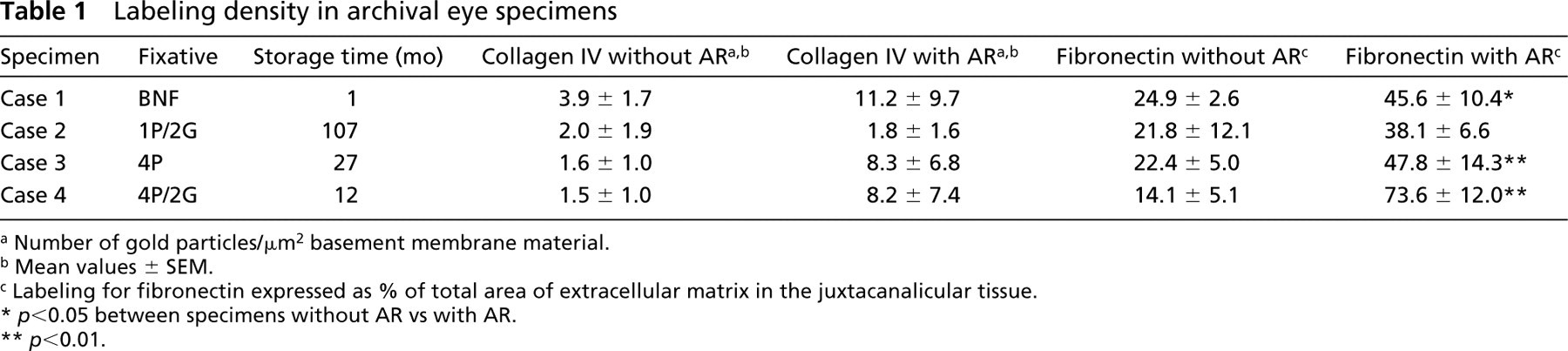
a Number of gold particles/μm2 basement membrane material.
b Mean values ± SEM.
c Labeling for fibronectin expressed as % of total area of extracellular matrix in the juxtacanalicular tissue.
∗ p < 0.05 between specimens without AR vs with AR.
∗∗ p < 0.01.
Materials and Methods
Tissue Samples
Five normal human eyes were removed at autopsy from five individuals (Mayo Institutional Review Board approval 992–99). The mean age was 47.5 ± 33.0 years. None had a history of ocular disease, ocular surgery, or any systemic disease that affected the eye. Causes of death included sepsis, brain death, cardiogenic shock, and motor vehicle accident. Four eyes were from a tissue archive collection and had been fixed in one of four fixatives: 4% paraformaldehyde (4P), 10% buffered neutral formalin (BNF), 4% paraformaldehyde/2% glutaraldehyde (4P/2G), or 1% paraformaldehyde/2% glutaraldehyde (1P/2G), all prepared in phosphate buffer. Storage times for these eyes ranged from 1 month to 9 years (Table 1). Eyes had been stored in fixative at room temperature (RT).
The fifth eye was obtained unfixed within 18 hr postmortem. It was dissected and portions were placed in four different fixatives. This was done to assess the effect of biological variability on labeling. Tissues from the same eye would be expected to be more similar in labeling than tissues from different individuals. The fixatives were 4P, 10BNF, 4P/2G, and 4% paraformaldehyde/0.1% glutaraldehyde (4P/0.1G). The last fixative did not match any of the fixatives from the eyes above but was chosen because it is often used for IEM fixation. Specimens from each fixative were stored for 3 months before processing and embedding. The 3-month period was chosen to study the effect of longer storage times, although it was a shorter storage period than that of most of the eyes under study.
Immunohistochemical Technique
Wedges of tissue from each eye, containing Schlemm's canal and the trabecular meshwork, were processed for LRWhite embedding using a method modified from Marshall et al. (1990). Specimens were dehydrated in 60% ethanol on ice for 15 min, placed in 70% ethanol for 60 min, and the temperature then lowered to −20C. Specimens were then immersed in 80% ethanol for 60 min, infiltrated in two 60-min changes of LR White resin, embedded in capped gelatin capsules, and polymerized at 55C overnight.
Thin sections (dark gold, ∼130 nm) were mounted on nickel grids coated with 1.5% parlodion prepared in a 1:1 solution of amyl acetate:acetone. Grids were dipped in this solution and allowed to dry for 30 min before sections were mounted. After sections were mounted, they were allowed to dry overnight on a 50C hotplate.
Multiple grids were made from each tissue block. Some grids were treated with AR before labeling, and others were labeled without AR. AR was done according to the method of Stirling and Graff (1995). AR grids were boiled in 0.01 M citrate buffer, pH 6.0, on a hotplate for 10 min, removed from the heat, and allowed to cool for 15 min in the citrate buffer. After AR, all grids were processed identically. For each eye, AR and non-AR grids were processed simultaneously, using the same reagents and dilutions of antibodies. Grids were first placed in blocking buffer (see below) for 15 min, then treated with primary antibody for 2 hr at RT. Grids were rinsed four times in PBS–Tween. Then secondary antibodies were applied for 1 hr at RT, followed by four rinses in PBS–Tween and four additional rinses in water. Grids were then dried overnight on a hotplate. Silver enhancing solution was prepared according to the manufacturer's instructions and grids were silver-enhanced for 2–4 min, rinsed in water, and allowed to dry. Sections were stained in aqueous uranyl acetate for 15 min followed by 4 min in lead citrate. Control sections included grids processed without primary antibody. Immunostaining was performed on tissue from two grids from each eye.
Micrographs were taken of the endothelial cells and underlying basal lamina of Schlemm's canal, trabecular beams with trabecular cells, and adjacent scleral tissue at ×25, 000 on a JEOL 1200 electron microscope (Peabody, MA). For the four archival specimens, two grids from one tissue block per eye were examined. For the fifth eye, in which comparisons were made within the same eye, one tissue block per quadrant was examined.
Antibodies and Reagents
The antibody to collagen IV was a goat polyclonal antibody to human and bovine Type IV collagen, supplied by Southern Biotechnologies (Birmingham, AL) and used at a 1:100 dilution. The fibronectin antibody was a rabbit polyclonal antibody to human fibronectin obtained from Sigma (St Louis, MO) and was used at a 1:50 dilution. Normal goat serum was obtained from Amersham Life Science (Piscataway, NJ). Secondary antibodies included rabbit anti-goat (5-nm gold particles) and goat anti-rabbit (5-nm gold particles) and were obtained from Amersham Life Science. Both were used at a 1:100 dilution. Silver enhancement was performed with the IntenSE M Silver Enhancement Kit from Amersham Life Science. LR White resin embedding kit was supplied by Ted Pella (Redding, CA). PBS–Tween, Tris buffer, ovalbumin, and glycine were obtained from Sigma. Dilutions of primary and secondary antibodies were made in PBS–Tween. Citrate buffer, 0.01 M, pH 6.0, was prepared according to the method of Stirling and Graff (1995). Blocking buffer consisted of PBS–Tween, 1% ovalbumin, 1% glycine, and 2% normal goat serum. All antibodies and blocking buffers were made in 10 mM PBS with .05% Tween (PBS-T). Nickel grids, 200-mesh thin bar from Ted Pella, were used.
Quantitation
Quantitation of labeling density was performed to assess changes after AR. For labeling of collagen IV, 10 micrographs at a final magnification of ×25, 000 were randomly selected from each specimen. A transparent grid measuring 150 mm by 20 mm (3000 mm2; total actual tissue area 4.8 μm2) was overlaid on each micrograph, centered on the basement membrane underlying the cells of Schlemm's canal. Both the inner and outer wall regions were examined. The total number of gold particles present overlying the basement membrane in these regions was counted and the density of labeling was expressed as number of gold particles/μm2 of basement membrane material.
Fibronectin was more diffuse in its labeling pattern than collagen IV, and a larger grid was used (240 mm by 200 mm; each grid square 10 mm × 10 mm, total area 48, 000 mm2; total actual tissue area 77 μm2). This grid was placed over the entire region adjacent to Schlemm's canal. On the inner wall, this encompassed the juxtacanalicular tissue, which extends about 10 μm from Schlemm's canal to the first trabecular lamellae. Label on the opposite side of Schlemm's canal, the “outer wall,” was also assessed. In contrast to collagen IV, in which label was predominantly in the basement membrane adjacent to the canal, fibronectin label was found in the basement membrane, sheath and tendon material, and other extracellular matrix components. Therefore, all of these were included in the area of possible fibronectin labeling. Fibronectin label occurred as clumps of six to eight gold particles rather than as individual particles as occurred with collagen IV. As a result, fibronectin label was expressed as the number of grid squares containing fibronectin-label/total number of grid squares containing extracellular matrix.
Data are expressed as mean ± SEM. Results were compared using a Student's two-tailed t-test for paired data. T-tests were subjected to the Bonferroni calculation (Neter et al. 1985) of multiple comparison to ascertain significance. Labeling counts from single micrographs that were greater than three times the mean of other micrographs were considered “outliers” and eliminated from final analysis. This involved only three of 306 micrographs.
Reproducibility of counts was determined by having a second observer count labeling of collagen IV on four specimens, both before and after AR (total of 78 micrographs counted by each).
Results
Collagen IV
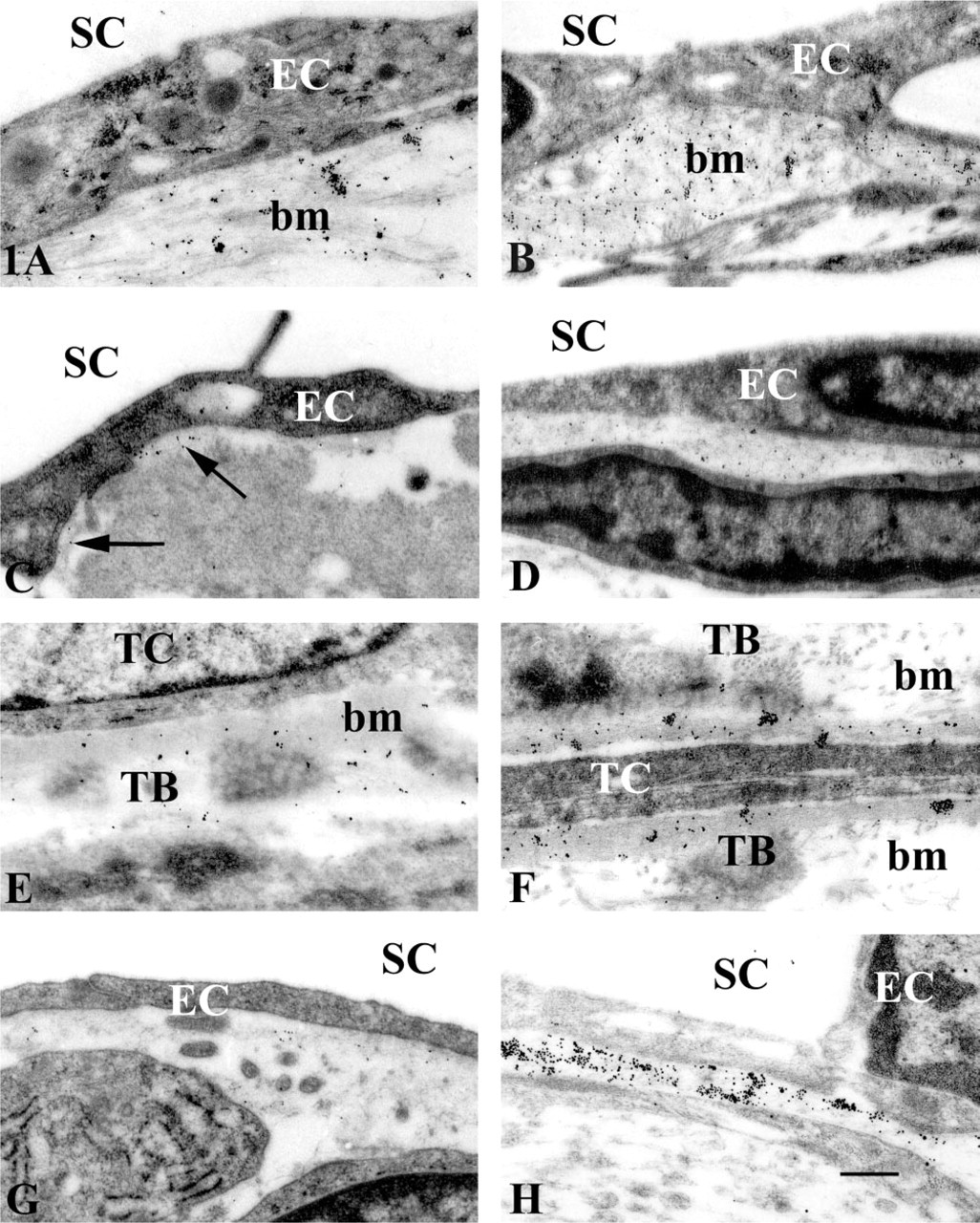
Qualitative Evaluation. Label was found in the basal lamina and adjacent basement membrane material of Schlemm's canal endothelial cells along both the inner wall (IW) and outer wall (OW) of Schlemm's canal (IW, Figures 1A and 1B; OW, Figures 1C and 1D). Label was also observed in the scattered patches of “amorphogranular material” adjacent to the cells in the juxtacanalicular region (Rohen 1983). In addition, label was found in the basement membrane of the trabecular lamellae, underlying the trabecular cells (Figures 1E and 1F). Labeling of collagen IV was seen as a diffuse pattern of gold particles distributed throughout the basement membranes (Figures 1B and 1D).
By subjective assessment, AR increased label for tissue fixed in all four fixatives and for all storage times. AR changed the labeling from sparse patches to a more continuous pattern in the basement membrane (compare Figures 1A and 1B). AR did not cause a new appearance of label in structures that were not labeled on non-AR grids. In tissues fixed in BNF, AR appeared to decrease the granularity and clumping of the label (compare Figures 1B and 1A). A slight amount of nonspecific background labeling was noted with AR over nuclei and in non-tissue areas, such as the lumen of Schlemm's canal.
Quantitative Evaluation. Tissues fixed in 4P/2G had the greatest increase in labeling density, with approximately five times more label than seen in non-AR grids. This increase was most noticeable in the tissue underlying Schlemm's canal (Figures 1G amd 1H). Although this was significant at p = 0.02 for this single comparison, it did not reach significance after the Bonferroni correction for multiple comparisons. AR also increased labeling density in tissues fixed in 4P and BNF, but to a lesser extent (Table 1).
Comparison Within the Same Eye. Neither the labeling pattern nor the type of extracellular matrix components that labeled changed with the different fixatives. Although labeling density increased after AR, no “new” components of the extracellular matrix demonstrated label that had not been found in non-AR tissue. Tissue in 4P/2G had the greatest increase in labeling density after AR, with labeling density six times higher than non-AR grids (p < 0.01; Table 2). This increase was similar to the increase seen in the four eyes above. Labeling density was increased about twofold after AR for the other fixatives, although this increase was not statistically significant (Table 2).
Fibronectin
Qualitative Evaluation. Fibronectin labeling was found adjacent to collagen fibers in the outer wall region of Schlemm's canal (Figures 2A and 2B). In the juxtacanalicular tissue, label was found in the sheath mate-membrane (bm). With AR. (
Immunogold labeling of collagen IV in the trabecular meshwork. Fixatives: 10% Buffered neutral formalin (
Labeling density: Comparison within the same eye
a Number of gold particles/μm2 basement membrane material.
b Mean values ± SEM.
c Four quadrants from same eye (S, superior; T, temporal; I, inferior; N, nasal). Labeling for fibronectin expressed as % of total area of extracellular matrix in the juxtacanalicular tissue.
∗ p < 0.05 between specimens without AR vs with AR.
∗∗ p < 0.01.
Fibronectin labeling occurred in aggregated focal clumps rather than in the diffuse pattern of the collagen IV label (Figures 2D and 2F). With AR, the most notable increase in labeling density occurred near collagen fibers in the outer wall of Schlemm's canal (Figures 2A and 2B) and the sheath material surrounding the elastic tendons (Figures 2C and 2D). This was true for all fixatives examined. Label for the central core region of the trabecular lamellae (Figures 2G and 2H) was increased in all fixatives, with the smallest increase noted with 1P/2GL.
Quantitative Evaluation. The greatest increase in labeling density was found for tissue fixed in 4P/2G, which was approximately five times greater than non-AR tissue (p < 0.01; Table 1). Label was also increased for other fixatives, with an approximate doubling of label after AR. Tissues fixed in 1P/2G demonstrated the smallest increase after AR.
Comparison Within the Same Eye. The labeling pattern and the type of extracellular matrix components that labeled did not change with the different fixatives nor after AR. Tissue in 4P/2G had the greatest increase in labeling density after AR, with labeling density six times higher than non-AR grids, similar to the increase seen in the four eyes above. Labeling density was increased about twofold after AR for the BNF and 4%P (p < 0.01; Table 2).
Control Sections
Sections processed without primary antibody were negative for label for collagen IV and fibronectin. When these negative controls were treated with AR, a small amount of nonspecific background label was present.
Reproducibility
Comparison of results from the two observers revealed that labeling densities were within 12% of each other (78 micrographs counted by both observers).
Discussion
Antigen retrieval increased the labeling of collagen IV and fibronectin of eye tissues after storage in a variety of fixatives and for differing lengths of time. Of importance, although the amount of label increased after antigen retrieval, no additional or “new” extracellular matrix components were labeled after AR. For most fixatives, labeling density generally doubled after AR. A larger increase was seen with 4P/2G, which showed an increase of five to six times that of non-AR grids. Without AR, labeling was best in non-glutaraldehydefixed tissue, whereas with AR labeling density increased to become generally similar among fixatives. The exception was 4P/2G, which had a marked increase in labeling. The comparison of fixatives from tissues within the same eye gave similar results. Comparison within the same eye minimizes the effect of biological variability on labeling, which can occur when tissues from different individuals are compared. Overall, these results indicate that the study of archival specimens, often collected in different fixatives over a period of years, is possible with AR methods.
Immunogold labeling of fibronectin in the trabecular meshwork. Fixatives: 10% Buffered neutral formalin (
With AR, tissue fixed in 4P/2G had best recovery of both collagen IV and fibronectin labeling. Our findings extend the results of other studies that used AR with low concentrations of fixative for shorter periods of time. Wilson et al. (1996) studied basal laminae and showed that AR increased labeling after microwave fixation. The results from the current study suggest that collagen IV and fibronectin labeling is enhanced by citrate AR, not only in freshly fixed tissue but also in archival tissues. A comparison of the labeling pattern and density we found for collagen IV with a previous study of the trabecular meshwork, which did not use archival tissues or AR, revealed similar labeling patterns and density of labeling (Marshall et al. 1990). Tissue in Marshall's study was fixed in either 4P or 4P with 0.2–1% glutaraldehyde. Use of the same antibody sources as Marshall et al. allowed comparison with that work.
LR White is often employed as the resin of choice to enhance antigenicity. Comparisons of LR White vs Epon-embedded tissues show increased labeling in LR White (Newman 1989; Wilson et al. 1996; Brorson, 1998). Because LR White resin allows the use of partial dehydration, epitopes that may have been extracted in low concentrations of fixatives (less than 2%) or high concentrations of ethanol are preserved. Although it has been reported that LR White sections may not require AR techniques (Newman et al. 1983), some antigens display low levels of labeling even in LR White. This was the case with the extracellular matrix antigens of the eyes in this study. Although the source of antibody influences labeling, as does the region of the antigen recognized by the antibody, sectioning properties of LR White may also play a role in preserving antigenicity. Whereas tissues crosslink with epoxies, LR White is believed to surround the tissue without crosslinking with it (Kellenberger et al. 1987). In sectioning LR White, cleavage planes may go around epitopes, whereas in epoxies the plane of section goes through epitopes, especially in large molecules (Kellenberger et al. 1987, Brorson, 1998).
In the current study, tissues fixed in a combination of 4% paraformaldehyde and 2% glutaraldehyde showed a greater increase in label than tissues fixed in lower concentrations of these fixatives. One explanation for this could be the fact that higher-concentration fixatives preserve more epitopes. On dehydration and embedding less washout occurs (Hayat 1970). When AR is employed on these tissues, more epitopes are present to be labeled. Another probable explanation of increased label after AR in higher paraformaldehyde/glutaraldehyde concentration fixatives may be the modulation of the crosslinking of the glutaraldehyde by paraformaldehyde. Paraformaldehyde is known to be reversible, whereas glutaraldehyde fixation is irreversible (Hayat 1981). In combination fixatives of paraformaldehyde and glutaraldehyde, a two-step reaction is believed to occur. Paraformaldehyde penetrates more rapidly and stabilizes proteins first, followed by crosslinking with glutaraldehyde (Hayat 1989). This suggests displacement or further cross-linking of epitopes by glutaraldehyde. Evidence for such modulation is the finding that the combined reaction of formaldehyde and glutaraldehyde creates a new set of reaction products not seen with either agent alone (Johnson 1986). Some evidence for this is shown by the greater effect of antigen retrieval we found for labeling of collagen IV and fibronectin in the higher concentration paraformaldehyde (4P/2G vs 1P/2G; Table 1, Case 4 vs Case 2).
Antigen retrieval can be used with LR White-embedded sections to increase the labeling of the extracellular matrix molecules collagen IV and fibronectin. AR increased the labeling density of tissue in each fixative without changing the overall pattern or distribution of label. Although each antigen is different, AR used with LRWhite is another tool to enable retrospective studies of rare archival tissue specimens.
Footnotes
Acknowledgements
Supported in part by National Institutes of Health research grant EY 07065, Research to Prevent Blindness, Inc. (New York, NY), the Bonner Glaucoma Research Fund (Princeton, NJ), and the Mayo Foundation (Rochester, MN).