
Abstract
The purpose of this investigation was to develop a method that could be used to estimate how damaging sodium ethoxide is to different antigens with respect to immunolabeling when epoxy sections are deplasticized. If we obtain weak labeling for an antigen on deplasticized epoxy sections, this might be caused by the damaging effect of the ethoxide solution. It is therefore interesting to develop a method to check if this really is the reason. Fibrin clots and tissues of human kidney and thyroid were embedded in LR White resin. Some thin sections from these specimen blocks were exposed to sodium ethoxide in the same way as epoxy sections are when being deplasticized. Other sections from the same blocks were not exposed to sodium ethoxide. Both categories of sections were immunogold-labeled with anti-fibrinogen, anti-thyroglobulin, anti-IgA, anti-IgG, or anti-IgM. The intensity of immunolabeling of sections treated with ethoxide was compared with the immunolabeling of corresponding sections that were not treated with ethoxide. No significant differences were found in immunolabeling for fibrinogen, IgA, IgG, and IgM. For thyroglobulin, the intensity was approximately 30% less in tissues that were exposed to sodium ethoxide. The practical significance of this method is that we easily can examine the degree to which a given antigen is affected by sodium ethoxide, which is the agent used for deplasticizing epoxy sections.
E
LR White sections are well suited for immunoelectron microscopy because the antigens protrude easily from the surface of the sections (Brorson and Skjørten 1996a; Kellenberger et al. 1987). LR White resin is not affected by sodium ethoxide because this resin has chemical qualities different from those of epoxy resin. LR White sections are therefore not deplasticized, and more epitopes are not exposed after treatment with sodium ethoxide. If destruction by sodium ethoxide is the reason for a given antigen to be negatively or weakly immunolabeled on deplasticized epoxy sections, one should obtain significantly weaker immunolabeling for LR White sections treated with sodium ethoxide than for untreated LR White sections. Our aim is to develop a method, based on the above chain of reasoning, that can estimate the damaging effect of sodium ethoxide to antigens on deplasticized epoxy sections. We have used fibrinogen, thyroglobulin, and immunoglobulin deposits in kidney as test antigens, because these occur abundantly and are therefore easy to work with as test antigens.
Materials and Methods
Materials
Antibodies. The antibodies used were rabbit anti-fibrinogen (1:200), rabbit anti-thyroglobulin (1:2000), rabbit anti-IgA (1:200), rabbit anti-IgG (1:200), and rabbit anti-IgM (1:400) (all from Dakopatts; Glostrup, Denmark).
Colloidal Gold Probe. The colloidal gold probe was goat anti-rabbit IgG (H + L) Auroprobe EM Gar G15 (15-nm gold) (Amersham; Little Chalfont, Bucks, UK). Goat anti-mouse IgG (whole)-gold conjugate (10 nm) was from Sigma (St Louis, MO).
Substrates. Thyroid tissue was obtained at autopsy; the cause of death was lymphoma. Kidney tissue was obtained from a needle biopsy taken at Ullevål Hospital. Fibrin clots were prepared by a method described earlier (Brorson et al. 1994).
The electron microscope used was a JEOL 1200 EX.
Procedures
The tissues were cut into small pieces (<1 mm), fixed in 4% paraformaldehyde in phosphate buffer, pH 7.3, at 4C, and washed for 30 min with 0.2 M cacodylate buffer, pH 7.3.
Tissues were embedded in LR White according to the following procedure. Dehydration was performed once in 70%, and twice in 96% ethanol. Infiltration was done in a mixture of LR White and 96% ethanol (1:1) for 2 hr, and in pure LR White for 5 hr. Embedding was done in gelatin capsules, size 1. Polymerization took place at 56C for about 40 hr.
Cutting. EM sections with pale gold interference color (70–100 nm) were cut with a diamond knife (Jumdi; Juniper Ultra Micro, Stockholm, Sweden) from 10 independent specimen blocks of LR White-embedded tissues on an LKB 2088 Ultrotome V. The EM sections were mounted on uncoated 200-mesh nickel grids. This was done for each of the tissues mentioned above.
Treatment with Ethoxide. The procedure was that of Mar and Wight (1988), subjected to minor revision. Some of the LR White sections were exposed to a mixture of saturated sodium ethoxide solution and 100% ethanol (1:1) for 10 min. This is the same procedure used for deplasticizing epoxy sections (Brorson and Skjørten, 1995, 1996b). Other LR White sections were exposed to prolonged treatment with sodium ethoxide for 40 min. Grids were then hydrated to water through a graded ethanol series (100%, 50%, distilled water twice).
Immunoprocedure. Immunogold labeling was done on grids for all LR White sections, whether they had been exposed to sodium ethoxide or not (Brorson et al. 1994). For the primary antibodies, the incubation was carried out overnight at 4C (fibrin clots, anti-fibrinogen; thyroid, anti-thyroglobulin; kidney, anti-IgA, anti-IgG, anti-IgM), and for the secondary immunoreagents the conditions of incubation were 1 hr at 22C. Secondary antibodies coupled to 15-nm gold particles were used for all tissues. To examine if more (or fewer) nonspecific sites were created by the sodium ethoxide treatment, we also incubated both ethoxide treated and untreated LR White sections with goat anti-mouse IgG (10 nm) (a nonspecific antibody-gold conjugate). After immunolabeling, the sections were stained with 5% uranyl acetate in 30% ethanol and with Reynolds’ lead citrate.
Immunostained EM sections were examined and photographed in the electron microscope. In all cases the exact magnification was determined with a replica grid (2160 lines/mm cross-ruled grating replica). The sections were photographed at a display magnification of X 15,000. Measurements and calculations from the photographs were done in the following way. Corresponding areas of antigens were examined on both ethoxide-treated sections and untreated sections for degree of labeling in terms of number of gold particles/μm2. An area of ≈ 1 μm2 of antigen was selected from a section of each of the 10 specimen blocks and was used for calculation of the number of gold particles/μm2.
Results
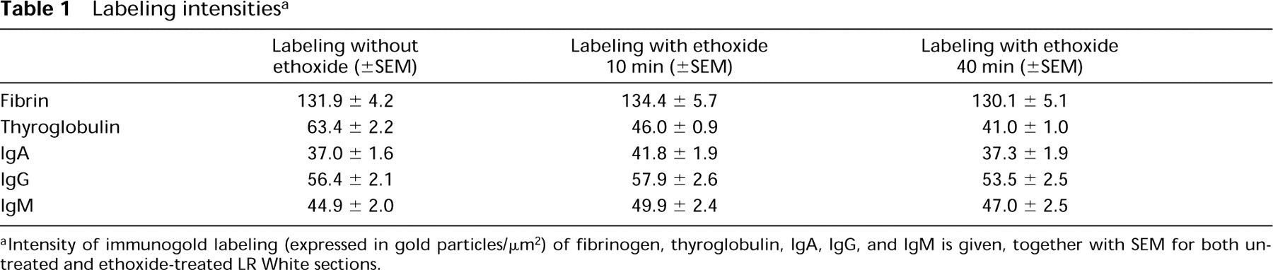
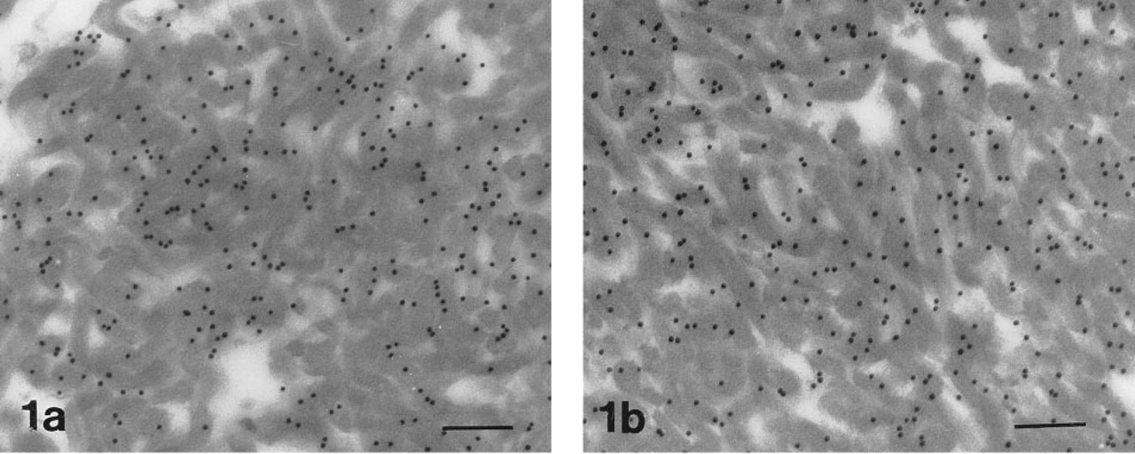
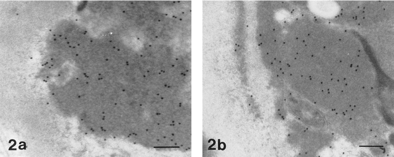
Values for the intensity of immunogold labeling for both ethoxide-treated and untreated LR White sections are given in Table 1. No significant differences in immunogold labeling were found for fibrinogen (p=0.78) (Figures 1A and 1B), IgA (p=0.14), IgG (p=0.73), and IgM (p=0.25) (Figures 2A and 2B) whether or not the LR White sections were exposed to sodium ethoxide (in reference to normal treatment with ethoxide for 10 min). For thyroglobulin (Figures 3A and 3B) there was a significant difference (p=0.0001); the intensity was about 30% less when sections were exposed to sodium ethoxide. There were no significant differences in immunolabeling when the sections were treated for 10 or 40 min with sodium ethoxide for any of the antigens examined (Table 1). The control with nonspecific antibody-gold conjugate did not indicate the presence of more (or fewer) nonspecific sites on the sections treated with sodium ethoxide (Table 2).
Labeling intensities a
a Intensity of immunogold labeling (expressed in gold particles/μm2) of fibrinogen, thyroglobulin, IgA, IgG, and IgM is given, together with SEM for both untreated and ethoxide-treated LR White sections.
Fibrin on LR White sections immunolabeled with anti-fibrinogen (15-nm gold particles were used as secondary probe).
Immune complex deposit in human kidney on LR White sections immunolabeled with anti-IgM (15-nm gold particles were used as secondary probe).
Thyroglobulin on LR White sections immunolabeled with anti-thyroglobulin (15-nm gold particles were used as secondary probe).
Unspecific labeling a
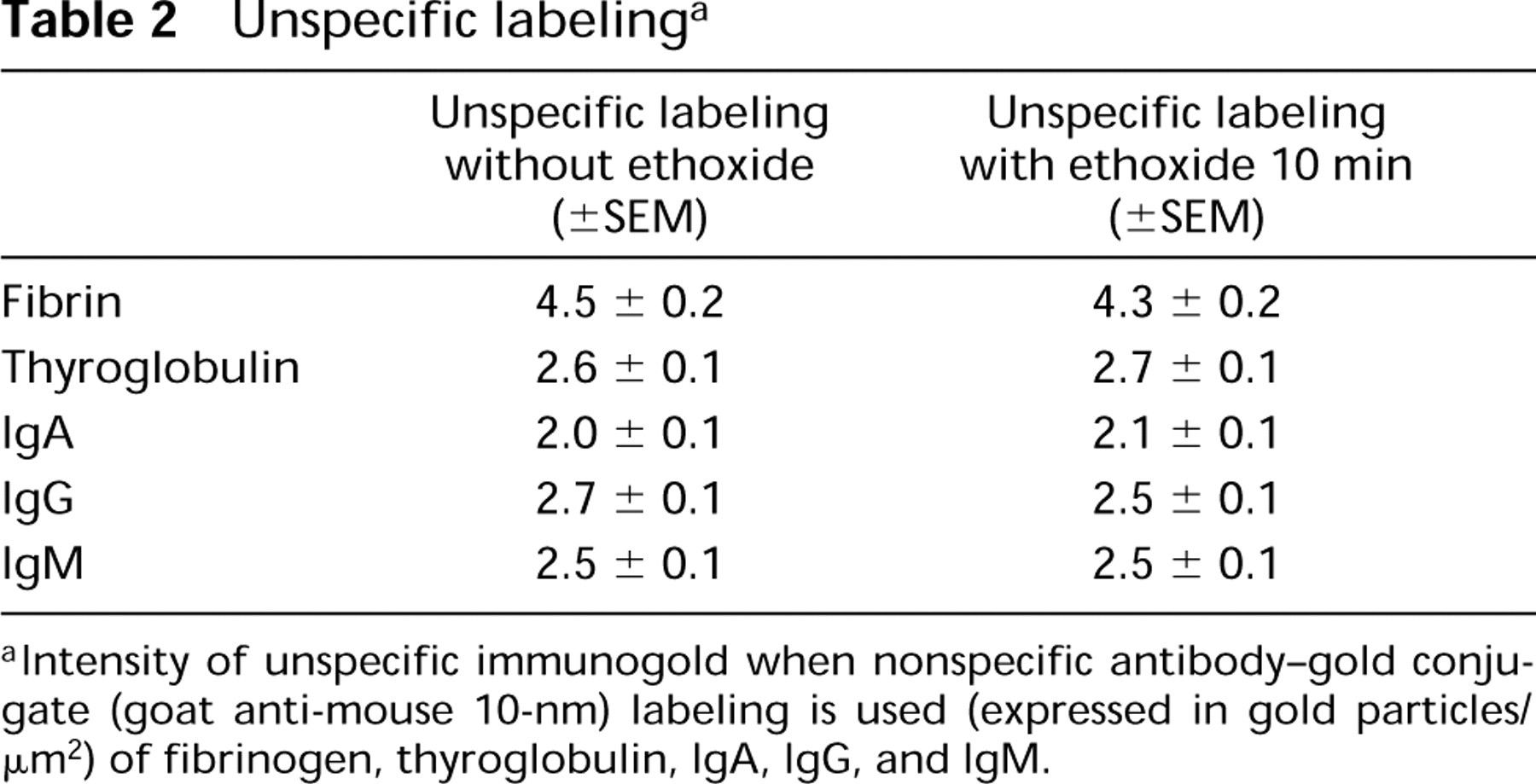
aIntensity of unspecific immunogold when nonspecific antibody-gold conjugate (goat anti-mouse 10-nm) labeling is used (expressed in gold particles/μm2) of fibrinogen, thyroglobulin, IgA, IgG, and IgM.
Discussion
As observed from the Results, the intensity of the immunogold labeling for most of the antigens tested was not negatively affected by sodium ethoxide. The immunolabeling was reduced only for ethoxide-treated thyroglobulin. We conclude from these results that sodium ethoxide does not reduce the immunolabeling of fibrinogen, IgA, IgG, and IgM, and that this strong basic agent has a certain reducing effect on the labeling of thyroglobulin, which corresponds to the impression of Brorson and Skjørten (1996b). If we had obtained a result in which the labeling for an antigen was negative on the sodium ethoxide-treated sections but clearly positive on the untreated sections, we would have had to conclude that the technique based on deplasticized epoxy section was not usable for that antigen. A result with approximately the same immunolabeling on ethoxide-treated LR White sections and untreated LR White sections means that if the immunolabeling on deplasticized epoxy section is not satisfactory, one must seek a reason for the discouraging result other than the sodium ethoxide solution.
A single 15-nm gold particle with multiple secondary antibody molecules attached to its surface could bind one or several primary antibody molecules. Therefore, each 15-nm gold particle could represent (be bound to) several individual epitopes. Because of this, one could in theory have a significant decrease in active epitopes on the ethoxide-treated sections and yet see no decrease in the number of gold particles compared to the untreated sections. Considering fibrinogen, IgA, IgG, and IgM, we can say that the sodium ethoxide does not damage the epitopes to such an extent that it influences the number of gold particles/μm2, but we cannot be sure that there is no damage to epitopes at all.
We can theoretically imagine antigens that are immunolabeled with significantly stronger intensity on ethoxide-treated LR White sections than on untreated LR White sections. In such cases, fixation bonds released by the action of sodium ethoxide may be the reason. Therefore, when the same intensity of immunogold labeling is observed for both ethoxide-treated and untreated ethoxide LR White sections, we cannot exclude that the reason is a combination of damage to the antigens and release of the fixation bonds.
In conclusion, we have developed an easy method that can be used to examine the degree to which a given antigen is affected by sodium ethoxide, which is the agent used for deplasticizing epoxy sections.