
Abstract
Plastic embedding has been used to localize various antigens in conjunction with immunohistochemistry. Peptide hormones have been among the antigens that have been studied extensively. Recent application of water-soluble plastics such as LR White and Lowicryl has extended the ranges of detectable antigens and enabled the observation of antigen–antigen or mRNA–antigen combinations. This review article deals with technical aspects, procedures, and applications in endocrine cells.
Keywords
I
In situ hybridization demonstrates mRNA, which indicates production of peptide hormones, and immunocytochemistry demonstrates the localization of peptides. A regulatory pathway via secretory granules and a constitutive pathway via secretory vesicles in the protein secretion are also discussed.
Technological Aspects of Plastic Embedding in the Postembedding Method
Plastic embedding was done first with the usual epoxy resin for detection of various antigens, including peptide hormones (Roth et al. 1978; Bendayan 1982; Childs and Unabia 1982). For the Epoxy resin, “etching” by hydrogen peroxide or periodate is mandatory for the antigenicity to be exposed on the surface of the polymerized plastic surface (Baskin et al. 1979a, b). Recently introduced LR White and Lowcryl are plastic materials that can be polymerized without high temperatures and can preserve antigenicity much better than the conventional plastic embedding materials, e.g., epoxy resin (Roth et al. 1978; Herrerma et al. 1987; Al-Nawab and Davies 1989; Bowdler et al. 1989; Smith et al. 1990; Itoh et al. 1996; Osamura et al. 1997). Without postfixation by osmic acid, these embedding media also can preserve ultrastucture adequately and satisfactorily. We have had satisfactory results for both ultrastucture and antigenicity in localizing large molecules such as IgA, carcinoembryonic antigen (CEA), and peptides. Although it has been claimed that conventional embedding, such as epoxy resin, can preserve antigenicity only in secretory granules, LR White and Lowycril can demonstrate a variety of substances with larger molecular size on various cell organelles such as the rough endoplasmic reticula, Golgi saccules, and secretory granules, and on secretory vesicles (Itoh et al. 1996; Osamura et al. 1997). Recently, the plastic embedding method has been used for in situ hybridization to localize DNA and mRNA. Matsuno et al. (1994a) have compared the preservation of mRNA by ISH between the pre-embedding method and the postembedding method, using LR White, and found that the postembedding method could be utilized for semiquantitative electron microscopic ISH despite the markedly deteriorated amount of mRNA caused by plastic embedding (Singer et al. 1989; Jirikowski et al. 1990; Le Guellec et al. 1990, 1991, 1992; Egger et al. 1994; Escaig-Haye et al. 1992; Gingras and Bendayan 1995; Morey et al. 1995; Matsuno et al. 1994a, b, 1995, 1996, 1998a, b,c).
Procedures for Immunoelectron Microscopy with Plastic Embedding
In general, the tissue fragments are fixed in 4% paraformaldehyde or 4% paraformaldehyde with 0.05% glutaraldehyde solution. Then the tissue is dehydrated and embedded in LR White or Lowycryl by the method described in the literature (Itoh et al. 1996; Osamura et al. 1997). This procedure is performed without osmification, which is believed to deteriorate antigenicity to a significant degree. Ultrathin sections cut with a diamond knife are attached to nickel grids. Immunohistochemical reaction is done by either a polycolonal or a monoclonal antibody. As a second reaction, gold-labeled protein A or gold-labeled antibody is used. We have been using the protein A–gold method. Colloidal gold particles of various sizes are available commercially. Frequently used gold particles are 10, 15, and 20 nm in diameter. To localize two different antigens on the same ultrathin section by the protein A–gold method, each reaction must to be done on opposite surfaces of the section. When the double staining is done by gold-labeled secondary antibody of different species, two reactions to detect different antigens can be done on the same surface of the ultrathin section (Bendayan 1982). Gold particles of 10, 15, or 20 nm in diameter can be easily discerned in single detection as well as double staining.
Procedure for Electron Microscopic In Situ Hybridization (EM-ISH) with Plastic Embedding
Postembedding EM-ISH of rat GH mRNA Using LR White Resin-embedded Tissues (Matsuno et al. 1994a, 1998c). Anterior pituitary tissues were embedded in LR White resin, after fixation at 4C overnight with 4% paraformaldehyde dissolved in PBS. The pituitary tissues were carefully placed at the bottom of gelatin capsules (Lilly Pharmaceuticals; Indianapolis, IN), which were filled with LR White resin and sealed. After polymerization at 50C for 24 hr in a vacuum oven, ultrathin sections were attached to nickel grids. Hybridization with a biotinylated antisense oligonucleotide probe for rat GH mRNA at a concentration of 1 ng/ml was carried out on the grid at 37C overnight after prehybridization at the same temperature for 30 min. After hybridization, the grid was dipped in 2 × SSC (saline-sodium citrate concentrate), 1 × SSC, and then 0.5 × SSC for 5 min each. Hybridization signals were developed for 30 min with 20-nm streptavidin–gold (British Biocell International, Cardiff, UK) diluted 1:50 in 1% bovine serum albumin (BSA)–PBS. After being dipped in PBS and distilled water and dried at room temperature, the grids were inspected under an electron microscope. The control experiments were hybridization studies with a sense probe or a scrambled sequence and without a probe.
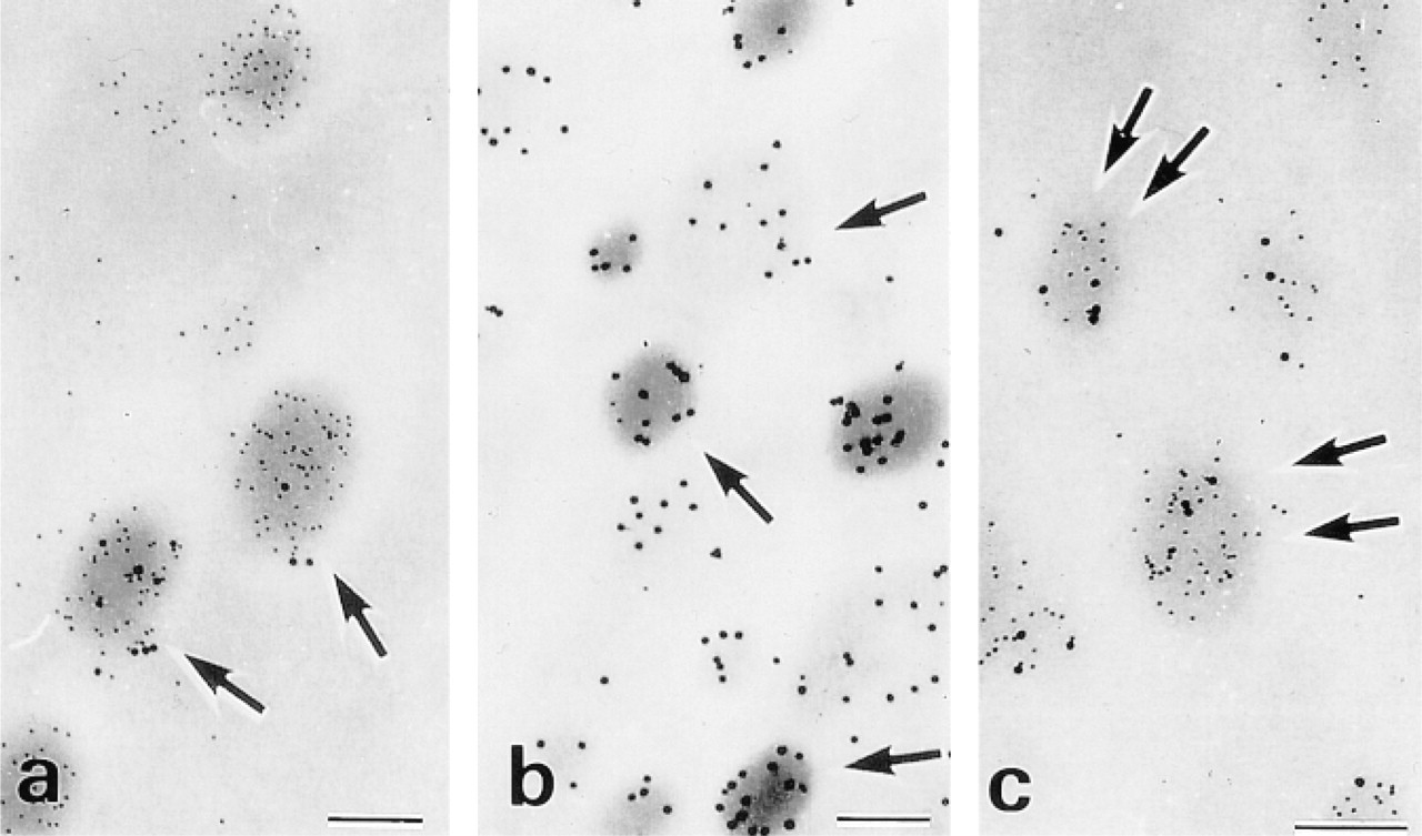
Hybridization signals for rat GH mRNA were localized on the polysomes of the rough endoplasmic reticulum (RER) using 20-nm streptavidin–gold (Figure 1a). A negative control experiment with a sense probe yielded no hybridization signals (Figure 1b).
Combined Immunohistochemistry and EM-ISH Using LR White Embedding (Matsuno et al. 1998a).
This procedure utilizes pre-embedding ISH for intracellular localization of GH mRNA and postembedding immunohistochemistry for identification of protein (GH). Anterior pituitary tissues were fixed overnight in 4% paraformaldehyde dissolved in PBS. After immersion in graded concentrations of sucrose dissolved in PBS (10% for 1 hr, 15% for 2 hr, 20% for 4 hr), tissues were embedded in OCT compound (Tissue-Tek; Miles Laboratories, Elkhart, IN). Six-μm-thick-sections were mounted on slides coated with 3-aminopropylmethoxysilane. After air-drying for 1 hr, tissue sections were washed with PBS for 15 min. Then they were treated with 0.1 mg/ml proteinase K (Boehringer Mannheim; Mannheim, Germany) at 37C for 30 min, followed by treatment for 10 min with 0.25% acetic anhydride in 0.1 M triethanolamine. The slides were washed with in 2 × SSC at RT for 3 min and then pre-hybridized at 37C for 30 min. Hybridization with biotinylated antisense oligonucleotide probe for rat GH mRNA at a concentration of 0.1 ng/ml was carried out at 37C overnight. After hybridization, the slides were washed at RT with 2 × SSC, 1 × SSC, and then 0.5 × SSC for 15 min each and the hybridization signals were detected with streptavidin–biotin–horseradish peroxidase, using Vectastain's ABC kit (Vector Laboratories; Burlingame, CA). The hybridization signals were developed with diaminobenzidine and H2O2 for 7 min. After osmification at RT for 1 hr and dehydration with ethanol, tissue sections were embedded in LR White resin using an inverted beam capsule, which was polymerized at 50C for 2 days. After polymerization, ultrathin sections were attached to nickel grids. The grids were floated in 0.1 M phosphate buffer (PB) containing the antibody against rat GH (monkey, diluted 1:100), supplied by the National Institute of Diabetes and Digestive and Kidney Diseases (NIDDK) (Bethesda, MD) at RT for 1 hr. The grids were washed with 0.1 M PB, and then floated in 0.1 M PB containing 20-nm protein A–colloidal gold (diluted 1:40; EY Laboratories, San Mateo, CA) at RT for 1 hr for visualization of the labeling. After being washed with 0.1 M PB and distilled water, the grids were dried at RT and observed under an electron microscope. The control experiment for pre-embedding EM-ISH consists of hybridization with a sense or scrambled sequence probe and hybridization without a probe. The immunohistochemical control experiment includes substitution of normal monkey serum for anti-GH antibody.
(
Hybridization signals of rat GH mRNA using a biotinylated antisense oligonucleotide probe were confirmed by light microscopy. No positive reactions for the mRNA were observed with a sense oligonucleotide probe. EM-ISH with an antisense probe for the mRNA revealed its diffuse localization on the polysomes of the entire RER (Figure 1c). Subsequent immunohistochemical staining using an anti-rat GH antibody and 20-nm protein A–colloidal gold identified rat GH protein on the secretory granules (Figure 1c). Colloidal gold signals for GH protein were distributed mainly on the secretory granules and were also identified in the cisternae of the RER (Figure 1c). EM-ISH with a sense probe for GH mRNA and an immunohistochemical control experiment substituting normal monkey serum for anti-GH antibody revealed no signals for GH mRNA and GH protein (Figure 1d).
Applications of LR White and Lowicryl Plastic in Postembedding Immunoelectron Microscopy
The Regulated Pathway and the Constitutive Pathway
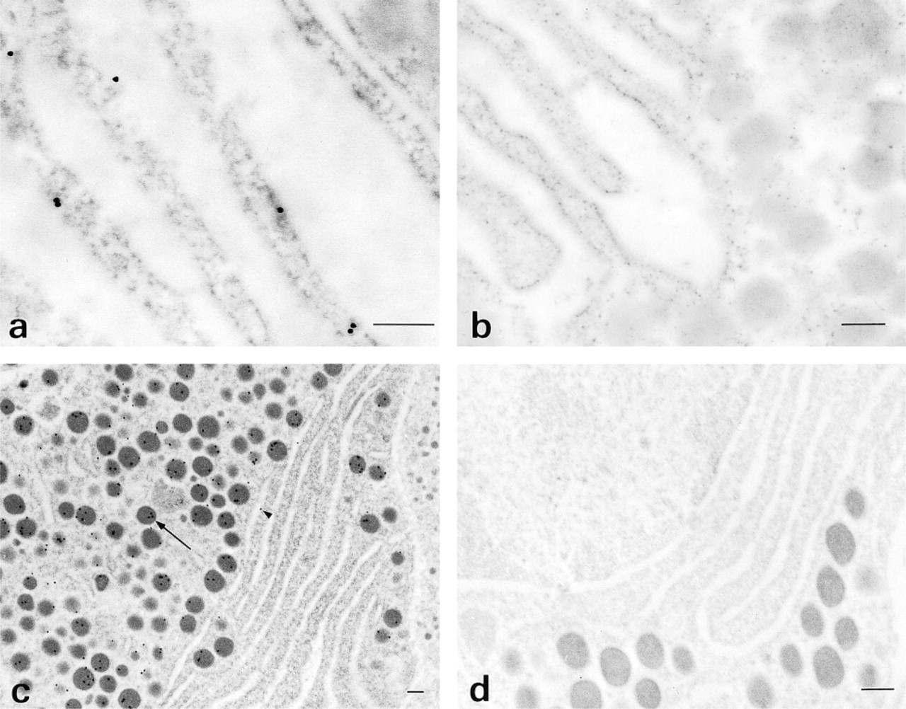
Embedding in LR White or Lowycryl has successfully demonstrated IgA in the kidney and carcinoembryonic antigen (CEA), as well as various peptide hormones, in tumors such as medullary carcinoma of the thyroid gland (MCT). By this method, in the same tumor cells of MCT, calcitonin (CT), a peptide hormone was localized exclusively in the secretory granules (SGs), and CEA was localized in the vesicles and the membrane (Figure 2a and 2b). It was also detected in the Golgi saccules. CT was also localized in the amyloid deposition, which is another characteristic finding in MCT. CT and CEA can be detected in the same tumor cells by the double immunoelectron microscopic staining decribed above (Figure 2c and 2d). These results suggest that CT and CEA can be secreted by different secretory pathways, a regulated pathway via secretory granules for CT and a constitutive pathway via secretory vesicles for CEA, (Osamura et al. 1997) It is suggested that the site of sorting may lie in the trans-Golgi networks (TGNW) (Kelly 1985; Griffiths and Simons 1986; Rambourg et al. 1988; Osamura et al. 1991; Tooze and Stinchcombe 1992; Farquhar and Palade 1998).
(
Processing of Prohormones and Secretory Granules
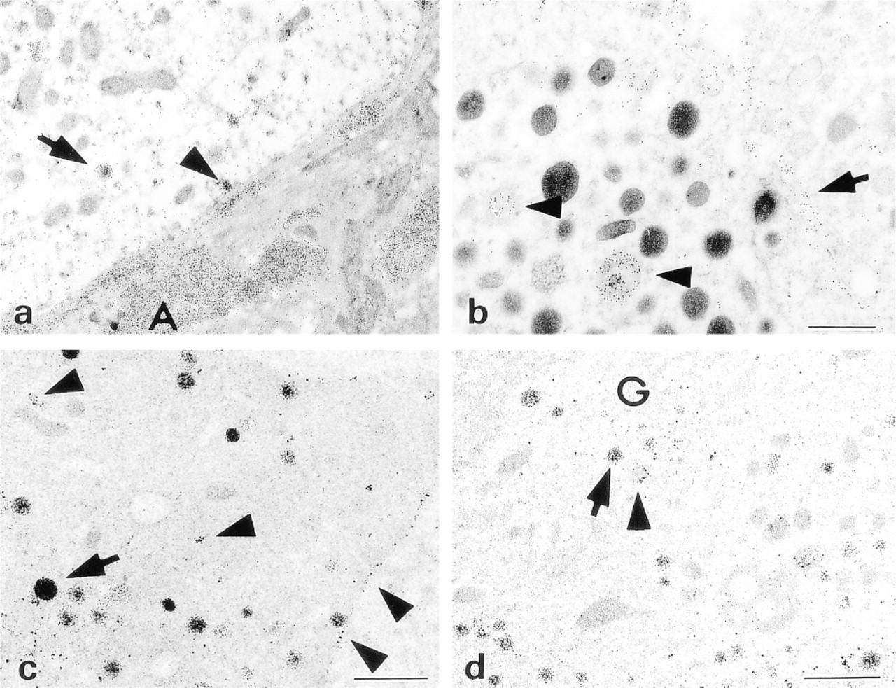
It is well known that peptide hormones are produced as larger precursor molecules, which are called prohormones. In the secretory process of endocrine cells, the prohormones are digested (designated as processing) into the corresponding bioactive peptides by specific proteolytic enzymes, now called prohormone convertases (PCs) (Cool et al. 1997; Steiner 1998). PCs are composed of six subtypes: PC1/3, PC2, PC4, PC5, PC6, and PC7. Among these, PC1/3 and PC2 function in the digestion of prohormones, such as proinsulin, proglucagon, and pro-opiomelanocortin (POMC). It has been found that proinsulin is processed into insulin by PC1/3 and PC2 and that proglucagon is processed into glucagon by only PC2. POMC is processed into ACTH by PC1/3 and further into MSH by PC2. In the rat pancreatic islets, postembedding immunoelectron microscopy using LR White demonstrated the co-localization of PC1/3 and PC2 with insulin in the same secretory granules (Figure 3a and 3b). On the other hand, PC2 is co-localized only with glucagon in the same secretory granules (Figure 3c) (Itoh et al. 1996). These findings indicate that the specific site for processing of the prohormones is the secretory granules. Therefore, it is indicated that the secretory granules in endocrine cells are essential not only for the regulatory pathway but also for the processing of prohormones.
Application of Plastic Embedding EM to In Situ Hybridization
Postembedding EM-ISH studies have been reported by several investigators (Le Guellec et al. 1990, 1991, 1992; Matsuno et al. 1994a, 1995, 1998c). Le Guellec and co-workers stated that no hybridization signals were observed in an EM-ISH study using Epon-embedded sections (Le Guellec et al. 1992). The postembedding EM-ISH study using Lowicryl embedding is not suitable for precise localization of hybridization signals on the polysomes of the RER because of frequent nonspecific signals in the ER cisternal space. In contrast, the postembedding EM-ISH study using LR White-embedded sections can provide well-preserved ultra-structure, visualization with higher resolution, and more precise localization of hybridization signals for rat GH mRNA on the polysomes of the RER.
Compared with the pre-embedding EM-ISH method, the postembedding EM-ISH method has difficulty in message preservation during polymerization of LR White at high temperature for an extended period of time, which leads to mRNA degradation (Matsuno et al. 1994a, 1995, 1998c). Another difficulty of the post-embedding EM-ISH method is a possible nonspecific affinity of colloidal gold particles. For elucidating the intracellular localization of free ribosomes, the pre-embedding EM-ISH method did not give adequate resolution. If the difficulties of the postembedding method stated above can be resolved, then this method using gold particles will be a promising technique for clarifying the intracellular localization of specific mRNA in free ribosomes. It is also expected that this method will be able to detect several different mRNAs simultaneously on the same RER using double staining with different-sized gold particles.
Immunoelectron microscopy shows co-localization of insulin (10-nm) and PC1/3 (20-nm) (
The intracellular spatial relationship between pituitary hormone and its message at the ultrastructural level is another of our major concerns. Previous publications on the ultrastructural simultaneous detection of mRNA and encoded protein, other than our previous reports (Matsuno et al. 1995, 1996, 1998a, b,c), utilized postembedding EM-ISH using colloidal gold particles for Lowicryl K4M- or LR White resin-embedded tissues (Singer et al. 1989; Escaig-Haye et al. 1992; Egger et al. 1994; Gingras and Bendayan 1995; Morey et al. 1995). In these postembedding EM-ISH studies, relatively frequent nonspecific reactions of colloidal gold particles used for the detection of mRNA were observed in the cisternae of the RER. We experienced the decreased message preservation in the postembedding EM-ISH studies using LR White-embedded tissues (Matsuno et al. 1994a, 1995, 1998c). To obtain specific hybridization signal detection and well-preserved messages, we have employed pre-embedding EM-ISH and subsequent immunoreaction for simultaneous ultrastructural identification of mRNA and encoded protein (Matsuno et al. 1996, 1998a, b,c). In a previous report, we utilized Epon resin for tissue embedding, which required etching using H2O2 or sodium periodate for retrieval of immunoreactivity (Matsuno et al. 1996, 1998b, c). In general, osmification and embedment in Epon resin are reported to decrease the immunoreactivity of the targeted protein, and the etching process using H2O2 or sodium periodate results in deosmification and shades off the signals of mRNA. To resolve these problems, we have recently used LR White resin for tissue embedment (Matsuno et al. 1998a). In LR White resin-embedded tissues, retrieval of immunoreactivity with H2O2 or sodium periodate is not required, and therefore the gradation of the signals of mRNA can be avoided. Ultrastructure, including intracellular organelles, is well preserved and definitely positive signals for both mRNA and protein are obtained. As shown in our previous reports using tissues embedded in Epon resin (Matsuno et al. 1994a, b, 1995, 1996, 1998b, c), somewhat heterogeneous electron density was observed in GH mRNA expression, which is distributed diffusely on the RER. In LR White resin-embedded tissues, GH mRNA is also distributed diffusely on the RER, its electron density being homogeneous (Matsuno et al. 1998a). The cause of this difference in electron density of heterogeneity and homogeneity cannot be determined but may be attributed to the characteristic difference of both resins. As the manufacturer stated, the hydrophilic and low lipid-solvent character of LR White resin serves for excellent visualization of membrane and cytosol structures and may therefore be helpful for the preservation of the reaction products in LR White resin-embedded tissues. The simultaneous ultrastructural identification of mRNA and encoded protein by combined immunohistochemistry and pre-embedding EM-ISH using LR White resin-embedded tissues can be a much better choice for pathophysiological studies of hormone secretion.
Conclusions
mRNA and peptide hormones have been successfully localized on ultrathin plastic sections, especially by using the water-soluble plastics LR White and Lowicryl. This technology is very useful for study of the cell biology of hormone-secreting cells, from production to the secretion of the peptides. In addition, this technique is expected to be applicable to other bioactive cells, because it can localize both large protein molecules and mRNA on ultrathin sections.