
Abstract
Background
Potassium ethylenediaminetetraacetic acid (K-EDTA) contamination of serum samples is a common cause of spurious electrolyte results, which may adversely affect patient care. The source of K-EDTA sample contamination is unknown since it is not caused by reverse order of draw. Other possible mechanisms are either direct transfer of blood from K-EDTA containing tubes to other tubes or syringe needle/top contamination when delivering blood into EDTA sample tubes before other sample tubes, but these have not been studied in clinical practice. We report on a quality improvement programme aimed at identifying the source of K-EDTA-contaminated samples.
Methods
We routinely measure EDTA in all serum samples with a potassium ⩾6.0 mmol/L. We identified individuals responsible for K-EDTA-contaminated samples (EDTA >0.15 mmol/L) and in close-to-real-time discussed their phlebotomy methods for the collection of these samples.
Results
Over four months, we investigated 96 EDTA-contaminated samples. Of these, we identified and interviewed 64 (67%) individuals responsible for contaminated samples; 52 (81%) doctors, 9 (14%) phlebotomists and 3 (5%) nurses. Fifty-two individuals recalled taking the sample and the phlebotomy method used. All used open phlebotomy methods.
Conclusions
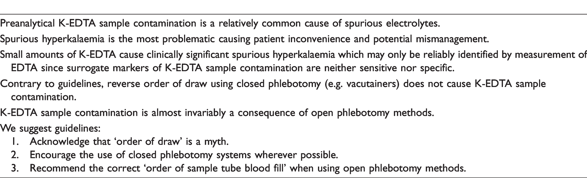
We report, for the first time, that K-EDTA sample contamination almost always, if not exclusively, occurs following open phlebotomy methods. Phlebotomy training and guidelines should, therefore, encourage use of closed systems as well as include and emphasize the importance of ‘order of blood sample tube fill’ when using open phlebotomy methods.
Background
Potassium ethylenediaminetetraacetic acid (K-EDTA) is a common blood sample tube anticoagulant. In vitro K-EDTA contamination of lithium heparin, gel-containing or plain tubes from K-EDTA containing tubes is a relatively common but often unrecognized cause of spurious hyperkalaemia, as well as pseudohypocalcaemia, pseudohypomagnesaemia, pseudohypozincaemia and pseudohypophosphatasia.1–5 These spurious results waste healthcare resources and, especially if unrecognized, may adversely affect patient care.4–7
Reverse order of draw using closed phlebotomy systems is widely accepted as a major cause of K-EDTA contamination based on an early report. 8 It has been postulated that this occurs by K-EDTA backflow into the venesection system by collecting blood first into K-EDTA tubes, which is subsequently dispensed into the following sample tubes. By measuring EDTA, we have definitively shown that reverse order of draw using various closed phlebotomy systems does not cause K-EDTA sample contamination.9,10 This is consistent with other similar studies using surrogate markers of K-EDTA contamination.11,12 Order of draw with closed phlebotomy systems, however, continues to be recommended based on common sense rather than an evidence base.7,13,14
In vitro experiments have indicated that K-EDTA contamination may occur either due to syringe needle/top contamination when delivering blood into EDTA sample tubes before clot activator gel or lithium heparin tubes15,16 or the direct transfer of blood from K-EDTA-containing tubes to other tubes. The contribution of these potential causes for K-EDTA contamination, which would be expected to occur only with open phlebotomy, has received scant attention and therefore not been studied in clinical practice.
We report on a quality improvement programme, aimed at identifying the causes of K-EDTA-contaminated samples in routine clinical practice with the purpose of planning its mitigation. We identified individuals responsible for K-EDTA-contaminated samples and in close-to-real-time discussed the phlebotomy methods used to obtain these samples.
Methods
We use the Sarstedt Safety Monovette system (Aktiengesellschaft & Co, Germany) with samples for serum collected into a Sarstedt serum/z4 gel tube. On arrival in the laboratory, serum gel tubes are centrifuged within 30 min, and separated serum is analysed for routine biochemistry on an Abbott Architect c16000 analyser (Abbott Laboratories, Chicago, USA).
K-EDTA contamination was detected by measuring EDTA, as previously described, 4 in all serum samples with a potassium ⩾6.0 mmol/L. Serum EDTA concentrations >0.20 mmol/L, between 0.15 and 0.20 mmol/L and <0.15 mmol/L are classified as contaminated, possibly contaminated and not contaminated, respectively. 4
Over a four-month period, hyperkalaemic serum samples with EDTA concentrations >0.15 mmol/L were identified, and sample requester details were obtained. This allowed identification of the clinician requesting the sample test through the laboratory management system. If the requester had not taken the sample, then the individual performing the venepuncture was identified by interrogating the request form.
Those identified were interviewed and the following were ascertained:
Job role. Recollection of taking the blood sample or having seen it taken. Recollection of the method of obtaining the sample. If not using a closed system, the reasons why. Usual method of obtaining blood.
Results
Over four months, 96 K-EDTA-contaminated samples were investigated: 58% on inpatients, 11% on outpatients and 31% on primary care patients. Of these, we were able to identify and contact 64 (67%) healthcare professionals who collected the contaminated samples: 52 (83%) doctors, 9 (14%) phlebotomists and 3 (5%) nurses.
Of the 64 contacted, 52 (81%) remembered taking the sample and the phlebotomy method they used. None of the K-EDTA-contaminated samples were obtained using a closed system. All 52 (100%) recollected taking the sample using open phlebotomy methods; 27 (54%) via needle and syringe, 13 (25%) using butterfly needles, 10 (19%) via arterial lines or stabs and two (4%) by neonatal heel pricks with the S-Monovette bottles being opened and syringe contents emptied into them. None decanted blood between tubes.
Four reasons were identified for using open phlebotomy techniques. These were convenience pertaining to equipment availability (n = 21), usual phlebotomy method for the individual (n = 16), difficulty in bleeding patients requiring the closed system to be abandoned in favour of a more amenable open system (n = 6) and critically unwell patients (n = 9). Critically unwell patients had either arterial blood samples taken due to poor venous access or blood samples taken from established arterial or central venous lines to avoid unnecessary venepuncture.
Discussion
This study indicates that K-EDTA contamination does not occur with closed phlebotomy systems, irrespective of venesection difficulty. These data are entirely consistent with in vivo studies reporting that reverse order of draw has no impact on sample integrity9–12 and in vitro simulation studies demonstrating that K-EDTA carryover does not occur with closed phlebotomy systems. 15
Our data strongly support the notion that K-EDTA contamination most commonly, if not exclusively, occurs when using open phlebotomy methods. Regardless of method employed, open systems represent a significant risk of K-EDTA contamination most likely due to the syringe tip or needle contamination with K-EDTA when delivering blood into K-EDTA sample tubes before other sample tubes. This is consistent with previous studies reporting that only small amounts of K-EDTA are needed to cause significant spurious hyperkalaemia,4,15,16 in vitro simulation experiments demonstrating K-EDTA contamination with syringe tip/needle contamination15,16 and the fact that the use of open phlebotomy techniques is relatively common. 17 Decanting of blood from K-EDTA sample tubes to other tubes was not evident in this study. We, however, were unable to evaluate all K-EDTA-contaminated samples due to either the inability to identify those responsible for generating K-EDTA contaminated samples, or those responsible were unable to recall the phlebotomy technique used. It is therefore possible, but unlikely, that some K-EDTA sample contamination may have occurred using closed phlebotomy systems.
Inconvenience of equipment placement and unfamiliarity with closed phlebotomy systems were the commonest cited reasons for using open phlebotomy methods. This highlights two areas for improvement locally; training in the use of closed collection systems and increasing the availability of closed phlebotomy systems. We, however, recognize that in practice, it may not always be possible or advisable to use closed phlebotomy systems. We, therefore, recommend that ‘order of sample tube blood fill’ when using open phlebotomy methods should follow that recommended for ‘order of draw’ with closed phlebotomy systems, i.e. culture/sterile tubes, plain tubes, gel tubes, sodium citrate tubes, lithium heparin tubes, EDTA tubes, and finally fluoride/EDTA tubes or fluoride/oxalate tubes to minimize sample contamination.
In summary (Table 1), our data support the notion that K-EDTA sample contamination is not a feature of closed phlebotomy systems. We report, for the first time, that K-EDTA contamination is almost always, if not exclusively, a feature of open phlebotomy methods, presumably due to syringe tip or needle contamination with K-EDTA when delivering blood into K-EDTA sample tubes before other sample tubes. The emphasis in phlebotomy training and guidelines should, therefore, be placed on using closed collection systems and when this is not feasible focus on the importance of ‘order of sample tube blood fill’ when using open phlebotomy methods. Although improved phlebotomy is the best strategy for preventing K-EDTA sample contamination, the laboratory needs to take an active role in robustly identifying K-EDTA contamination to reduce its adverse impact on patient care.
Preanalytical K-EDTA sample contamination; take home messages.
Footnotes
Declaration of conflicting interests
The author(s) declared no potential conflicts of interest with respect to the research, authorship, and/or publication of this article.
Funding
The author(s) received no financial support for the authorship and/or publication of this article.
Ethical approval
Registered Quality Improvement Project. Permission to publish data granted by Caldicott Guardian.
Guarantor
RG.
Contributorship
All designed the study. UA identified subjects, conducted interviews, collected data, analysed data, reviewed the literature and wrote the first draft. All authors critically reviewed the article and approved the final version of the article.