
Abstract
Annexin V, which recognizes the phosphatidylserine of apoptotic cells, was conjugated to crosslinked iron oxide (CLIO) nanoparticles, a functionalized superparamagnetic preparation developed for target-specific magnetic resonance imaging (MRI). The resulting nanoparticle had an average of 2.7 annexin V proteins linked per CLIO nanoparticle through disulfide bonds. Using camptothecin to induce apoptosis, a mixture of Jurkat T cells (69% healthy and 31% apoptotic) was incubated with annexin V–CLIO and was applied to magnetic columns. The result was an almost complete removal of the apoptotic cells (>99%). In a phantom MRI experiment, untreated control cells (12% apoptotic cells, 88% healthy cells) and camptothecin-treated cells (65% apoptotic cells, 35% healthy cells) were incubated with either annexin V–CLIO (1.0, 0.5, and 0.1 μg Fe/mL) or with unlabeled CLIO. A significant signal decrease of camptothecin-treated cells relative to untreated cells was observed even at the lowest concentration tested. Unmodified CLIO failed to cause a significant signal change of apoptotic cells. Hence, annexin V–CLIO allowed the identification of cell suspensions containing apoptotic cells by MRI even at very low concentrations of magnetic substrate. Conjugation of annexin V to CLIO affords a strategy for the development of a MRI imaging probe for detecting apoptosis.
Introduction
Apoptosis or programmed cell death is an essential feature of normal tissue homeostasis and tissue differentiation. Excessive apoptosis occurs in disease states including AIDS, neurodegenerative disorders (e.g., Alzheimer's disease), myelodysplastic syndromes like aplastic anemia and thalassemia, progressive heart failure, chronic hepatitis, and transplant rejection [1], [2]. On the other hand, tumor growth is often associated with insufficient apoptosis, and in vivo imaging of apoptosis induction would be extremely useful in monitoring the effects of chemotherapeutic drugs [3], antihormonal therapeutics [4], or antiangiogenic therapies [5]. The ability to image apoptosis may provide real-time information on the spatial distribution of apoptosis and permit the noninvasive and informative characterization of pathological processes in a wide variety of disease states.
An often used approach for imaging apoptosis involves the protein annexin V, which binds to extracellular phosphatidylserine, an early marker of apoptosis [6]. 99mTc has been attached to annexin V and used to image apoptosis in vivo for such conditions as tumor treatment [7] and imaging of neonatal hypoxic brain injury [8]. 99mTc annexin V is currently undergoing clinical evaluation in the US and Europe for its efficacy in monitoring cardiac rejection [9], [10].
Magnetic resonance imaging (MRI) has the advantage over scintigraphic imaging of being able to detect apoptotic cells and cell clusters at much higher spatial resolutions. One approach to the detection of targeted molecules by MRI is accomplished by attaching biomolecules to superparamagnetic nanoparticles, which have very high relaxivities, high numbers of iron atoms per biomolecule, and can be detected at nanomolar amounts of particle using clinical MRI systems [11]. Recently, the C2 domain synaptotagamin, which, like annexin V, binds to phosphatidylserine, has been attached to a superparamagnetic iron oxide nanoparticle and used to image the response of a tumor to chemotherapy [12]. However, these images were obtained at a dose of 20 mg Fe/kg (equivalent to 1400 mg Fe/70 kg person), which exceeds the usual limit of iron administration (about 300 mg/person, or one unit of blood), established to avoid iron overload related toxicity. The high doses of the synaptotagamin nanoparticle for contrast may reflect the uptake of large amounts of the injected nanoparticle by phagocytes of the reticuloendothelial system, which rapidly accumulate high concentrations of nanoparticles after intravenous administration [13–15].
We therefore undertook to synthesize a magnetic nanoparticle for use as a MRI probe using annexin V as a targeting biomolecule and aminated crosslinked iron oxide (amino-CLIO) as a magnetic nanoparticle. Unlike synaptotagamin, annexin V is in clinical use, and, in addition, the amino-CLIO nanoparticle has a coating of crosslinked dextran. The dextran blocks the binding of opsonins, which leads to poor recognition by phagocytes. In mice, amino-CLIO has a blood half-life of 647 ± 35 min [16]. For use as a MRI agent, a long blood half-life is an advantage because a prolonged vascular phase affords the reagent time to react with specific sites on target cells and minimizes competitive uptake by phagocytes.
Here, we report on the synthesis of an annexin V–CLIO nanoparticle, a reagent that utilizes a long blood half-life of a parent amino-CLIO nanoparticle. Annexin V–CLIO was used to magnetically separate apoptotic cells and to detect apoptotic cells by MRI.
Materials and Methods
The general scheme for the synthesis of annexin V–CLIO is shown in Figure 4. Amino-CLIO, a superparamagnetic iron oxide nanoparticle with a coating of crosslinked dextran, was synthesized and reacted with N-succinimidyl 3-(2-pyridyldithio) propionate (SPDP). Amino-CLIO and SPDP have been used to conjugate biomolecules like peptides [17], oligonucleotides [18], transferrin [19], and an antibody to E-selectin [20]. Annexin V was reacted with SATA to provide a protected sulfhydryl group [20]. After deprotection of the thioether group, the thiolated annexin V was reacted with 2-pyridyl disulfide–CLIO to yield annexin V–CLIO.
Synthesis of Amino-CLIO
A dextran-coated monodispersed iron oxide nanoparticles (MION) colloid was prepared, crosslinked with epichlorophydrin, and reacted with ammonia to yield NH2-functionalized CLIO at high yields [17], [19].
Synthesis of 2-Pyridyl Disulfide-Derivatized CLIO
NH2–CLIO (0.67 mL, 10 mg Fe in Na-citrate pH 8) was added to 0.83 mL PBS (pH 7.4) and then reacted with 0.5 mL of 60 mM SPDP (Molecular Biosciences, Boulder, CO) dissolved in DMSO. After reaction for 2 hr at room temperature (RT), low-molecular-weight impurities were removed by filtration through Sephadex G25F columns equilibrated with 20 mM Na-citrate buffer, pH 8. To determine the number of 2-pyridyl disulfide groups on the 2-pyridyl disulfide–CLIO, 50 mL of the solution was added to 75 μL of DTT (20 mM) in PBS, pH 7.4. After 30 min at RT, a microconcentrator with a 30-kDa cutoff (Amicon, Beverly, MA) was used to separate the product of the reduction, pyridine-2-thione, from iron. The concentration of pyridine-2-thione was determined using an extinction coefficient of 8100 M−1 cm−1 at 343 nm. On average, thirty-five 2-pyridyl disulfide groups were introduced per CLIO. Iron was determined spectrophotometricaly, and the particle number was calculated assuming 2,064 Fe atoms per particle [21]. 2-Pyridyl disulfide–CLIO was stored at 4°C and was stable. R1 and R2 of annexin V–CLIO were measured using a 0.47-T tabletop relaxometer, and size was determined by light scattering as described [17], [19].
Preparation of Annexin V
Annexin V was purified from the annexin V expressing E. coli clone ACL3 (E. coli strain BL21 (DE3), containing plasmid pET12a.PAPI) obtained from Jonathan F. Tait according to the published method [22]. A culture of this annexin V cell line was grown in 500 mL Terrific Broth (TB) with 50 μg/mL Kanamycin, 37°C, 200 rpm, overnight. After centrifugation for 10 min at 2500 × g at 4°C, the cells were washed with 500 mL 20 mM triethanolamine, pH 7.2, 150 mM NaCl and spun down under the same conditions. The cells were then resuspended in 500 mL 20 mM triethanolamine, 10 mM CaCl2. Ten portions of this solution were sonicated in 50-mL tubes, each 6 × 1 min using a Fisher scientific dismembranator 60 set at 8 W, on ice, followed by 20 min of centrifugation at 22,500 × g at 4°C. The precipitate was resuspended in 60 mL 20 mM triethanolamine, pH 7.2, 20 mM EDTA, on ice, and after that, it was centrifuged for 20 min at 22,500 × g and 4°C. The supernatant was dialyzed (membrane cutoff 12–14 kDa) against 20 mM triethanolamine, pH 8.0, at 4°C overnight and then filtered through a 0.45-μm filter. The filtrate was applied to HiTrap Q column (Pharmacia, Piscataway, NJ), and annexin V was eluted with a 100-mL gradient from 0 to 1 M NaCl containing 20 mM triethanolamine, pH 8.0 (elution at approximately 0.22 M NaCl). Fractions were pooled based on SDS–PAGE [23] and then concentrated and dialyzed against 0.1 M bicarbonate, pH 8.0, with centriprep 10 columns (3000 × g, 4°C). Purity, assessed by SDS–gel electrophoresis, was 97% (Figure 5).

Magnetic removal of apoptotic T cells. Before magnetic separation, the mixture contained 69% healthy and 31% apoptotic cells (left). After incubation with annexin V–CLIO and application to a magnetic separation column, the apoptotic cells were almost completely removed (M2 = 0.81%).
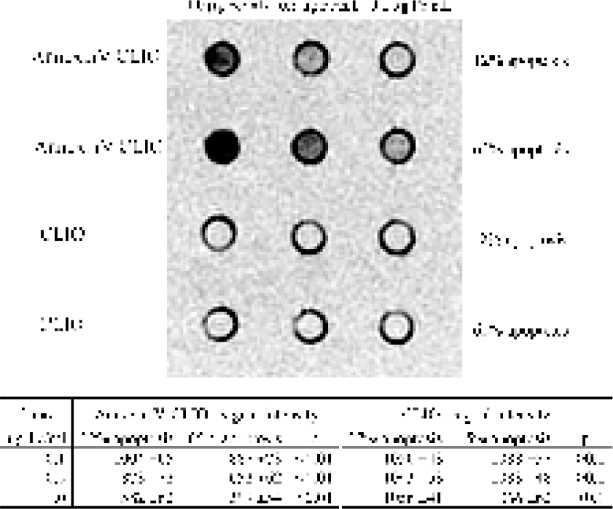
MRI of apoptotic cell with annexin V–CLIO. Camptothecin-treated Jurkat T cells (65% apoptotic cells) had a significant decrease in signal intensity to non-campothecin-treated cells (12% apoptotic cells). Amino-CLIO failed to change the signal intensity of control or camptothecin-treated cells. Both nanoparticles were employed at 1, 0.5, and 0.1 μg/mL. TR = 3,000; TE = 25.

T2 relaxation times of CLIO and annexin V–CLIO for three different CLIO concentrations (see Figure 2). For camptothecin-treated cells (65% apoptotic cells) incubated with annexin V–CLIO, the T2 relaxation times are significantly shorter compared with nontreated controls (12% apoptotic cells). Nonlabeled CLIO did not shorten the T2 times significantly.

Synthesis of annexin V–CLIO. Annexin V was purified from the ACL3 Escherichia coli clone. By reaction with SATA protected SH groups were introduced and then activated with hydroxylamine. Amino-CLIO was modified with SPDP and was finally coupled with the activated annexin V.

SDS–PAGE of purified annexin V. Lane 1 shows the purified annexin V. Lane 2 shows a pure commercially available annexin V. Lane 3 shows protein size markers.
Introduction of SH Groups on Annexin V and Synthesis of Annexin V–CLIO
Sulfhydryl groups were introduced on annexin V by treatment with SATA. One milliliter of annexin V (5 mg protein, in 0.1 M sodium bicarbonate, pH 8.0) was incubated with 32 μl SATA (1 mM) for 2 hr at RT. Then, 100 μl hydroxylamine (25 mM hydroxylamine, 10 mM EDTA) was added to deprotect the SH group, and the mixture was incubated for 40 min at RT. Low-molecular-weight compounds were removed using 10-mL spin columns filled with BioGel P6 Gel Medium (BIO-RAD), in 0.1 M sodium bicarbonate, pH 8.6.
To the activated annexin V, 1.32 mL 2-pyridyl disulfide–CLIO (3.72 g Fe/l, 20 mM Na-citrate buffer, pH 8) was added and incubated overnight at 4°C. Unbound proteins were removed by using a Millipore Stirred cell 8050 (Amicon Bioseparators) with a 300-kDa cutoff membrane. The resulting nanoparticles had an average of 2.7 annexin V proteins per CLIO particle, determined by iron and protein concentration as described [17], [19].
Cell Culture
To test the ability of annexin V to selectively label apoptotic cells, the following experiments were performed. Apoptosis was induced in Jurkat T lymphoma cells (Clone E6-1, ATCC no. TIB-152) by treatment with camptothecin. The Jurkat T cells were grown in RPMI 1640 medium (Cellgro, with
Magnetic Cell Fractionation
Camptothecin-treated apoptotic Jurkat cells and untreated control cells were pooled to give a mixture containing 69% healthy and 31% apoptotic cells determined by flow cytometry. After washing and 15 min of incubation with annexin V–CLIO (1 μg Fe/mL) and annexin V FITC in BB, a part of the mixture was applied to MACS magnetic separation columns (Miltenyi Biotec). The mixture and the nonmagnetic flow through were analyzed by flow cytometry. FACS analyses were performed with a FACS Calibur (Becton Dickinson) according the manufacturer's instructions.
MRI of Cells
After inducing apoptosis with camptothecin, 4 × 10⁷ Jurkat T cells were sedimented for each tube imaged. First, cells were washed in BB. A portion of the cells was used for flow cytometry to assess the fraction of apoptotic Jurkat cells. The treated and nontreated cells for MRI were incubated for 20 min with 0.1, 0.5, and 1.0 μg Fe/mL annexin V–CLIO or unlabeled CLIO–NH2, respectively. Thereafter, samples were washed three times with BB and were finally precipitated in 250-μl tubes. The supernatant was almost completely removed except 1 mm overlay. The tubes were placed into a water bath at RT for MRI. MRI was performed with a clinical 1.5-T superconducting magnet (Signa 5.0; GE Medical Systems, Milwaukee, WI) using a 5-in. surface coil. The imaging protocol consisted of a spin-echo sequence (SE; TR 3,000 msec, variable TE 15–160 msec). The 1.5-mm imaging slice was carefully placed to avoid partial volume effects. At a field of view of 8 × 8 cm and a 256 × 256 imaging matrix, each voxel had a size of 0.146 mm3. Signal intensity (SI) measurements of cell pellets were obtained by manual placement of regions of interest (ROIs) of 1.5 mm in diameter in the center of the cell pellets. Errors are the standard deviation of the pixels measured using ImageJ software from NIH. ROIs were also placed on the surrounding water to determine the overall homogeneity of the image. To obtain T2 values, signal intensities were plotted against the 12 different TEs, which showed the differences in T2 decay time of each pellet as described. Curve fitting for exponential decay allowed the calculation of T2 relaxation times (SI = A × e(−TE/T2) + B, where SI = signal intensity, TE = echo time, A = amplitude, and B = offset) for each incubation concentration of the conjugate. T2 relaxation times were computed from 12 different TEs (15–160 msec).
Results
Here, we report on the development of a magnetic nanoparticle achieved by conjugating annexin V to amine-functionalized magnetic nanoparticles (amino-CLIO). Annexin V–CLIO had an average of 2.7 annexin V proteins per CLIO particle, based on measurements of the protein and iron concentrations. The average particle size was 52.9 ± 1.0 nm, measured by light scattering. The relaxivities were R1 = 21.3 (mM s)−1 and R2 = 45.3 (mM s)−1 (0.47 T, 38 C).
The functionality of the annexin V–CLIO nanoparticle was verified by its ability to magnetically separate apoptotic from healthy cells. Before separation, a mixture of Jurkat T cells containing 69% healthy and 31% apoptotic cells [determined by fluorescence-activated cell sorter (FACS) analysis, Figure 1] was incubated with annexin V–CLIO and annexin V–FITC. After application to magnetic separation columns, the apoptotic cells were almost completely removed from the nonmagnetic flow through (eluate: 0.81% apoptotic cells; retained: > 99% apoptotic cells, Figure 1).
To test the ability of annexin V–CLIO to detect apoptotic cells by MRI, a phantom MRI experiment was performed as shown in Figure 2, which shows one of the 12 MR images (TR/TE of 3,000/25) used to estimate T2. Untreated control Jurkat T cells (ca. 12% apoptotic cells) and camptothecin-treated cells (ca. 65% apoptotic cells) were incubated with either annexin V–CLIO or with unlabeled CLIO (1.0, 0.5, and 0.1 μg Fe/mL BB). Cells were washed, precipitated, and then used for MRI. For all concentrations of annexin V–CLIO, a stronger decrease in signal intensity resulted for camptothecin-treated samples compared to untreated cells using a range of T2-weighted imaging sequences (Figure 2). Nonlabeled CLIO failed to cause a considerable decrease in the signal intensity of both camptothecin-treated and nontreated samples.
Incubation with nonlabeled CLIO did not notably decrease the T2 relaxation times for apoptotic and control cells in any concentration (nontreated control samples had slightly lower T2 times—higher uptake or binding). In contrast, a substantial drop of T2 relaxation times could be observed for the samples incubated with annexin V–CLIO. For all annexin V–CLIO concentrations, the T2 relaxation times of the camptothecin-treated samples (65% apoptotic cells) were substantial shorter compared with the non-camptothecin-treated controls (12% apoptotic cells). All T2 data points were derived from curve fits out of 12 different TEs with R2 values greater than .95. We have shown that T2 determinations by MRI have coefficients of variation of less than 5% [11].
Discussion
One of the major ways of determining apoptosis is achieved through the use of proteins, like annexin V, which bind to phosphatidylserine, a lipid that becomes externalized as one of the early events in apoptosis [6]. Annexin V was first purified from placenta [24], but it can now be expressed and purified from bacteria [25]. FITC-labeled annexin V is widely used for quantifying apoptotic cells by FACS analysis or visualizing apoptotic cells by microscopy.
To show the ability of annexin V–CLIO to act as an MR contrast agent, a phantom experiment with Jurkat T cell pellets was performed (Figures 2 and 3). The incubation of cells with annexin V–CLIO caused a signal decrease, which was sufficient to distinguish between cell pellets containing 12% (not camptothecin treated) versus 65% (camptothecin treated) apoptotic cells even at a nanoparticle concentration as low as 0.1 μg Fe/mL (about 1.8 μM Fe). This concentration was about 100 times lower than a synaptotagmin I nanoparticle that was used to label cells in vitro in a similar fashion [12]. In contrast, unmodified amino-CLIO did not cause a substantial signal decrease in all concentrations. Moreover, the non-camptothecin-treated cells had slightly lower T2 relaxation times, which could be due to a higher uptake activity of nonapoptotic cells.
We describe the conjugation of annexin V to amino-CLIO. Since the only naturally occurring cysteine in annexin V is chemically unreactive [26], we introduced an additional SH group by the reaction of the protein with N-succinimidyl S-acetylthioacetate (SATA) following treatment with hydroxylamine (Figure 4). In the future, additional control of the site of reaction between annexin V and the nanoparticle maybe achieved through the use of annexin V where site-directed mutagenesis has produced a single N-terminal reactive cysteine distal to the phosphatidylserine binding site [26].
In conclusion, an annexin V–CLIO nanoparticle was synthesized and shown to be useful in separating apoptotic cells from healthy cells. By MRI, annexin V–CLIO allowed the identification of cell suspensions containing a high percentage of apoptotic cells. Annexin V–CLIO, which is potent at very low doses of iron in vitro, may prove to be a useful MR contrast agent for imaging apoptosis in vivo.
Footnotes
Acknowledgments
We acknowledge Hye Won Kang, Anna Moore, and Nikolai Sergeyev for helpful discussions.