
Abstract
Background:
Indirect inguinal hernias are fundamentally the result of failure of closure of the processus vaginalis (PPV). In the laparoscopic era, there have been many reports describing the internal view of inguinal hernias and contralateral patent PPV. However, there has been no report of describing a duplicated patent PPV.
Objective:
This report describes an anatomical variant of the internal ring in a child diagnosed with a bilateral duplicated PPV who presented with a right-side indirect inguinal hernia.
Case:
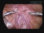
A 16-month-old male presented with an incarcerated right inguinal hernia. Manual reduction was effective, and 2 days later, laparoscopic hernia repair was performed. Under general anesthesia, a 5-mm optical port was inserted into the abdomen and a 30° 5-mm camera was introduced. The other two 3-mm instruments were placed along the lateral border of the rectus muscle. In the laparoscopic view, the edematous and thickened internal ring was found showing evidence of incarceration on the right side. Another hole was found close to the internal ring, which had the same direction and a shared wall of the inguinal canal. On the contralateral side, there was also a duplicated patent PPV. For hernia repair, the right-side internal ring was completely divided at the internal ring level and ligated intracorporeally. The duplicated PPV on the contralateral side was also repaired using the same method.
Results:
The operative time was 28 minutes and there were no complications. The patient was followed up for 24 months, during which time, there was no recurrence.
Conclusions:
Laparoscopic hernia repair in children has the additional benefit of enabling the accurate diagnosis of a hernia and the detection of any unexpected anatomical variants.
No competing financial interests exist.
Runtime of video: 3 mins 23 secs


Get full access to this article
View all access options for this article.
Supplementary Material
Please find the following supplemental material available below.
For Open Access articles published under a Creative Commons License, all supplemental material carries the same license as the article it is associated with.
For non-Open Access articles published, all supplemental material carries a non-exclusive license, and permission requests for re-use of supplemental material or any part of supplemental material shall be sent directly to the copyright owner as specified in the copyright notice associated with the article.