
Abstract
Introduction:
Femoral hernia (FH) is extremely rare in children, comprising 0.1%–0.9% of all inguinal region hernia repairs, as reported in the prelaparoscopic era. 1,2 In centers where inguinal hernia (IH) is treated laparoscopically, FH was diagnosed in 1.1%–2.8%, 1,2 suggesting a higher actual prevalence. Many cases are diagnosed perioperatively or discovered only when an IH recurrence occurs. 3 We propose a laparoscopic approach to close the femoral ring, according to McVay's principles. 4
Materials and Methods:
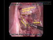
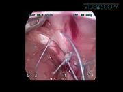
A 4-year-old boy was referred to our service for IH. His parents and the pediatrician observed a recurring bulging in the right inguinal region. On examination, the bulging appeared below and laterally to the pubic tubercle while the boy was straining, but quickly reduced spontaneously. The child was scheduled for a laparoscopic reparative operation of a FH. A 5-mm 30° scope placed in the umbilicus, two 3.5-mm trocars placed in both flanks, and CO2 insufflation at 8 mm Hg were used. On inspection, the right lower abdominal wall seemed normal. The deep inguinal ring was closed, excluding an IH. The region medially to the external iliac vein (EIV) seemed undisturbed but receded easily with light pressure, causing a visible bulging externally, exactly where the hernia was observed. The peritoneum covering the receding area and the preperitoneal fatty tissue were removed using monopolar hook and blunt dissection. A triangular space was revealed, limited laterally by the EIV, superior-medially by the transversus abdominis and the conjoint tendon, and inferior-medially by the rim of the superior pubic ramus covered by Cooper's ligament. We placed three nonabsorbable braided 2-0 sutures, with 16-mm needle, approximating the transversus and conjoint tendon margin to Cooper's ligament. The opening was completely obliterated. The peritoneum was closed with continuous sutures. The anatomy on the contralateral side was normal. The operating time was 50 minutes.
Results and Conclusions:
Recovery from surgery was uneventful. The child was discharged on the first postoperative day. Follow-up clinical examinations at 1 week, 1 month, and 6 months postoperatively revealed no pathology and the child resumed normal activities. The laparoscopic approach allows a more accurate diagnosis of FH and a direct repair of the femoral ring.
No competing financial interests exist.
Runtime of video: 3 mins 51 secs

Keywords
Get full access to this article
View all access options for this article.
References
Supplementary Material
Please find the following supplemental material available below.
For Open Access articles published under a Creative Commons License, all supplemental material carries the same license as the article it is associated with.
For non-Open Access articles published, all supplemental material carries a non-exclusive license, and permission requests for re-use of supplemental material or any part of supplemental material shall be sent directly to the copyright owner as specified in the copyright notice associated with the article.