
Abstract
Introduction:
Near infrared fluorescence imaging with indocyanine green (ICG) is an adjunct technology to facilitate intraoperative assessment of renal vascularization and tissue perfusion. We present our surgical technique of robot-assisted partial nephrectomy (RAPN) with selective arterial clamping using near infrared fluorescence imaging.
Materials and Methods:
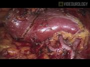
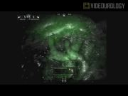
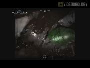
A 37-year-old male presented with a 4 cm right lower pole renal mass (RENAL nephrometry score 8a). Near infrared fluorescence imaging was used to aid in selective arterial clamping during RAPN. The procedure is performed with a standardized technique using the Da Vinci surgical system with the integrated Firefly™ Fluorescence imaging. After the main renal artery, vein, and renal tumor have been identified, defatted, and the margins scored, the hilar microdissection is performed. Selective clamping of higher order tumor-feeding artery/ies is carried out with mini Bulldog clamps. ICG (5–7.5 mg) is injected intravenously to assess the renal vasculature and adequacy of tumor ischemia before resection, essentially performing an intraoperative renal arteriogram. Areas of tissue perfusion fluoresce green when viewed under near infrared light, while areas of ischemia appear dark. After ischemia is confirmed, the renal mass is excised with cold sheers. The resection bed is closed with a combination of a running barbed suture, TissueLink™, Floseal®, and a sliding weck clip renorrhaphy technique. An additional dose of ICG is then given to evaluate perfusion of renal parenchyma after completion of the renorrhaphy.
Results:
The total operative time was 145 minutes, estimated blood loss was 75 mL, and warm ischemia time was 18 minutes. There were no intraoperative or postoperative complications. The patient was discharged home after a 1.5-day hospital stay. Pathological analysis demonstrated a pT1a papillary type I renal cell carcinoma, Fuhrman grade 2, with negative surgical margins. Discharge creatinine was 1.1 mg/dL (preoperative 1.0 mg/dL) and eGFR was 92 mL/minute/1.73 m3 (preoperative 102 mL/minute/1.73 m3). In our entire RAPN with selective arterial clamping using near infrared fluorescence imaging experience, we have noted that large tumors that cross Brodel's line or in cases of complex hilar tumors, the selective clamping approach involves clamping both the posterior and anterior arterial branches. However, this technique appears most successful in tumors that are entirely anterior or posterior to the Brodel's line or are hilar in location. We advise that robotic surgeons near the beginning of the learning curve of near infrared fluorescence imaging-enhanced RAPN start by clamping secondary arterial branches and moving up to higher order branches as he or she becomes more comfortable.
Conclusion:
Our technique of RAPN with selective arterial clamping utilizing near infrared imaging is presented. This technology provides real-time intraoperative arteriogram to confirm selective ischemia. Further studies are needed to confirm our findings and show whether its use can improve postoperative renal functional outcomes.
Runtime of video: 6 mins 46 secs
Keywords
Get full access to this article
View all access options for this article.