
Abstract
Introduction:
There are currently no good real-time methods to measure renal ischemic damage during hilar clamping. In this video we describe a novel technique for monitoring renal ischemia during hilar clamping using near-infrared tissue oximetry.
Materials and Methods:
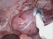
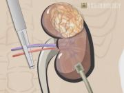
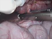
After obtaining approval from the Institutional Animal Care and Use Committee, we performed continuous tissue oxygen measurements on six renal units of Yorkshire swine per protocol. Pneumoperitoneum to 10 mm Hg was achieved, three trocars were placed, and the kidney was mobilized laparoscopically. The ViOptix T.Ox™ Tissue Oximeter (ViOptix Inc., Fremont, CA) probe was secured to the kidney using a harness fashioned from umbilical tape. A clamp was used to occlude the renal artery and vein. Local tissue oxygen saturation measurements were obtained (up to a depth of 10 mm) and recorded at 4-sec intervals at baseline, during hilar clamping, and for 5 min after unclamping the renal vasculature. Clamp times and sequence were varied in each renal unit.
Results:
Near-infrared tissue oximetry reflected baseline tissue oxygen saturation levels followed by an ischemic drop levels during hilar clamping and then a return to pre-clamp levels.
Conclusions:
This video demonstrates a novel technique for monitoring renal ischemia during hilar clamping in a porcine model. Tissue oximetry may be a useful tool for assessing intraoperative renal ischemia during partial nephrectomy. Further studies are necessary to determine if local tissue oximetry correlates with long-term renal function.
No competing financial interests exist.
Runtime of video: 4 mins
Get full access to this article
View all access options for this article.