
Abstract
Introduction:
Laparoscopic nephron-sparing surgery has been practiced for management of different renal pathologies. 1 Pelvicaliceal disruptions encountered during this exercise have been conventionally tackled by separate calicorrhaphy apart from renorrhaphy. We present a video demonstration of our technique of laparoscopic heminephrectomy without separate calicorrhaphy.
Methods:
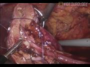
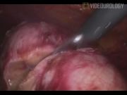
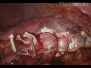
Patients were evaluated in detail, including presenting complaints and blood profile. Computed tomography scan was performed to define the tumor pathology. Patients were planned for laparoscopic extirpation through transperitoneal access. Renal mobilization was carried out following the plane between Gerotas' fat and the renal capsule except around the tumor where a generous cuff of peritumoral fat was preserved. Hilar dissection was conducted with delineation of the renal pedicle. We practiced nonselective hilar clamping. To ensure tumor extirpation with an adequate surgical margin, a decision of heminephrectomy was taken. The line of resection was mapped with hook electrocautery. Hilar clamping was then undertaken. Heminephrectomy was performed following the predemarcated line of resection. The remnant renal bed was fulgurated using laparoscopic spatula and monopolar electrocautery. Full-thickness single-layer renorrhaphy was conducted using No. 1-0 polyglactin suture in a continuous fashion. Tension in suture line was maintained by applying sequential Hem-o-lok clips. No separate tissue sealant or surgicel bolster was employed. Despite that major pelvicaliceal disruptions were discernible, no separate calicorrhaphy was undertaken. No additional ureteral catheter or stenting was undertaken. Specimen was retrieved in entrapment bag. Operative and postoperative events were recorded. Ultrasonography evaluation was carried out immediately and 1 week after drain removal to rule out any significant urinoma. Follow-up contrast imaging was performed to assess the pelvicaliceal integrity.
Results and Discussion:
Between January 2009 and January 2010, six cases of laparoscopic heminephrectomy were performed using the same technique. Mean age was 42.67 years. Mean body–mass index was 23.4 kg/m2. Two patients were women and four men. Mean tumor size was 4.8 cm. All neoplasms were predominantly endophytic. Left lower half involvement was observed in two cases, left upper half in one, right upper half in one, and right lower half in two cases. Mean warm ischemia time, mean blood loss, and mean surgical duration was 23.67 minutes, 266.67 mL, and 191.67 minutes, respectively. All patients recovered uneventfully. Mean time to drain removal was 3.3 days. Mean time to discharge was 4.2 days. No patients reported any delayed bleeding or urinoma. Histopathology revealed clear cell renal malignancy in all. Negative surgical margins were appraised in all (100%). Mean follow-up duration was 9.67 months. Till last follow-up, all patients were disease free. Follow-up imaging demonstrated satisfactory function of the remnant renal unit with preserved pelvicaliceal integrity in all. Single-layer tension renorrhaphy provides durable hemostasis and pelvicaliceal control. Separate pelvicalicorrhaphy portends the risk of prolongation of the warm ischemia incurred during nephron sparing surgery, and this may be conveniently avoided by this technique. Ureteral stent- or catheter-related morbidity could also be avoided in these situations.
No competing financial interests exist.
Runtime of video: 7 mins 7 secs
Get full access to this article
View all access options for this article.