
Abstract
Abstract
Introduction:
Paragangliomas are commonly found in the skull base/neck region and abdomen. We present a unique case of paraganglioma found in the paraspinous thoracic region and method of safe resection.
Methods:
Patient data and information were collected using electronic medical record system and intraoperative video was edited using iMovie.
Results:
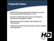
The patient is a 26-year-old woman who presented with a left pheochromocytoma to an outside institution but had persistent catecholamine excess after resection. Further work-up with genetic testing and GA-68 DOTATATE/PET was performed. She was discovered to have a succinate dehydrogenase D mutation. Imaging revealed multiple skull base tumors and a thoracic paraganglioma. Since the thoracic tumor was thought to be the most likely to be catecholamine secreting, it was targeted for resection. She was blocked preoperatively with doxazosin and then underwent video-assisted thoracoscopic resection.
Conclusions:
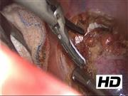
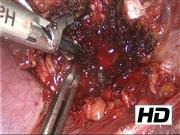
Technically, paragangliomas are removed similar to schwannomas through ensuring separation of the blood supply before manipulation to minimize risk to catecholamine release into systemic circulation. We review the comprehensive laboratory and imaging work-up for a patient with a hereditary paraganglioma-pheochromocytoma syndrome. Our technique for a minimally invasive thoracic paraganglioma resection is highlighted.
No competing financial interests exist
.
Runtime of video: 9 mins 24 secs
Patient Consent: The investigations were performed after approval by a local human investigations committee or institutional review board, and after obtaining informed consent from a patient (or other responsible individuals).
Get full access to this article
View all access options for this article.