
Abstract
Introduction:
Thyroid cancer is the most common endocrine cancer, and its incidence is increasing worldwide over the past three decades. Prognosis for early stage differentiated cancers is good, but poorly differentiated/anaplastic cancers portend a poor prognosis. 1 Most effective mouse models developed to study thyroid cancer involve xenograft models (injecting human thyroid cancer cell lines into an immunocompromised mouse) or genetically engineered models. 2 Our goal is to develop a minimally invasive syngeneic mouse model by injecting mouse generated thyroid cancer cell lines using ultrasound guidance into the thyroid of immunocompetent mice. 3 This type of model would more accurately represent the human condition and could allow us to study disease progression and novel therapeutics.
Materials and Methods:
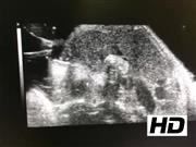
Animal protocols have been approved by the Institutional Animal Care and Use Committee at our institution. We utilized 16 mice, B6 strain, from Jackson laboratories. We planned to inject mouse thyroid cancer cell lines in a set of immunocompetent mice. The mice would receive the TBP3743 cell line (PDTC/ATC, BrafV600E/WT and p53−/−), 4 at a concentration of 1000 cells per 5 μL or 10,000 cells per 5 μL. We used the Vevo 2100 high-frequency animal ultrasound machine for imaging. The mice underwent inhalational anesthesia with isoflurane in oxygen and were placed on the rail system. Hair was removed from the neck using a chemical depilatory cream, warmed gel was placed, and the ultrasound probe (MS550S linear array transducer) was lowered into position to observe the thyroid. Using a Hamilton syringe with a 30 gauge needle, the cells were injected into the right thyroid lobe under direct ultrasound observation. The mice were monitored weekly by clinical examination (recording mouse weights and visual examination for neck mass) and neck ultrasound to monitor for tumor growth.
Results:
Six of eight mice in the TBP3743 10,000 cell group exhibited tumor growth within 14 days after injection. In five of the mice, the tumor was growing in the right thyroid lobe. In one mouse, the tumor was growing lateral to the thyroid in the sternocleidomastoid muscle. The remaining two mice had no signs of tumor growth. Seven of eight mice in the TBP3743 1,000 cell group exhibited tumor growth within 4 weeks after injection. One mouse had no evidence of tumor growth. Tumor growth was defined as growth of any mass in or near the thyroid lobe in the right neck. Tumor location was ultimately confirmed during thyroid lobectomy and/or necropsy.
Conclusions:
We have effectively performed orthotopic injections of the TBP3743 cell line through ultrasound guidance in a small group of mice. Our ultimate goal is to use this model to study disease progression by performing thyroid lobectomy and external beam radiation on these mice to study locoregional recurrence and metastatic spread, and then study other potential treatment options (novel drug treatments, thyroid hormone suppression, etc.). This technique of ultrasound-guided orthotopic injections of thyroid cancer cell lines in a syngeneic mouse model represents a simple model that does not require surgery for orthotopic cancer cell injection.
No competing financial interests exist.
Funding Information: This study was supported by 1S10OD018156-01 grant award, titled “Small Animal Ultrasound Imager - Vevo 2100,” and the Mary Rossick Kern and Jerome H. Kern endowment.
Runtime of video: 5 mins 25 secs

Get full access to this article
View all access options for this article.