
Abstract
Abstract
Introduction:
The preoperative identification of parathyroid adenomas is a mandatory prerequisite to perform a minimally invasive approach to primary hyperparathyroidism (PHPT). To preoperatively localize an adenoma in case of PHPT, our strategy is to combine technetium (99mTc) sestamibi (MIBI) scintigraphy with CT scan with contrast injection and 3D reconstruction. We use the VR-Render® 3D reconstruction software (Visible Patient, Strasbourg, France), which allows for a virtual 3D observation of the cervical anatomy. However, this reconstruction does not make it possible to observe normal parathyroid glands. The suspicion of a multifocal lesion makes the minimally invasive exploration more difficult and calls for a conventional approach.
Materials and Methods:
In the given case, 99mTc-MIBI scintigraphy showed a hyperfixation in the superior right parathyroid region. The preoperative 3D reconstruction allows to identify three potential targets, including a suspected superior right parathyroid adenoma. The cervical approach is performed according to the technique described by Miccoli and colleagues 1 with the use of a 2 cm incision. The cervical exploration is made possible using the MIVAP kit (Karl Storz, Tuttlingen, Germany).
Results:
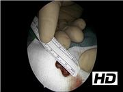
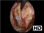
The cervical exploration is methodical and starts with the search for the right upper parathyroid gland once the inferior thyroid artery and the recurrent nerve have been identified. This gland corresponds to the MIBI scintigraphy image. The gland is easily found as it was perfectly identified preoperatively. The adenoma is resected. The right lower parathyroid gland is found in an orthotopic position. The right cervical exploration is continued anteriorly to the jugulocarotid gutter and the target is found in the shape of an anthracosic lymph node, which is resected. The exploration is pursued to the left. The recurrent nerve and the inferior thyroid artery are identified. The left parathyroid gland is identified in an orthotopic position, encapsulated in the adipose tissue forming the structure identified during 3D reconstruction. In principle, the left upper parathyroid gland is searched for to complete the cervical exploration.
Conclusion:
The minimally invasive exploration allows for the exploration of all parathyroid sites if necessary. Preoperative imaging usually helps to identify one target only. In our video, the presence of two other potential localizations shown during 3D reconstruction led us to dissect all parathyroid locations. The cervical exploration was as complete as the one performed with a conventional approach.
No competing financial interests exist.
Runtime of video: 9 mins 43 secs
Get full access to this article
View all access options for this article.