
Abstract
Humic substances (HSs) are products of biochemical transformations of plant and animal residues that make up a major fraction of the organic carbon of soil and aquatic systems in the environment. Because radioisotopes occur in the Earth's crust and because the entire biosphere is continuously exposed to cosmic radiation, ionizing radiation continually interacts with HSs. This chronic irradiation could have a significant ecological impact. However, very few publications are available that address possible consequences of chronic exposure of HSs to ionizing radiation from terrestrial and cosmic sources. This study was conducted to investigate possible impacts of exposure of HSs to ionizing radiation.
Dried humic acid (HA) or its associated aqueous solution (in 0.1 M Na2CO3) were exposed to absorbed γ-radiation in high doses of 1–90 kGy using a 60Co source. Following the γ-ray exposures, a secondary, ultraweak radiation emanation with wavelengths in the spectral range λ = 340–650 nm was recorded as a long-lived chemiluminescence (CL) from the aqueous solutions; however, the CL was not observed after irradiating dry HA.
Absorption spectra (for λ = 240–800 nm) of irradiated solutions indicated that polymerization/degradation processes were operating on the HA macromolecules. The effect of specific CL enhancers (luminol and lucigenin) on the intensity and kinetics of the CL implicated the participation of reactive oxygen species and free radicals in the CL and polymerization/degradation processes. For the range of absorbed doses used (1–10 kGy), the intensity of the induced CL was nonlinearly related to dose, suggesting that complex radical formation mechanisms were involved.
INTRODUCTION
Humic substances (HSs) are products of the biochemical transformations of plant and animal residues and they make up a major fraction of the organic carbon of soil and aquatic systems. Their reactivity in the environment primarily depends on their functional-group chemistry and macromolecular structures. However, their exact structures are unknown. The presence of aromatic and semiquinone moieties as well as an extended π-electron system makes HSs a target for all kinds of electromagnetic radiation. The HSs absorb all wavelengths associated with the solar energy spectrum and also contribute significantly to the albedo at the surface of the Earth. Both UVB and UVC light in the presence of oxygen and water gradually degrade HSs thereby having long-term ecological implications (Drozd et al., 1997; Slawinski et al., 1978; Auger and Richard, 1996; Gorski et al., 1996). The UV-induced, oxidative degradation of HSs is accompanied by a secondary radiation emanation called chemiluminescence (CL) (Slawinski et al., 1978; Gorski et al., 1996).
Degradation products of HSs can be a source of energy, carbon, and nitrogen for soil and aquatic microorganisms. CO2 released in the degradation processes probably contributes to the greenhouse effect (Drozd et al., 1997).
Probably HSs are also efficient absorbers of ionizing radiation, although, at the time of this writing, few publications are available on the possible interaction of HSs with ionizing radiation (Orlov et al., 1997; Goraczko, 2000). However, there are no data in the literature on the dose-dependent γ-radiation and induced secondary emission. These substances in the dry state are very resistant both to UV (Slawinski et al., 1978) and ionizing radiation (Goraczko and Slawinski, 2004). The only other substances that have an elemental composition, molecular weight, and physicochemical properties similar to HSs are melanins. Although their interaction with ionizing radiation is of medical interest (Lukiewicz and Ablewicz-Strzalkowska, 1974), the possible secondary CL induced by γ- or X-rays has not been investigated. Earlier studies have focused only on proteins, nucleic acids, and several polymers (Sapezhinsky et al., 1980; Sapezhinsky, 1983).
Because radioisotopes occur throughout Earth's crust and because the biosphere is under continuous exposure to cosmic radiation, ionizing radiation permanently interacts with HSs. Such long-term interactions may have ecological significance; however, there are few publications related to this topic. Here we report for the first time the observation in the visible-spectral range of ultraweak luminescence (secondary radiation) induced in basic solutions of humic acids (HAs) by high doses of γ-radiation.
In our preliminary research note (Goraczko and Slawinski, 2004), we presented results on γ-radiation-induced secondary radiation (radioluminescence) of an alkali-soluble fraction of HSs, the so-called HA. Our investigation has focused on the kinetics and spectral characteristics of this luminescence, the associated dose-response relationship, and the enhancement of the ultraweak luminescent signals by highly efficient chemiluminogenic probes. Because there is a lack of published data on this topic, we applied a broad range of doses (1–90 kGy). Although such high doses cannot be received from cosmic or terrestrial radiations, they are attainable from technical sources or in the cases of nuclear power station accidents or nuclear weapon tests.
MATERIALS AND METHODS
In our experiments, a commercial preparation of HA (Merk, Darmstadt, Germany) soluble only in alkaline solution was used. Its elemental composition was as follows: 52.96% C, 4.60% H, 2.91% N, 39.53% O, and 0.38% ash (minerals). The color coefficient Q400/600 = 5.62 (OD400 nm/OD600 nm).
Irradiation Procedure
An aerated solution of HA (200 mg HA in 1000 cm3 of 0.1 M Na2CO3) was irradiated in a glass vessel, in a RChM-γ-20 (Co-60) device (Techsnabexport, Moscow). The system was calibrated with a Fricke dosimeter. The absorbed doses were 1, 2, 5, 10, 40, 50, 60, and 90 kGy. These large absorbed doses required irradiation times from 40 min to 4 hr. Aliquots (15 ml) of irradiated solutions transferred from the stock solution were placed into liquid scintillation, Beckman vials, and the γ-radiation-induced ultraweak CL was measured in a scintillation counter (Beckman LS-100C) after each 12–15-min cycle and several days after irradiation.
The dead time between end of irradiation and first measurement was about 5 min. The LS-100C was equipped with an S-11-type photocathode PMT-sensitive in the spectral range 320–600 nm. The counting (sampling) time was 10 s. Nonirradiated empty scintillation vials, vials containing Na2CO3 + HA solutions, and vials containing Na2CO3 solutions alone were used as controls.
Spectral Measurements
We determined the intensity and spectral characteristics of the emitted γ-ray-induced secondary luminescence from both HA and the solvent Na2CO3. The following absorbed radiation doses were applied using the previously indicated γ-radiation source: 1, 2, 3, 4, and 5 kGy. After irradiation, samples were frozen and solidified in carbon dioxide. The freezing procedure was necessary to stop or slow the radical processes taking place within the irradiated samples. Just before taking spectral measurement, the samples were warmed to 15°C. The range of the luminescence spectral intensity was divided using 17 cutoff filters (Figure 1). The wavelengths of the emitted radiation spanned the range 350–700 nm.

Scheme of the SPC—cutoff measuring system: HVS, high voltage supplier; C, measurement cell; P, photomultiplier M12FQC51 (Zeiss, Germany); F, filters; PA, preamplifier; D, diaphragm; A, amplifier; AC, automatic counter C570.
Spectral distributions were determined both for irradiated HA + Na2CO3 solutions and for Na2CO3 solutions irradiated alone. The counting time was 10 s for each filter. The spectra were evaluated at two time points: immediately after defrosting of samples and again 15–20 min later.
The absorption spectra for the control and irradiated HA solutions were measured over the wavelength range 240–800 nm, using a Beckman DV-7000 spectrophotometer.
CL-Enhancing Probes
To enhance the weak CL signal and acquire information about the chemical intermediates and the mechanisms involved in the secondary emission, hydrogen peroxide and luminol (3-aminophthalhydrazide, Sigma) probes were added in 10 μM concentrations to the solutions after irradiation and to nonirradiated control solutions.
RESULTS
Luminescence Intensity and Kinetics: Absorbed Dose Dependence
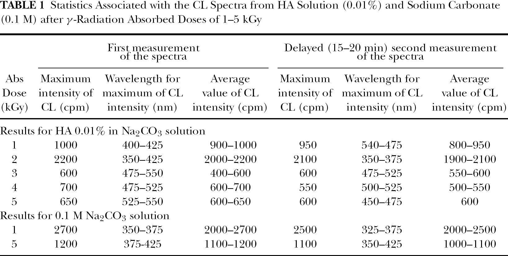
HA in dry form that was γ-irradiated with absorbed doses of 10–90 kGy did not reveal statistically significant luminescence. Only after dissolving the irradiated, dry HA in 0.1 M nonirradiated Na2CO3 aqueous solution was a trace of luminescence observed. This suggests that the deposition of high amounts of γ-radiation energy (i.e., large absorbed doses) in HA does not generate long-lived radicals or trapped electrons at concentrations high enough to produce luminescence measurable through the detection system and experimental conditions used. For nonirradiated, empty vials, and sodium carbonate, the average noise (i.e., N-value) was 28 ± 8 cps; but an average N-value of 15 ± 4 cps was observed for nonirradiated solutions of HA in 0.1 M Na2CO3, which gave a weak but distinct decaying luminescence signal lasting about 50 min (Figure 2). For 1–5 kGy absorbed γ-radiation doses, the intensity of the CL was only significantly elevated during the first 20–50 min of the kinetic processes (Figure 3). Luminescence from separate, γ-irradiated compounds is shown in Figure 4 as an example.

Kinetics of the single-photon counting rate from nonirradiated samples: (1) HA + sodium carbonate solutions; (2) sodium carbonate only; and (3) empty vial (BG, background).

CL kinetics data obtained after γ-irradiation of 0.02% HA in 0.1 M Na2CO3 solution. Absorbed doses were 1, 2, 3, 4, or 5 kGy. Time after irradiation is in minutes.

A comparison of the CL kinetics from irradiated 0.1 M Na2CO3 solution alone (curve 2), after irradiating 0.1 M Na2CO3 + 0.01% HA (curve 1), BG represents background (curve 3). The γ-radiation absorbed dose was 1 kGy. Time after irradiation is in minutes
For high doses of γ-radiation (i.e., 10–90 kGy), only flat, residual low-intensity luminescence kinetics of the irradiated system were recorded (not shown). The variable complexities of the shapes of ICL = f(D) functions suggested that different absorbed doses caused different kinetics and also that the mechanism associated with the luminescence process is likely complex.
Spectra of Luminescence
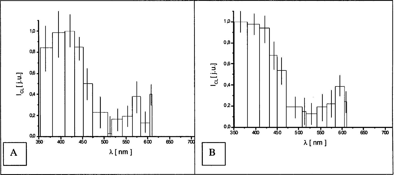
As can be seen from Table 1 and Figures 5–9, the range of luminescence-associated wavelengths related to the irradiated HA spanned the range 400–650 nm for absorbed radiation doses of 1, 2, 3, 4, and 5 kGy. The height of rectangles in Figures 5–9 gives the mean value for the relative CL intensity. The width of rectangles corresponds to the difference in wavelength between two successive filters. The length of vertical segments gives the maximum experimental error. Spectra were corrected for the spectral sensitivity of the photomultiplier, but not for the self-absorption of the solutions.
Statistics Associated with the CL Spectra from HA Solution (0.01%) and Sodium Carbonate (0.1 M) after γ-Radiation Absorbed Doses of 1–5 kGy

CL spectra of HA in Na2CO3 solution after irradiation with 1 kGy of γ-radiation: (A) first measurement immediately after defrosting; (B) second measurement taken 15 to 20 min later.

CL spectra of HA in Na2CO3 solution after irradiation with 2 kGy of γ-radiation: (A) first measurement immediately after defrosting; (B) second measurement taken 15–20 min later.

CL spectra of HA in Na2CO3 solution after irradiation with 3 kGy of γ-radiation: (A) first measurement immediately after defrosting; (B) second measurement taken 15–20 min later.

CL spectra of HA in Na2CO3 solution after irradiation with 4 kGy of γ-radiation: (A) first measurement immediately after defrosting; (B) second measurement taken 15–20 min later.

CL spectra of HA in Na2CO3 solution after irradiation with 5 kGy of γ-radiation: (A) first measurement immediately after defrosting; (B) second measurement taken 15–20 min later.
After an absorbed dose of 2 kGy, the range of the HA-associated wavelengths were in the more narrow range 350–425 nm. Similar results were also found for Na2CO3 (i.e., 350–375 nm after 1 kGy and 325–425 nm after 5 kGy). From these spectra, it can be concluded that increasing the absorbed dose caused a shift of the range of the HA emissions with longer wavelengths being involved after the higher doses. This effect was less pronounced for irradiated Na2CO3.
The emission levels of irradiated solution of Na2CO3 after 1 kGy (Figure 10A) and 5 kGy (Figure 10B) are higher than for the HA solution after irradiation with 1, 3, or 5 kGy. This nonlinear phenomenon cannot be explained at this time.
CL spectra for 0.1 M Na2CO3 solution after γ-irradiation: (A) absorbed dose was 1 kGy; (B) absorbed dose was 5 kGy.
The sodium carbonate Na2CO3 emits light in the wavelength range 350–400 nm (violet-blue range). However, the HA solutions emit light in a longer wavelength range: 400–500 nm (blue-green range).
From the spectral measurements, it appears that the luminescence level of Na2CO3 was near 2700 cpm after 1 kGy and near 1200 cpm after 5 kGy. Again, this is a nonlinear effect. For HA solution, after exposure to 1, 3, 4, and 5 kGy absorbed doses, lower luminescence intensities were observed in comparison with irradiated pure Na2CO3.
The levels of luminescence of the HA solutions were in range 600–900 cpm. The only exception was the HA irradiated by 2 kGy. This sample had the same characteristic of intensity as pure Na2CO3. Wavelengths of the emissions were in the range 350–400 nm, and the intensity of the luminescence was higher—about 2000 cpm.
To better understand the kinetics of induced emission, the delayed (15–20 min) spectra were measured, both for HA and Na2CO3 (Figures 5B–9B). After comparison of these spectra immediately after defrosting with the corresponding spectra after the indicated delay, the average shift of the maxima λmax (465–475 nm) was observed. This shift reflects changes in the luminescence intensity and spectrum with time.
Experimental uncertainties associated with the spectra generated are too large to extract the exact structure of the spectra. The most reliable endpoints therefore are spectral shifts and cutoff regions of the maximum.
Based on the data generated, we hypothesize the following schema based on the notion that primary γ-radiolysis products of an aqueous solution of Na2CO3 quickly react with HA, yielding electronically excited products X*, with simultaneous quenching of a certain fraction of the X*:

The abbreviation ROS stands for reactive oxygen species. The possible reaction
Effect of CL Enhancers after Different Times and Absorbed Doses of γ-Irradiation
The compound 5-amino-2,3-dihydrophthalazine-1,4-dione (luminol, L) is a chemiluminescent that is widely used for detecting ROS. Luminol can be oxidized by 1-e− and 2-e− oxidants:

The emitter is 3-aminophthalate (λmax = 425 nm and the quantum yield ≈0.02) (Mueller and Arnhold, 2001).
Addition of micromolar concentrations of L to irradiated solution of Na2CO3 or HA+ Na2CO3 brings about very strong CL with the signal-to-noise (S/N) ratio ≥5000. I = f(t) curves at 1–5 kGy clearly show a consecutive character of CL kinetics (Figure 11). This may suggest formation of reactive oxygen species containing higher amounts of H2O2, OH•, OOH•, 1O*2, and ROO• than superoxide anion O2−.

CL kinetics from 0.02% HA in 0.1 M Na2CO3 solution irradiated with different absorbed γ-radiation doses after adding 10 μM luminol. Numbers associated with the curves match the radiation doses (1, 1 kGy; 2, 2 kGy; 3, 3 kGy; 4, 4 kGy; 5, 5 kGy). Time after irradiation is in minutes.
From the ascending part of the I = f(t) curves, it follows that the chemiluminescent reactions are slowly developing (i.e., substrates are still found in the irradiated solutions for such reactions). A proportionality between the absorbed dose D and the initial CL intensity is also seen.
Analogous kinetic curves for 40–90 kGy doses reveal only a flat, residual CL with S/N ≅ 40 and a reciprocal relationship between D and I (not shown). The rate of radioluminescent processes is probably high enough to consume all reactants: HA, Na2CO3, and H2O/O2, and the time of irradiation necessary to achieve high doses is too long to observe all of the kinetics. A fast flow system would be required to measure these fast processes in the initial phase of irradiation. H2O2 (10 μM) does not affect the intensity of CL for all doses of irradiation.
Optical Absorbance
After γ-irradiation, HA solutions changed their color from dark brown to pale yellow. After the higher absorbed doses used, the change of color was more pronounced, which may be explained by the absorbance or transmittance at λ = 254, 400, and 600 nm (Figure 12). The basic indicators of the degree of degradation of HA are color coefficients Qi/j, where Q is the ratio of absorbance at respective wavelength i/j. The Qi/j value reflects the degree of condensation of the aromatic moieties of HA macromolecules and correlate with the chromophore and/or auxochrome concentration, molecular weight, and the intrinsic free radical concentration (Drozd et al., 1997).

Absorption spectra for the HA solution after administering γ-radiation absorbed doses of 1–5 kGy.
Calculated values of Qi/j for λ = 270, 400, and 600 nm indicate (Table 2) a degradation of long wave-length-absorbing chromophores leading to simpler aliphatic compounds. A smaller value of Qi/j observed for the lower doses and shorter irradiation times is probably related to competitive secondary reactions such as cross-linking of subunits and formation of higher polymerized products.
Changes in the Color Coefficient Qi/j of Irradiated Solutions of Humic Acid as a Function of Absorbed Dose

DISCUSSION
Natural radioisotopes occur in very small amounts in all organisms and ecosystems. The entire biosphere is under constant exposure to cosmic radiation. Small radiation doses are associated with both radiation sources. Such very low doses of ionizing radiation might have direct hormetic effects (Luckey, 1991; Goraczko, 1996). Moreover, an indirect mechanism may operate, namely Cerenkov radiation. It is known that any charged particle that moves at a speed (vn) higher than the speed of light (cn) in an environment with the refraction index (n) (i.e., β = vn/cn > 1) produces Cerenkov radiation. In the case of high-energy charged particles such as electrons from K-40 and P-32, the threshold energy E0 in water (n = 1.33) for Cerenkov radiation is 0.26 MeV. Exposure of water or aqueous solutions of biologically important compounds to cosmic or terrestrial radiation leads to excitations in the UV and visible radiation ranges in addition to Cerenkov radiation. The excitation of bacterial and yeast suspensions by β-emissions from added P-32 (producing Cerenkov radiation) results in fluorescence with a spectral distribution similar to that of mitogenetic radiation. Biologically important molecules such as tryptophan, DNA, RNA, and lysozyme are similarly excited, yielding their characteristic fluorescence (Barenboim and Domanski, 1971; Quickenden and Que Hee, 1971). These secondary radiations are delayed fluorescence or CL and can influence biochemical and physiological processes in a nonlinear way by the mechanism called “dark photobiochemistry” (Cilento, 1988). Therefore, one can speculate that the high specific energy of the secondary radiation plays an important role (Kozlov and Magaladze, 1991).
Other authors also stated that living systems exposed to γ-radiation at low doses were found to emit secondary radiation of very low intensity for an hour after cessation of γ-irradiation. Knowledge of this secondary radiation stimulated the development of biological detectors. Various biological detectors have been used used: unicellular organisms such as ciliate cells (Colpoda, Paramecia) and algae (Chlorella, Scemedesmus, Dactylococcopis), plant seeds, fresh-cut grass, and animal samples (fresh-cut wool fibers, fresh human blood, insect body, native hen egg albumen, and chicken embryos). (Kuzin, 1977; Surkenova and Kuzin, 2000) and Kozlov (Kozlov and Magaladze, 1991) hypothesize that it is the formation of the chromatin polaritons that leads to the emission of secondary bioradiation.
The results obtained in our model experiments may be relevant to the indirect stimulatory effects of low doses of γ-radiation. These effects may be limited to HA macromolecules and/or to the nearest (vicinal) environment of HA.
γ-Radiation-induced ROS and electronic excited states can initiate postradiation prolonged oxidative degradation processes. Such processes have been observed after UV irradiation of HA (Gorski et al., 1996).
The blue part of the visible CL and particularly the UV light may influence microbial and plant rhizosphere physiological processes. Depending on the dose of such radiation, one can expect stimulatory or inhibitory effects on the rate of cell division and enzymatic activity.
Of particular interest are low doses of γ-radiation, less than 1 kGy. The recorded intensity of the UV emission (λ < 400 nm) is underrated because of two factors:
HAs are much more efficient quenchers than generators of CL (Chmura and Slawinski, 1997).
At low doses of γ-irradiation, the oxidative degradation is less advanced and the absorbency of HA solution is still high enough to absorb the UV part of the secondary radiation. Because the reported spectra were not corrected for the self absorption and quenching of HA solution, the real CL intensity at λ < 400 nm has to be much higher. It is amazing that there is almost a complete lack of research on the interaction of ionizing radiation with HAs—an important ubiquitous substances. We hope our work will stimulate new research in this direction.
CONCLUSIONS
Irradiation of alkaline solutions of HA by high-energy γ-quanta initiates prolonged secondary emission (i.e., CL in the spectral region 320–600 nm). HAs in dry form irradiated with 1–90 kGy do not reveal statistically significant CL. Only doses lower than 10 kGy generate very weak short CL signals.
Combinations of substances, irradiated and nonirradiated, indicate that all components of the reaction system—H2O, O2 dissolved, Na2CO3, and HA—contribute to the observed delayed CL and its very complex mechanisms. Reactive oxygen species, especially of H2O2, OH•, and O*2−, are involved in CL as the luminol probe indicated.
γ-irradiation of HA induces oxidative degradation of their macromolecules to smaller and more acidic, hydrophilic fragments, which is shown by the increase in values of the optical indices Q260/400 and Q400/600.
One can expect that the secondary radiation (CL) is much stronger in the UV region than that recorded, since the spectra were not corrected for the self-absorption of HA solutions and the CL-quenching property of HA.